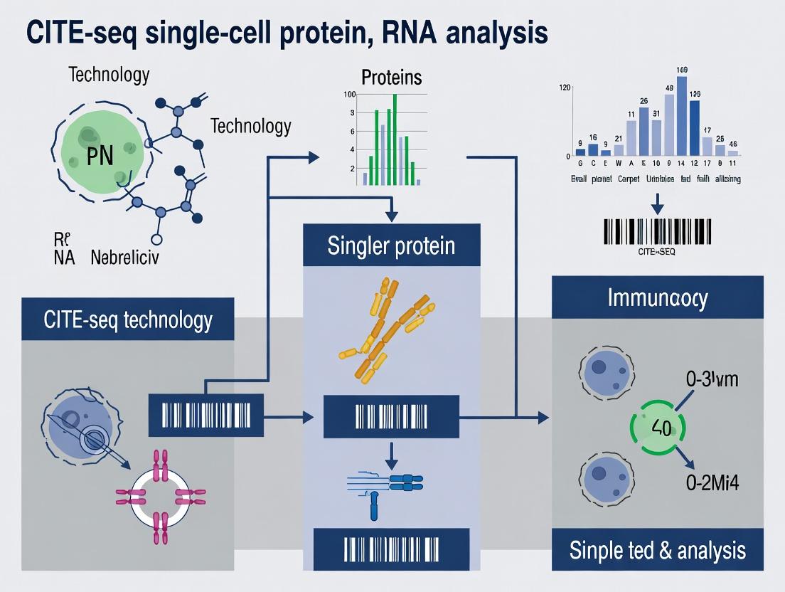
CITE-seq Demystified: A Complete Guide to Simultaneous Single-Cell RNA and Protein Profiling
This comprehensive guide for researchers, scientists, and drug development professionals explores CITE-seq (Cellular Indexing of Transcriptomes and Epitopes by Sequencing), the groundbreaking technique for simultaneous measurement of RNA and cell...
CITE-seq Demystified: A Complete Guide to Simultaneous Single-Cell RNA and Protein Profiling
Abstract
This comprehensive guide for researchers, scientists, and drug development professionals explores CITE-seq (Cellular Indexing of Transcriptomes and Epitopes by Sequencing), the groundbreaking technique for simultaneous measurement of RNA and cell surface proteins at single-cell resolution. We cover the foundational principles of how oligonucleotide-labeled antibodies bridge proteomics and transcriptomics, detail the end-to-end workflow from sample preparation to data analysis, and provide practical troubleshooting strategies. The article critically evaluates CITE-seq against other multimodal methods, discusses validation benchmarks, and highlights its transformative applications in immunology, oncology, and therapeutic development. This guide serves as a strategic resource for implementing and optimizing CITE-seq to unlock deeper insights into cellular identity and function.
What is CITE-seq? Core Principles of Multi-Omic Single-Cell Analysis
The advent of single-cell multimodal technologies, particularly CITE-seq (Cellular Indexing of Transcriptomes and Epitopes by Sequencing), has revolutionized cellular phenotyping. By simultaneously quantifying RNA expression and surface protein abundance in thousands of individual cells, researchers overcome the limitations of unimodal analysis. This integrated approach is imperative because RNA and protein levels are often discordant due to post-transcriptional regulation, differing half-lives, and technical artifacts. Simultaneous measurement provides a more accurate, comprehensive, and functional view of cell identity, state, and function, which is critical for elucidating disease mechanisms, identifying novel biomarkers, and developing targeted therapies.
Application Notes: Key Insights from Multimodal Analysis
Multimodal CITE-seq experiments consistently reveal critical biological insights obscured by single-modality approaches.
Table 1: Comparative Data from a Representative CITE-seq Study in Immune Oncology
| Metric | RNA-Seq Only | CITE-seq (RNA + Protein) | Implication |
|---|---|---|---|
| Cell Type Resolution | Identified 8 major immune clusters | Identified 15 distinct immune subsets, including rare populations | Protein markers resolve transcriptionally similar but functionally distinct states. |
| Discordance Rate | N/A | ~30% of genes show poor correlation (r<0.5) with their protein product | Highlights importance of direct protein measurement for surface markers. |
| Activation State Detection | Moderate confidence based on cytokine gene expression | High confidence via CD69, HLA-DR protein co-expression | Direct protein readout confirms functional cell states more reliably. |
| Drug Target Identification | Potential targets: 12 | Prioritized, high-confidence targets: 5 | Co-expression of target RNA and protein ensures relevance for antibody-based therapies. |
Detailed Experimental Protocols
Protocol 1: CITE-seq Library Preparation (10x Genomics Workflow)
This protocol outlines the simultaneous capture of transcriptome and surface protein data from single-cell suspensions.
Key Research Reagent Solutions:
| Item | Function | Example/Note |
|---|---|---|
| TotalSeq Antibodies | Oligo-tagged antibodies for protein detection | Pool of ~200 antibodies against surface epitopes. Pre-titrate. |
| Viability Dye | Exclusion of dead cells | e.g., LIVE/DEAD Fixable Near-IR Stain. |
| Cell Staining Buffer | Buffer for antibody incubations | PBS with 0.04% BSA. |
| Single Cell 3' GEM Kit | Creates Gel Bead-In-Emulsions for barcoding | 10x Genomics v3.1. |
| Chromium Controller | Microfluidic device for single-cell partitioning | Essential hardware. |
| SPRIselect Beads | Size selection and clean-up of cDNA libraries | Beckman Coulter. |
| Index Kit | Sample indexing for multiplexing | 10x Genomics Dual Index Kit. |
Procedure:
- Cell Preparation: Generate a single-cell suspension with >90% viability in cold cell staining buffer. Count cells.
- Antibody Staining: Incubate 0.5-1 million cells with the pre-titrated TotalSeq antibody cocktail (in 50-100µL volume) for 30 minutes on ice. Protect from light.
- Wash: Wash cells twice with 1-2mL of cell staining buffer to remove unbound antibodies. Resuspend in buffer at 700-1200 cells/µL.
- GEM Generation & Barcoding: Combine cells, Master Mix, and Gel Beads on a Chromium Chip B. Run on the Chromium Controller. Within each GEM, poly-adenylated RNA and antibody-derived oligos are reverse-transcribed, each acquiring a unique cell barcode and a unique molecular identifier (UMI).
- cDNA & Library Construction: Break emulsions, recover cDNA. Amplify cDNA via PCR. The product is then split for separate library constructions:
- Gene Expression Library: Fragmented and sequenced-ready libraries are generated from the cDNA amplicon using standard Illumina adapters.
- Antibody-Derived Tag (ADT) Library: A separate PCR is performed on the cDNA amplicon using primers specific to the constant regions of the TotalSeq antibodies. This enriches the antibody-derived tags for sequencing.
- Library QC & Sequencing: Quantify libraries (Qubit, Bioanalyzer). Pool Gene Expression and ADT libraries at an appropriate molar ratio (typically 9:1) and sequence on an Illumina system. Recommended sequencing depth: 20,000-50,000 reads/cell for gene expression; 5,000-10,000 reads/cell for ADTs.
Protocol 2: Data Processing & Multimodal Analysis (Seurat Pipeline)
This protocol details the bioinformatic integration of RNA and protein data.
Procedure:
- Demultiplexing & Alignment: Use
Cell Ranger(10x Genomics) orkb-pythonto demultiplex raw sequencing data, align reads to a combined reference (transcriptome + antibody oligo sequences), and generate feature-barcode matrices. - Initial Object Creation in Seurat: Load the RNA and ADT matrices. Create a Seurat object with the RNA data, then add the ADT matrix as a second assay (
"ADT").
QC & Normalization: Filter cells based on RNA/ADT UMIs and mitochondrial percentage. Normalize assays independently:
Feature Selection & Dimensionality Reduction: Identify variable features for RNA. Scale data and run PCA on RNA assay. Use the RNA PCA to find neighbors and construct a shared multimodal nearest-neighbor graph.
- Clustering & Visualization: Perform graph-based clustering on the multimodal neighbor graph. Run UMAP for visualization, which will now be informed by both RNA and protein data.
- Integrated Analysis: Identify differentially expressed features (genes or proteins) across clusters. Visualize protein expression on RNA-derived clusters (and vice-versa) to validate and refine population definitions.
Visualizations
CITE-seq Experimental Workflow
Multimodal Data Analysis Pipeline
Application Notes
CITE-seq enables the simultaneous quantification of single-cell transcriptomes and surface protein abundance, revolutionizing multimodal single-cell analysis. This technology bridges a critical gap in immunology, oncology, and drug development by linking gene expression with functional protein markers.
Key Applications:
- Comprehensive Immune Profiling: Precisely define immune cell states and subsets (e.g., memory T cells, activated B cells) by correlating transcriptomic signatures with canonical protein markers (CD3, CD19, CD45RA).
- Cancer Microenvironment Analysis: Decipher tumor-immune interactions by characterizing malignant cells (via intracellular transcriptomes) and the surrounding immune infiltrate (via surface proteins like PD-1, CTLA-4).
- Drug Mechanism of Action: Assess the impact of therapeutic candidates (e.g., checkpoint inhibitors, CAR-T therapies) on both the transcriptional program and surface proteome of target cells in preclinical models.
- Cell Surface Biomarker Discovery: Identify novel protein markers associated with specific transcriptional states, accelerating target identification for diagnostic and therapeutic development.
Quantitative Performance Metrics: Recent benchmarking studies (2023-2024) provide the following typical performance data for CITE-seq experiments:
Table 1: Typical CITE-seq Performance Metrics
| Metric | Typical Range | Notes |
|---|---|---|
| Cells Recovered | 5,000 - 20,000 per lane (10x Genomics) | Depends on cell viability and loading concentration. |
| Antibodies per Panel | 20 - 200+ | Larger panels require more extensive titration and compensation. |
| Reads per Cell (RNA) | 20,000 - 50,000 | Sufficient for robust transcriptome detection. |
| Reads per Cell (ADT) | 5,000 - 20,000 | Higher reads improve sensitivity for low-abundance proteins. |
| Background Signal (ADT) | 1-5% of cell hashing/multiplexing | Minimized by thorough antibody cleanup and buffer optimization. |
| Multiplexing Capacity | 8-16 samples (with CellPlex/Hashtags) | Enables experimental pooling, reducing batch effects and costs. |
Detailed Experimental Protocol
Protocol: CITE-seq Library Preparation for Single-Cell RNA and Surface Protein
Principle: Cells are first labeled with a panel of monoclonal antibodies conjugated to DNA oligonucleotides (Antibody-Derived Tags, ADTs). The labeled cells are then co-encapsulated with barcoded beads in microfluidic droplets, where both cellular mRNA and antibody-associated ADTs are reverse-transcribed, incorporating a shared cellular barcode. Separate libraries for gene expression (GEX) and surface protein (ADT) are prepared from the same cDNA pool.
I. Pre-Experiment Preparation: Antibody Conjugate Panel
- Antibody Titration: Titrate each antibody-oligo conjugate on relevant cell lines or primary cells. Use a serial dilution (e.g., 1:50 to 1:1600) to determine the optimal signal-to-noise ratio.
- Panel Balancing: Combine titrated antibodies into a master mix. The final concentration of each antibody should be near its saturating concentration as determined by titration.
- Antibody Cleanup (Critical): Remove unbound oligos using a size-exclusion filter (e.g., 100 kDa MWCO). Resuspend in cell staining buffer (PBS + 0.5% BSA + 2mM EDTA).
II. Cell Staining and Preparation
- Cell Harvest & Viability: Harvest cells, wash twice in cold PBS + 0.5% BSA. Assess viability (>90% is ideal). Count cells.
- Fc Receptor Blocking: Incubate cells with Fc block (human/mouse) in staining buffer for 10 minutes on ice.
- Antibody Labeling: Centrifuge cells, resuspend in the prepared CITE-seq antibody panel master mix. Incubate for 30 minutes on ice, protected from light.
- Washing: Wash cells 3x with ample cold staining buffer to remove unbound antibodies.
- Final Resuspension: Resuspend the stained, washed cell pellet in cold PBS + 0.5% BSA. Pass through a 35-70 µm cell strainer. Keep on ice until loading. Target concentration: 700-1200 cells/µL.
III. Single-Cell Partitioning & Library Construction (10x Genomics Platform)
- Follow the manufacturer's protocol for the Chromium Next GEM Single Cell 5' v3 kit, which captures the 5' end of transcripts and is compatible with feature barcoding (ADTs).
- Critical Step: During the master mix preparation, include the Feature Barcode reagents that will amplify the ADT sequences.
- Load the stained cell suspension onto the chip for partitioning.
- After GEM generation and RT, the recovered cDNA will contain both gene expression and ADT sequences, share the same cellular barcode.
IV. Library Amplification & Sequencing
- cDNA Amplification: Amplify cDNA per kit instructions.
- Library Split: The amplified cDNA is used as input for two separate library constructions:
- Gene Expression Library: Follow standard fragmentation, size selection, and sample index PCR.
- ADT Library: Perform a separate PCR using primers specific to the constant region of the ADT oligonucleotides and the P5/P7 flow cell adapters. Use 8-12 PCR cycles.
- Library Quantification & Pooling: Quantify both libraries by qPCR or bioanalyzer. Pool the GEX and ADT libraries at an optimal molar ratio. Typical starting ratios range from 9:1 (GEX:ADT) to 4:1, but this must be empirically adjusted based on the panel size and desired read depth.
- Sequencing: Run on an Illumina sequencer. Recommended sequencing depths: ≥20,000 GEX reads/cell and ≥5,000 ADT reads/cell.
Visualizations
Title: CITE-seq Experimental Workflow
Title: Computational Analysis Pipeline
The Scientist's Toolkit: Essential Research Reagents & Materials
Table 2: Key Reagents and Solutions for CITE-seq
| Item | Function | Key Consideration |
|---|---|---|
| Antibody-Oligo Conjugates | Target-specific detection of surface proteins. Commercially available (BioLegend, BD) or custom-conjugated. | Require extensive titration and panel balancing to minimize background. |
| Cell Staining Buffer (PBS + 0.5% BSA + 2mM EDTA) | Preserves cell viability, reduces non-specific antibody binding during staining and washes. | Must be nuclease-free and cold. |
| Fc Receptor Blocking Reagent | Blocks non-specific antibody binding to Fc receptors on immune cells. | Species-specific (e.g., human TruStain FcX). |
| Single-Cell 5' Kit w/ Feature Barcoding (10x Genomics) | Provides all reagents for partitioning, RT, and library prep for both RNA and ADTs. | Must use the 5' kit, not the 3', to capture ADT sequences. |
| Size-Exclusion Filters (100 kDa MWCO) | Critical for removing unbound oligos from the antibody cocktail post-cleanup. | Reduces background signal dramatically. |
| Single-Cell Barcoded Beads | Deliver cell barcode, UMI, and RT primers to each droplet. | Part of the commercial kit. Quality control is essential. |
| SPRIselect Beads (Beckman Coulter) | For post-amplification cDNA and library size selection and clean-up. | Ratios are critical for optimal size selection. |
| High-Sensitivity DNA Assay (e.g., Qubit, Bioanalyzer) | Accurate quantification of cDNA and final libraries prior to sequencing. | Essential for determining optimal GEX:ADT library pooling ratios. |
| Cell Multiplexing Oligos (e.g., CellPlex, Hashtags) | Allow sample pooling prior to partitioning, reducing batch effects and cost. | Require separate antibody staining and optimization. |
Within the CITE-seq (Cellular Indexing of Transcriptomes and Epitopes by Sequencing) framework, oligonucleotide-tagged antibodies enable the simultaneous quantification of cell surface protein expression and transcriptome profiling at single-cell resolution. This technology conjugates monoclonal antibodies to DNA barcodes, which are co-detected alongside cellular mRNAs via next-generation sequencing. This application note details the underlying principles, protocols, and critical reagents for implementing this core technology.
Mechanism of Action
Oligonucleotide-tagged antibodies bind to specific cell surface antigens via their Fab regions. The conjugated DNA tag, typically containing a PCR handle, a unique barcode sequence, and a poly(A) tail, is then released, captured, and reverse-transcribed. The resulting cDNA is amplified and sequenced in parallel with cellular cDNA derived from mRNA, allowing for digital counting of both protein and RNA molecules from the same cell.
Signaling Pathway and Workflow
Diagram 1: CITE-seq Antibody Binding & Detection Workflow
Key Quantitative Data
Table 1: Typical Performance Metrics for CITE-seq Experiments
| Parameter | Typical Range | Notes |
|---|---|---|
| Number of Antibodies per Panel | 10 - 200+ | Limited by barcode diversity and spectral overlap. |
| Oligo Tag Length | 60 - 120 bp | Includes constant regions and unique barcode. |
| Recommended Cell Input | 5,000 - 100,000 cells | Optimized for 10x Genomics platforms. |
| Antibody Staining Concentration | 0.25 - 2 µg/mL | Must be titrated per antibody. |
| Sequencing Saturation (Protein) | > 80% | Often higher than RNA due to lower diversity. |
| Background Signal (Negative Control) | < 0.1% | Defined by isotype control antibody counts. |
| Correlation with Flow Cytometry (r) | 0.85 - 0.99 | Validates protein detection accuracy. |
Detailed Experimental Protocols
Protocol 1: Conjugation of Antibodies with Oligonucleotide Tags
This protocol is for in-house conjugation of purified monoclonal antibodies.
Materials: Purified antibody (non-lyophilized, 0.5-1 mg/mL), SM(PEG)24 crosslinker (Thermo), Reduced oligo (5' Thiol-C6-S-S), Zeba Spin Desalting Columns (7K MWCO), PBS (no azide).
Method:
- Antibody Reduction: Dialyze 100 µg of antibody into conjugation buffer (PBS, pH 7.2). Add 100-fold molar excess of Tris(2-carboxyethyl)phosphine (TCEP) and incubate at 37°C for 2h to reduce inter-chain disulfides.
- Desalting: Pass reduced antibody through a desalting column equilibrated with PBS to remove TCEP.
- Oligo Activation: Reduce the disulfide bond on the thiol-modified oligo using a 10-fold molar excess of TCEP for 1h at room temperature. Purify using a NAP-5 column.
- Conjugation: Mix reduced antibody and activated oligo at a 1:10 molar ratio. Add SM(PEG)24 crosslinker (50-fold molar excess over antibody). Incubate overnight at 4°C with gentle rotation.
- Purification: Purify the conjugate using size-exclusion HPLC or FPLC to separate conjugated antibody from free oligo and crosslinker. Aliquot and store at 4°C.
Protocol 2: Cell Staining for CITE-seq
This protocol precedes single-cell RNA-seq library preparation on platforms like 10x Genomics.
Materials: Single-cell suspension, Fc Receptor Blocking Solution (Human TruStain FcX), Cell Staining Buffer (CSB: PBS + 0.5% BSA + 2mM EDTA), Oligo-tagged antibody cocktail, Hashtag antibody (optional).
Method:
- Cell Preparation: Wash cells twice with cold CSB. Count and assess viability (>90% recommended).
- Fc Block: Resuspend up to 1x10^6 cells in 100 µL CSB containing Fc block. Incubate on ice for 10 minutes.
- Antibody Staining: Add pre-titrated, pooled oligo-tagged antibody cocktail. Final volume: 100-200 µL. Incubate on ice for 30 minutes, protected from light.
- Washing: Wash cells 3 times with 2 mL of cold CSB. Centrifuge at 300-500 rcf for 5 min at 4°C.
- Resuspension: Resuspend stained cell pellet in the appropriate volume of CSB for target cell loading concentration (e.g., 1000 cells/µL). Keep on ice until loading onto the single-cell platform.
- Proceed immediately with the standard single-cell RNA-seq protocol (e.g., 10x 3' v3.1). The oligo tags will be co-captured with polyadenylated mRNA.
Protocol 3: Data Analysis Workflow for Protein Counts
Diagram 2: CITE-seq Data Processing Pipeline
Method:
- Barcode Counting: Use tools like
CITE-seq-CountorCell Ranger(v7.0+) with a custom reference containing antibody barcode sequences. Input: R1 (cell+UMI) and R2 (antibody barcode) FASTQ files. - Quality Control: Filter out cells based on total RNA counts, protein counts, and percentage of counts from negative control antibodies.
- Normalization: Apply centered log-ratio (CLR) transformation to the protein-derived antibody tag count matrix:
clr(x) = ln[x_i / g(x)], whereg(x)is the geometric mean of counts for that cell. - Integrated Analysis: Use the normalized protein expression as an additional modality in standard single-cell analysis pipelines (e.g., Seurat's
FindClusterson a weighted nearest neighbor graph combining RNA and protein).
The Scientist's Toolkit
Table 2: Essential Research Reagent Solutions for CITE-seq
| Item | Function & Rationale |
|---|---|
| Purified Monoclonal Antibodies | High-affinity, carrier-protein-free antibodies are essential for efficient, specific oligo conjugation and staining. |
| Custom DNA Oligonucleotides | Contain a constant PCR handle, a unique barcode (6-15 nt), and a poly(dA) tail for capture/RT. Must include a thiol modification for conjugation. |
| Homobifunctional Crosslinkers (e.g., SM(PEG)n) | Covalently link reduced antibody cysteines to thiolated oligos while maintaining antibody affinity. |
| Single-Cell 3' RNA-seq Kit (e.g., 10x Genomics) | Provides the gel beads, partitioning oil, and enzymes for co-encapsulation and processing of cells, mRNA, and antibody tags. |
| Cell Hashing Antibodies (e.g., Totalseq-A/B/C) | Oligo-tagged antibodies against ubiquitous surface antigens (e.g., CD298) enable sample multiplexing and doublet detection. |
| Fc Receptor Blocking Reagent | Critical for reducing nonspecific binding of conjugated antibodies, lowering background signal. |
| Protein Normalization Controls | Include isotype control antibodies (negative) and antibodies against highly expressed proteins (positive) for data QC and normalization. |
| Data Analysis Software (Seurat, Scanpy, CITE-seq-Count) | Specialized packages for demultiplexing, normalizing, and performing integrated analysis of multimodal single-cell data. |
Within CITE-seq (Cellular Indexing of Transcriptomes and Epitopes by Sequencing), simultaneous measurement of single-cell RNA and surface protein expression is predicated on three interdependent technical pillars. This protocol details the application notes for designing antibody panels, constructing the ADT library, and integrating sequencing workflows to support a broader thesis on multi-modal single-cell analysis for drug discovery and biomarker identification.
Antibody Panel Design: Application Notes & Protocol
Core Principles
Panel design requires balancing biological goals with technical constraints. The primary objective is to select antibodies that provide maximal, non-redundant biological information on the cell types and states of interest.
Protocol: Step-by-Step Panel Design
Step 1: Define Biological Objectives
- Identify key cell populations and protein markers essential for the research thesis (e.g., immune cell profiling for oncology drug development).
- Prioritize markers that resolve overlapping clusters in transcriptomic data alone.
Step 2: Antibody Selection & Validation
- Source: Use commercially available, clone-validated TotalSeq antibodies (BioLegend) or similar conjugated products. Ensure antibodies are validated for CITE-seq.
- Validation Requirement: Confirm specificity and signal-to-noise ratio using known positive and negative control cell lines via flow cytometry prior to CITE-seq use.
- Titration: Perform small-scale titrations to determine the optimal antibody concentration (typical range: 0.5–2 µg per million cells). Aim for a staining index >5.
Step 3: Panel Size and Composition
- Size: Panels typically range from 10-200 antibodies. Consider sequencing depth and cost.
- Composition: Include "housekeeping" proteins (e.g., CD45 for immune cells) for quality control and "isotype controls" to assess non-specific binding.
- Barcode Balancing: Ensure antibody-derived tag (ADT) barcodes have balanced nucleotide composition to minimize sequencing bias. Use manufacturer-provided barcode balance information.
Step 4: Conjugation & Barcode Assignment
- If using custom conjugation, follow manufacturer protocols (e.g., from BioLegend or BD Biosciences) to attach oligonucleotide tags to purified antibodies.
- Assign barcodes from the TotalSeq library, avoiding sequence homology that could cause cross-hybridization.
Research Reagent Solutions
| Item | Function in CITE-seq |
|---|---|
| TotalSeq Antibodies | Pre-conjugated antibodies with unique DNA barcodes. Core reagent for protein detection. |
| Cell Staining Buffer | PBS-based buffer with Fc receptor blocking agent to reduce non-specific antibody binding. |
| Hashtag Antibodies | Antibodies conjugated to distinct barcodes for sample multiplexing, enabling pooled processing. |
| BSA (0.04% in PBS) | Used in washing steps to minimize cell loss and non-specific adhesion. |
| Viability Dye (e.g., LIVE/DEAD) | Distinguishes live from dead cells to prevent poor-quality data from lysed cells. |
ADT Library: Construction and Quality Control
The ADT library consists of the pooled, barcoded antibodies used in the experiment. Its construction is critical for data quality.
Protocol: ADT Library Preparation
Materials: Titrated antibody stocks, cell staining buffer, low-bind microcentrifuge tubes.
- Pool Creation: Combine each titrated, barcoded antibody into a single, master "ADT Cocktail" in a low-bind tube. Final concentration of each antibody should be at its determined optimal staining concentration.
- Aliquot and Store: Aliquot the master cocktail to avoid freeze-thaw cycles. Store at 4°C (short-term) or -80°C (long-term) with appropriate carrier protein (e.g., BSA).
- QC by Flow Cytometry: Validate the pooled cocktail's performance on a small aliquot of control cells. Compare staining patterns to individual antibody stains.
Quantitative ADT Data Metrics
Table 1: Key QC Metrics for ADT Library Performance
| Metric | Target Value | Purpose |
|---|---|---|
| Staining Index (Median) | >5 | Measures separation between positive and negative populations. |
| Background (Isotype Ctrl Signal) | < 50 UMIs | Indicates level of non-specific binding. |
| ADT Library Complexity | > 90% of antibodies detected | Ensures successful inclusion of all panel antibodies. |
| Correlation with FACS | R² > 0.85 (for known markers) | Validates protein measurement accuracy. |
Sequencing: Strategy and Data Generation
Sequencing must capture both the cDNA (RNA) and ADT (antibody) libraries, which are often prepared with distinct indices.
Protocol: Combined RNA+ADT Sequencing
Library Preparation:
- Following single-cell partitioning (10x Genomics Chromium), cDNA and ADT-derived amplicons are generated in separate PCR reactions.
- ADT Amplification: Amplify ADT library using ~15-18 PCR cycles with primers specific to the constant regions of the oligonucleotide tags.
- Library Quantification: Quantify both cDNA and ADT libraries using fluorometry (Qubit). Assess size distribution via Bioanalyzer/Tapestation.
- Pooling: Pool cDNA and ADT libraries at an optimal molar ratio. A typical starting ratio is 9:1 (RNA:ADT) by moles, but this requires optimization.
Sequencing Configuration: Table 2: Typical Sequencing Configuration for 10x Genomics 3' CITE-seq
| Library Type | Read Type | Cycles | Recommended Depth (per cell) |
|---|---|---|---|
| RNA (cDNA) | Read 1 | 28 | 20,000-50,000 reads |
| i7 Index | 10 | ||
| i5 Index | 10 | ||
| Read 2 | 90 | ||
| ADT | Read 1 | 24 | 5,000-10,000 reads |
| Custom i7 Index* | 10 | ||
| Read 2 | 20 |
*ADT libraries often use a custom sample index read (SI) in place of i5.
Integrated CITE-seq Experimental Workflow
A comprehensive protocol from cell preparation to data analysis.
Protocol: Full CITE-seq Experiment
Part A: Cell Staining with ADT Library
- Harvest and wash cells in cold cell staining buffer. Count and assess viability (>90% target).
- Fc Block: Resuspend cell pellet (up to 10^6 cells) in 50 µL buffer containing Fc block. Incubate 10 mins on ice.
- Antibody Staining: Add predetermined volume of ADT cocktail. Incubate for 30 mins on a rotator at 4°C.
- Wash: Wash cells 2-3 times with 1-2 mL of cell staining buffer. Pellet at 300-400 rcf for 5 mins.
- Resuspend in PBS + 0.04% BSA at desired concentration for single-cell platform loading.
Part B: Single-Cell Partitioning & Library Prep
- Load stained cells onto the single-cell platform (e.g., 10x Genomics Chromium) per manufacturer's instructions, targeting desired cell recovery.
- Generate cDNA and ADT libraries following the platform's protocol and the sequencing strategy above.
Part C: Data Analysis (Brief Overview)
- Demultiplexing: Use
Cell Ranger(10x) orCITE-seq-Countto generate separate feature-barcode matrices for RNA and ADT. - ADT Normalization: Apply centered log-ratio (CLR) transformation to ADT counts per cell:
clr(x) = ln[ (x_i) / g(x) ], whereg(x)is the geometric mean of ADT counts for that cell. - Integrated Analysis: Use Seurat or similar to perform WNN (Weighted Nearest Neighbor) analysis, combining RNA and protein data for clustering and visualization.
Visualizations
Cellular Indexing of Transcriptomes and Epitopes by Sequencing (CITE-seq) enables simultaneous measurement of single-cell transcriptomes and surface protein abundance. The core technological innovation is the use of oligonucleotide-tagged antibodies, known as Antibody-Derived Tags (ADTs). The primary computational output of a CITE-seq experiment is a unified cell-by-feature matrix that combines gene expression counts (from cDNA) and ADT counts (from antibody-derived oligonucleotides). This multi-modal data matrix is foundational for deriving integrated insights into cellular identity, state, and function, accelerating drug target discovery and biomarker identification in immunology and oncology.
The final analyzed data is typically represented in two key, aligned matrices. The rows represent the same set of single cells (barcodes), ensuring perfect cellular correspondence.
Table 1: Unified CITE-seq Data Output Structure
| Matrix Type | Feature Type | Measurement | Typical Dimensions (Cells x Features) | Primary Analytical Use |
|---|---|---|---|---|
| Gene Expression Matrix | mRNA transcripts | RNA-seq derived UMI counts | ~5,000-10,000 x ~15,000-30,000 | Transcriptomic clustering, differential expression, pathway analysis. |
| ADT Count Matrix | Surface proteins | Antibody-derived UMI counts | ~5,000-10,000 x ~20-200 | Protein abundance validation, cell surface phenotyping, corroborating clusters. |
| Unified Matrix (Combined) | mRNA + Protein | Normalized, co-embedded counts | ~5,000-10,000 x (Genes + ADTs) | Multi-modal dimensionality reduction (WNN, totalVI), integrated cell typing. |
Table 2: Key Preprocessing & Normalization Metrics
| Data Modality | Common Normalization Method | Typical Library Size Factor | Critical QC Metric | Purpose |
|---|---|---|---|---|
| Gene Expression (RNA) | LogNormalize (Seurat) or SCTransform | Median RNA counts per cell | % mitochondrial reads | Removes cell-to-cell technical variation, identifies stressed cells. |
| ADT Counts (Protein) | Centered Log Ratio (CLR) | Median ADT counts per cell | Staining background (neg. control) | Normalizes protein abundance independently, reduces ambient noise. |
| Integrated Data | Weighted Nearest Neighbors (WNN) | N/A | Modality weight per cell | Computationally fuses modalities for joint analysis. |
Detailed Experimental Protocol: CITE-seq Library Preparation
This protocol outlines the key steps for generating the primary output matrices, adapted from current methodologies.
Part A: Cell Staining and Barcoding
- Prepare Single-Cell Suspension: Generate a viable single-cell suspension in PBS + 0.04% BSA. Pass through a 35-40 µm cell strainer. Perform a cell count and viability assessment (e.g., Trypan Blue).
- Stain with CITE-seq Antibodies: Incubate 0.5-1 million cells with the titrated panel of DNA-barcoded antibodies (TotalSeq or similar) for 30 minutes on ice in the dark. Use a 1:100 to 1:200 initial dilution in a 50-100 µL volume.
- Wash Cells: Wash cells twice with 2 mL of PBS + 0.04% BSA to remove unbound antibodies. Centrifuge at 300-500 rcf for 5 minutes at 4°C.
- Resuspend for Partitioning: Resuspend the stained cell pellet at a target concentration of 700-1,200 cells/µL in the appropriate buffer for the chosen partitioning system (e.g., 10x Genomics Chromium).
Part B: Single-Cell Partitioning & cDNA Synthesis
- Generate Gel Bead-In-Emulsions (GEMs): Load the cell suspension, partitioning reagents, and Single Cell 3' Gel Beads (10x Genomics v3.1/v4) onto a Chromium Chip. The system co-partitions each cell with a uniquely barcoded gel bead and lysis buffer in a single oil droplet.
- Reverse Transcription & cDNA Amplification: Inside each GEM, cells are lysed, and poly-adenylated mRNA and antibody-derived oligonucleotides hybridize to the bead's poly(dT) primers. Reverse transcription creates barcoded cDNA. Post-GEM cleanup, the cDNA is amplified via PCR (12-14 cycles).
- Size Selection & Quality Control: Purify the amplified cDNA using SPRIselect beads (e.g., 0.6x-0.8x ratio). Analyze quality and yield via Bioanalyzer (Agilent) or TapeStation (Agilent).
Part C: ADT & Gene Expression Library Construction
- ADT Library Construction (Separate Indexing PCR):
- Use 10-25% of the total amplified cDNA as input for a separate PCR reaction to enrich the antibody-derived tags.
- PCR Setup: cDNA, P5 and sample index (SI) primers, TruSeq Read 2 primer, PCR mix.
- Cycling Conditions: 98°C for 45s; 10-14 cycles of (98°C for 20s, 65°C for 30s, 72°C for 20s); 72°C for 1 min.
- Clean up with SPRIselect beads (0.8x ratio).
- Gene Expression Library Construction (Fragmentation & Indexing):
- Use the remaining ~75-90% of cDNA for standard single-cell 3' gene expression library prep.
- Fragment, A-tail, ligate adaptors, and index via sample index PCR per manufacturer's protocol (e.g., 10x Genomics).
- Clean up with SPRIselect beads (0.8x ratio).
- Library QC & Sequencing:
- Quantify both libraries using qPCR (KAPA Library Quantification Kit) and assess size distribution (Bioanalyzer).
- Sequencing: Pool libraries. Sequence the ADT library with ~5,000-10,000 reads per cell (Read 1: cell barcode/UMI, i7: sample index, Read 2: ADT barcode). Sequence the Gene Expression library with standard depth (~20,000-50,000 reads/cell).
Visualization of the CITE-seq Workflow & Data Integration
Title: CITE-seq Experimental & Computational Workflow
Title: Unified CITE-seq Data Matrix Structure
The Scientist's Toolkit: Key Research Reagent Solutions
Table 3: Essential Materials for CITE-seq Experiments
| Item | Function & Role in Generating Primary Output |
|---|---|
| DNA-barcoded Antibody Panel (TotalSeq) | Core reagent. Antibodies conjugated to a unique oligonucleotide barcode, enabling protein detection via sequencing. Defines the ADT feature space. |
| Single Cell 3' GEM Kit (10x Genomics) | Provides gel beads, partitioning oil, and enzymes for cell barcoding, RT, and cDNA amplification. Generates the cell x gene expression matrix foundation. |
| Dual Index Kit (10x Genomics) | Provides unique sample indexes for multiplexing. Allows pooling of samples during library prep, sequenced separately via the i5/i7 indices. |
| SPRIselect Beads (Beckman Coulter) | For size selection and clean-up of cDNA and final libraries. Critical for removing primer dimers and optimizing library quality. |
| NextSeq 2000 P3 Reagent Kit (Illumina) | High-output sequencing kit. Provides the depth and read length required for simultaneous profiling of gene expression (150bp paired-end) and ADTs (50bp single-end). |
| Cell Staining Buffer (PBS/BSA) | Preserves cell viability and prevents non-specific antibody binding during the staining step, reducing background noise in the ADT matrix. |
| Bioanalyzer High Sensitivity DNA Kit (Agilent) | For quality control of cDNA and final libraries. Assesses fragment size distribution and confirms absence of contamination. |
| Cell Ranger (10x Genomics) & Seurat (R) | Primary software pipelines. Cell Ranger demultiplexes sequencing data and produces the initial count matrices. Seurat is the standard for downstream normalization, integration, and WNN analysis. |
CITE-seq (Cellular Indexing of Transcriptomes and Epitopes by Sequencing) is a multimodal single-cell technology that enables the simultaneous quantification of transcriptomic (RNA) and proteomic (cell surface protein) information from the same cell. This application note details its role in advancing single-cell research within drug development and immunology by providing a unified view of cellular identity and function.
Key Advantages:
- Defined Cell States: Resolves ambiguity in cell type clustering from scRNA-seq alone by integrating highly specific protein markers.
- Functional Insights: Correlates transcriptional activity (e.g., activation pathways) with surface protein expression (e.g., immune checkpoints, cytokine receptors).
- Discovery Power: Identifies novel cell subsets defined by unique RNA-protein combinations.
- Therapeutic Relevance: Directly profiles pharmacologically relevant protein targets alongside their transcriptional networks.
Quantitative Performance Metrics:
Table 1: Representative Performance Data from CITE-seq Experiments
| Metric | Typical Range | Notes |
|---|---|---|
| Cells Recovered | 5,000 - 20,000 per lane (10x Genomics) | Depends on cell viability and loading concentration. |
| Antibodies per Panel | 10 - 200+ | Limited by barcode diversity and spectral overlap. |
| Protein Detection Sensitivity | Higher than transcript detection for low-abundance targets | Antibody affinity provides strong signal amplification. |
| Transcripts per Cell | 20,000 - 100,000+ | Comparable to standard scRNA-seq workflows. |
| Data Concordance (Protein vs. RNA) | High for surface proteins; low for intracellular proteins | Validates specificity; confirms RNA-protein correlation is target-dependent. |
Experimental Protocols
Protocol 1: Core CITE-seq Workflow for PBMCs
I. Research Reagent Solutions Toolkit
Table 2: Essential Materials for CITE-seq
| Item | Function | Example (Supplier) |
|---|---|---|
| TotalSeq Antibodies | Oligo-tagged antibodies for protein detection. | TotalSeq-B/C/D (BioLegend) |
| Single-Cell 3' GEM Kit | Generves Gel Bead-In-Emulsions for barcoding. | Chromium Next GEM Kit (10x Genomics) |
| Cell Staining Buffer | Buffer for antibody staining without affecting viability. | Cell Staining Buffer (BioLegend) |
| Viability Dye | Distinguishes live/dead cells during staining. | Zombie NIR Fixable Viability Kit (BioLegend) |
| Magnetic Cell Separation Beads | For cell type enrichment/depletion. | CD4+ T Cell Isolation Kit (Miltenyi) |
| Single-Cell Compatible Lysis Buffer | Part of RT mix; lyses cells and inactivates enzymes. | Included in 10x GEM Kit |
| SPRIselect Beads | For post-cDNA amplification clean-up and size selection. | SPRIselect (Beckman Coulter) |
| Indexing Kit | Adds sample indexes for multiplexing. | Dual Index Kit TT Set A (10x Genomics) |
II. Detailed Staining and Library Preparation
A. Cell Preparation & Antibody Staining
- Harvest & Wash: Isolate PBMCs via density gradient centrifugation. Wash cells twice in cold Cell Staining Buffer.
- Viability Staining: Resuspend cell pellet (~1x10^6 cells) in 100 µL buffer. Add 1 µL of viability dye, incubate for 15 minutes at RT in the dark. Wash with 2 mL buffer.
- FC Receptor Block: Resuspend pellet in 100 µL buffer with human Fc receptor blocking reagent (optional but recommended). Incubate for 10 minutes on ice.
- Surface Protein Staining: Add pre-titrated TotalSeq antibody cocktail directly to cells. Incubate for 30 minutes on ice in the dark.
- Wash: Wash cells thoroughly 3x with 2 mL buffer to remove unbound antibodies.
- Resuspension & Counting: Resuspend in PBS + 0.04% BSA. Pass through a 35 µm strainer. Count using an automated cell counter. Adjust concentration to 700-1200 cells/µL.
B. Single-Cell Partitioning & Library Construction
- GEM Generation: Load cells, Master Mix, and Gel Beads onto a Chromium chip. Run on a Chromium Controller to generate single-cell GEMs.
- Reverse Transcription: Within each GEM, polyadenylated mRNA and antibody-derived oligos (ADTs) are captured by barcoded beads and reverse-transcribed.
- cDNA & ADT Amplification: Break emulsions. Amplify cDNA via PCR. The amplified product contains both gene expression (GEX) and ADT libraries.
- Library Separation: Perform a post-cDNA cleanup with SPRIselect beads. The supernatant contains the ADT library; the beads contain the GEX cDNA.
- GEX Library Construction: Process bead-bound cDNA per 10x protocol: fragmentation, end-repair, A-tailing, adaptor ligation, and sample index PCR.
- ADT Library Construction: To the supernatant, add a separate PCR mix with primers specific to the ADT constructs and a distinct sample index.
- Library QC & Sequencing: Pool libraries at an appropriate molar ratio (e.g., GEX:ADT = 9:1). Sequence on an Illumina platform (GEX: ~20,000 reads/cell; ADT: ~5,000 reads/cell).
Data Analysis & Integration
Protocol 2: Basic Data Processing with Seurat
- Create a Seurat Object: Load the GEX (filtered feature-barcode matrix) and ADT (count matrix) data. Initialize a Seurat object with the RNA data.
- Add ADT Assay: Add the ADT counts as a new "ADT" assay. Normalize ADT data using centered log-ratio (CLR) transformation.
- Standard RNA Analysis: Perform standard SCTransform normalization, PCA, and clustering on the RNA assay.
- Multimodal Clustering: Use the
FindMultiModalNeighborsfunction (Weighted Nearest Neighbors) to build a graph integrating PCA from RNA and CLR-transformed ADT data. Perform clustering on this integrated graph. - Visualization & Analysis: Visualize integrated clusters on UMAP. Plot canonical protein markers (e.g., CD3E-ADT, CD19-ADT) overlaid on RNA-derived clusters to validate and refine cell type annotations.
Visualization of Workflows and Pathways
Diagram 1: CITE-seq Core Workflow
Diagram 2: Multimodal Data Integration & Analysis
CITE-seq Protocol: Step-by-Step Workflow and Cutting-Edge Applications
This Application Note details the CITE-seq (Cellular Indexing of Transcriptomes and Epitopes by Sequencing) pipeline, a method for simultaneous quantification of single-cell RNA and surface protein expression. Framed within a broader thesis on multimodal single-cell analysis, this protocol enables researchers in immunology, oncology, and drug development to gain a unified view of cellular identity and function.
Key Research Reagent Solutions
Table 1: Essential Materials for CITE-seq Experiments
| Item | Function |
|---|---|
| Antibody-Derived Tags (ADTs) | Oligonucleotide-labeled antibodies that bind to specific cell surface proteins. Each tag contains a unique barcode for quantification via sequencing. |
| Single-Cell 3’ or 5’ Gene Expression Kit | Provides reagents for Gel Bead-in-emulsion (GEM) generation, reverse transcription, and cDNA amplification for transcriptome library construction. |
| Feature Barcoding Kit | Contains additives and primers for the specific amplification of ADT-derived cDNA, separate from the transcriptome-derived cDNA. |
| Cell Staining Buffer | A buffer containing Fc receptor blocking agents to reduce nonspecific antibody binding during the ADT staining step. |
| Viable Single-Cell Suspension | High-viability (>90%) cells prepared in a compatible buffer (e.g., PBS + 0.04% BSA). Cell number and quality are critical for success. |
| Dual Index Kit | Provides unique sample indices for multiplexing during the final library construction step. |
| SPRIselect Beads | Used for size selection and clean-up of cDNA and final sequencing libraries. |
| Next-Generation Sequencing Platform | Compatible with Illumina short-read sequencing (e.g., NovaSeq, NextSeq). |
Detailed Experimental Protocol
Cell Preparation and Antibody Staining
- Cell Harvest & Wash: Prepare a single-cell suspension from tissue or culture using standard dissociation protocols. Wash cells twice in cold cell staining buffer.
- Fc Block: Resuspend cell pellet in an appropriate volume of staining buffer containing a human or mouse Fc receptor block. Incubate on ice for 10 minutes.
- ADT Staining: Add a pre-titrated, validated panel of TotalSeq or similar oligonucleotide-conjugated antibodies to the cell suspension. Mix gently and incubate on ice for 30 minutes in the dark.
- Wash: Wash cells 3 times with ample cold staining buffer to remove unbound antibodies.
- Count & Resuspend: Perform a viability count. Resuspend cells at the target concentration (e.g., 700-1,200 cells/µL) in PBS containing 0.04% BSA. Filter through a 35 µm cell strainer.
Single-Cell Partitioning & Library Construction
- Load Chromium Chip: Mix stained cells with Master Mix and load onto a Chromium Next GEM Chip along with Gel Beads and Partitioning Oil according to the Chromium Next GEM Single Cell 5’ or 3’ Kit protocol.
- GEM Generation & RT: Single cells, Gel Beads (containing barcoded oligonucleotides), and reagents are co-partitioned into oil droplets (GEMs). Within each GEM, reverse transcription occurs, adding a cell-specific barcode and unique molecular identifier (UMI) to cDNA from both mRNA and antibody-derived oligonucleotides.
- Post-RT Cleanup & cDNA Amplification: Break emulsions, pool GEMs, and clean up cDNA with SPRIselect beads. Amplify cDNA via PCR.
- cDNA Size Selection: Perform a double-sided SPRIselect bead cleanup to select cDNA of the appropriate size range.
Feature Barcode Library Construction
- ADT Enrichment PCR: Perform a separate PCR reaction on an aliquot of the amplified cDNA using primers specific to the constant region of the antibody-derived tags (ADTs). This enriches the ADT-derived cDNA.
- Library Construction for ADT: Add sample index (i7) and partial adapter sequences via a second PCR. Clean up with SPRIselect beads.
- Gene Expression (GEX) Library Construction: Construct the standard gene expression library from the remaining cDNA according to the manufacturer's protocol, adding sample indices (i7 and i5).
Library Quantification, Pooling & Sequencing
- QC: Assess library quality and fragment size using a Bioanalyzer or TapeStation.
- Quantify: Precisely quantify libraries using qPCR (recommended).
- Pool Libraries: Pool the ADT and GEX libraries from the same sample at an appropriate molar ratio (typically a 1:10 to 1:20 ADT:GEX ratio). For multiplexing, pool samples based on quantified molarity.
- Sequence: Load onto an Illumina sequencer. Recommended sequencing depths are ~20,000 reads/cell for GEX and ~5,000 reads/cell for ADT data.
Data Processing & Analysis
- Demultiplexing: Use
cellranger multi(10x Genomics) orCITE-seq-Countto demultiplex samples and generate feature-barcode matrices. - Alignment & Counting: Align GEX reads to a reference genome and ADT reads to the barcode whitelist, generating UMI-count matrices for RNA and protein.
- Downstream Analysis: Import paired matrices into analysis environments (R/Seurat, Python/Scanpy) for normalization, clustering, and integrated analysis.
Table 2: Typical CITE-seq Experimental Parameters and Output Metrics
| Parameter | Typical Range / Value |
|---|---|
| Cell Input Recommendation | 5,000 - 20,000 cells per sample |
| Target Cell Recovery | 50-65% of loaded cells |
| Recommended ADT Panel Size | 10 - 200 antibodies |
| Sequencing Depth (GEX) | 20,000 - 50,000 reads per cell |
| Sequencing Depth (ADT) | 2,000 - 10,000 reads per cell |
| Median Genes per Cell | 1,000 - 3,000 (varies by cell type) |
| Median ADTs per Cell | ~90% of panel detected |
| Doublet Rate | ~0.8% per 1,000 cells loaded |
CITE-seq Experimental Workflow
CITE-seq Data Analysis Pipeline
1. Introduction In CITE-seq (Cellular Indexing of Transcriptomes and Epitopes by Sequencing) experiments, simultaneous detection of surface proteins and mRNA in single cells is achieved. The fidelity of protein detection via antibody-derived tags (ADTs) is exceptionally sensitive to sample quality. Non-viable cells exhibit increased nonspecific antibody binding and aberrant RNA profiles, leading to data artifacts. Therefore, rigorous sample preparation focused on cell viability is the critical first step for generating high-quality, multiplexed data. This protocol details the preparation and viability assessment of cell suspensions from tissues and culture for optimal CITE-seq.
2. Key Considerations & Quantitative Benchmarks Successful CITE-seq requires starting samples that meet stringent viability criteria. The table below summarizes the quantitative benchmarks for sample preparation.
Table 1: Quantitative Benchmarks for CITE-Seq Sample Preparation
| Parameter | Optimal Target | Minimum Acceptable | Measurement Method |
|---|---|---|---|
| Cell Viability | >90% | >80% | Flow cytometry (PI/DAPI), trypan blue, AO/PI stain. |
| Cell Concentration | 700-1,200 cells/µL | 500-1,500 cells/µL | Automated cell counter. |
| Debris/Doublet Rate | <10% | <15% | Flow cytometry (FSC-A/SSC-A, FSC-H/FSC-W). |
| Antibody Staining Index | >3 (Clear positive/negative separation) | >2 | Flow cytometry median fluorescence intensity (MFI) ratio. |
| RIN (RNA Integrity Number) | ≥8.5 (cultured cells) | ≥7.0 | Bioanalyzer/TapeStation (if bulk RNA checked). |
3. Detailed Protocol: Sample Preparation & Viability Staining
A. Materials: Research Reagent Solutions Table 2: Essential Reagents for Sample Preparation
| Reagent/Material | Function | Example/Notes |
|---|---|---|
| Viability Dye (e.g., Cisplatin, PI, DAPI) | Distinguishes live/dead cells for sorting or filtering. | Fixable viability dyes (cisplatin) are compatible with downstream fixation. |
| Fc Receptor Blocking Reagent | Reduces nonspecific antibody binding. | Human: Human TruStain FcX; Mouse: anti-CD16/32. |
| Cell Staining Buffer | Preserves viability during staining. | PBS with 0.5-2% BSA or FBS, 2mM EDTA. |
| DNase I | Reduces clumping in delicate samples (e.g., nuclei). | Added during tissue dissociation or resuspension. |
| RBC Lysis Buffer | Removes red blood cells from dissociated tissues. | Ammonium-Chloride-Potassium (ACK) lysis buffer. |
| 40µm Cell Strainer | Removes cell aggregates and debris. | Pre-wet with staining buffer. |
| Automated Cell Counter | Provides accurate concentration & viability. | Systems using trypan blue or AO/PI fluorescence. |
B. Step-by-Step Workflow
- Sample Harvest: Harvest cultured cells using gentle dissociation (e.g., enzyme-free dissociation buffer). For tissues, use a validated, rapid mechanical and enzymatic dissociation protocol optimized for your tissue type to minimize stress.
- Wash & Filter: Centrifuge cells (300-400 x g, 5 min, 4°C). Resuspend pellet in cold cell staining buffer. Pass through a pre-wet 40µm cell strainer.
- Count & Assess Viability: Take an aliquot for counting using an automated cell counter with AO/PI or equivalent viability stain. If viability <80%, proceed to Step 4 (Viability Enrichment).
- Viability Staining (Live-Cell Selection):
- Resuspend up to 1x10^6 cells in 100µL of cold staining buffer.
- Add recommended volume of Fc block. Incubate 10 min on ice.
- Add a fixable viability dye (e.g., 1:1000 dilution of 1mM cisplatin). Incubate for 5 min on ice, protected from light.
- Quench reaction with 5x volume of cold staining buffer. Centrifuge.
- Optional - Dead Cell Removal: If viability is suboptimal, use a magnetic dead cell removal kit per manufacturer's instructions. Alternatively, proceed to FACS.
- Fluorescence-Activated Cell Sorting (FACS) for Maximum Purity:
- Resuspend viability-stained cells in sorting buffer (PBS + 2% FBS + 1mM EDTA).
- Sort the viable (cisplatin-negative) population using a 100µm nozzle under low pressure.
- Collect sorted cells into a tube containing collection medium (staining buffer + 10% FBS).
- Final Preparation for CITE-seq:
- Count sorted/enriched cells. Adjust concentration to ~1000 cells/µL in cold staining buffer.
- Proceed immediately to antibody staining for CITE-seq.
4. Diagram: CITE-seq Sample Preparation & Viability Gating Workflow
Title: Workflow for Viable Cell Preparation in CITE-seq
5. Diagram: Impact of Viability on CITE-seq Data Quality
Title: Viability Impact on Protein & RNA Data Quality
Within the broader thesis on CITE-seq (Cellular Indexing of Transcriptomes and Epitopes by Sequencing) for single-cell multimodal analysis, Step 2 is critical. This step bridges the gap between cellular proteomics and transcriptomics by enabling the simultaneous detection of surface proteins and mRNA. The quality of antibody conjugation and the precision of titration directly determine data specificity, signal-to-noise ratio, and the validity of correlative findings between protein expression and RNA sequencing data. Imperfect staining leads to erroneous biological conclusions, undermining the integrative power of CITE-seq.
Conjugation Strategies for Oligonucleotide-Antibody Tags
The conjugation of antibodies to oligonucleotide tags (Antibody-Derived Tags, ADTs) is the cornerstone of CITE-seq. The chosen strategy impacts stability, binding efficiency, and lot-to-lot consistency.
Conjugation Chemistry Comparison
Table 1: Comparison of Common Oligonucleotide-Antibody Conjugation Strategies
| Conjugation Strategy | Chemistry Involved | Key Advantages | Key Limitations | Optimal Use Case |
|---|---|---|---|---|
| Succinimidyl Ester (NHS) - Maleimide | Amine-to-Sulfhydryl (NH2-SH) linkage. NHS ester reacts with lysine amines on antibody, maleimide reacts with thiol-modified oligo. | High efficiency, well-established protocol, good stability. | Potential interference with antibody binding site if lysines are critical. Requires reduction of antibody disulfides. | Standard conjugations for well-characterized antibodies. |
| Click Chemistry (e.g., SPAAC, CuAAC) | Strain-promoted or copper-catalyzed azide-alkyne cycloaddition. Antibody and oligo are separately modified with azide/alkyne. | Bioorthogonal, minimal interference with antibody function, high specificity. | Can be more expensive. CuAAC requires copper catalyst removal. | For sensitive antibodies or when site-specificity is paramount. |
| Enzymatic Ligation (e.g., Sortase, Transglutaminase) | Enzyme-mediated peptide/ligand transfer. Enzyme recognizes specific sequence on antibody Fc, attaches oligo with complementary motif. | Site-specific, preserves antibody activity, homogeneous conjugates. | Enzyme cost, sequence requirements may need antibody engineering. | For generating highly reproducible, clinical-grade conjugates. |
| Streptavidin-Biotin Bridge | Non-covalent high-affinity binding. Biotinylated antibody binds streptavidin-conjugated oligo. | Flexible, allows signal amplification. Very simple. | Large complex size may cause steric hindrance. Potential for non-specific binding. | For rapid pilot experiments or when direct conjugation is not feasible. |
Protocol: NHS-Maleimide Conjugation (Standard Method)
Materials:
- Purified monoclonal antibody (without carrier protein), 0.5-1 mg in PBS.
- Sulfhydryl-modified oligonucleotide (CITE-seq ADT sequence, 5' or 3' thiol).
- SATA (N-Succinimidyl S-Acetylthioacetate) or 2-Iminothiolane (Traut's Reagent).
- Sulfo-SMCC (Sulfosuccinimidyl 4-(N-maleimidomethyl)cyclohexane-1-carboxylate).
- Zeba Spin Desalting Columns, 7K MWCO.
- PD-10 Desalting Columns.
- Ellman's Reagent (DTNB) for thiol quantification.
Procedure:
- Antibody Thiolation: a. Buffer-exchange antibody into PBS (pH 7.2) using a Zeba column. b. Add a 10-20 molar excess of Traut's Reagent (for lysine amines) or use SATA (follow deacetylation protocol). Incubate 1 hour at room temperature. c. Pass reaction through a fresh Zeba column equilibrated with PBS to remove excess reagent. Use immediately.
Oligonucleotide Maleimide Activation: a. Dissolve sulfhydryl-oligo in degassed PBS. b. Add a 50-fold molar excess of Sulfo-SMCC. Incubate for 1 hour at RT, protected from light. c. Purify using a NAP-5 column equilibrated with degassed PBS.
Conjugation: a. Mix activated antibody (thiols) with maleimide-activated oligo at a 1:5 molar ratio (Ab:Oligo). b. React overnight at 4°C, with gentle agitation, under inert atmosphere.
Purification & Validation: a. Purify conjugate using size-exclusion chromatography (FPLC/SEC) or HPLC. b. Analyze by SDS-PAGE (stained for protein and nucleic acid) and HPLC to confirm conjugation efficiency (>90% desired). c. Quantify concentration via A280 (antibody) and A260 (oligo). Aliquot and store at -80°C.
Antibody Titration and Panel Validation
Titration is essential to determine the optimal antibody concentration that maximizes signal while minimizing background and non-specific binding.
Quantitative Data from Titration Experiments
Table 2: Exemplar Titration Data for a CD45-CITE-seq Antibody Conjugate
| Antibody Conjugate Conc. (ng/µL) | Median ADT Counts (Cell Population) | Signal-to-Background Ratio* | % of Cells Above Background Threshold | Recommended Use |
|---|---|---|---|---|
| 0.1 | 125 | 1.8 | 15% | Insufficient signal. |
| 0.5 | 980 | 5.2 | 65% | Suboptimal for rare populations. |
| 1.0 | 2,450 | 12.1 | 95% | Optimal working concentration. |
| 2.0 | 2,800 | 11.5 | 96% | Slight increase in background. |
| 5.0 | 3,100 | 8.3 | 97% | High background, wasted reagent. |
| FMO Control | 203 | - | - | Defines background threshold. |
*S/B = (Median Positive Pop.) / (Median FMO Control).
Protocol: Titration on a Cell Line or Primary Cells
Materials:
- Single-cell suspension (≥ 1x10^5 cells per condition).
- Titrated antibody conjugates (e.g., 0.1, 0.5, 1, 2, 5 ng/µL).
- Fc Receptor Blocking Reagent (e.g., Human TruStain FcX).
- Cell Staining Buffer (PBS + 0.5% BSA + 2mM EDTA).
- FMO (Fluorescence Minus One) control for each marker.
- Hashtag antibodies (for multiplexed titration) optional.
- Viability dye.
Procedure:
- Cell Preparation: Block cells with Fc block for 10 min on ice.
- Staining Master Mix: Prepare separate staining reactions for each concentration. Include a total cell number control and an FMO for the target antibody.
- Incubation: Add titrated antibodies to cells. Incubate for 30 min on ice, protected from light.
- Washing: Wash cells 2x with 2 mL cold cell staining buffer.
- Analysis: a. If using hashtags: Pool all titration samples and a separate aliquot of unstained cells. Proceed to CITE-seq library prep and sequencing. b. If pre-sequencing validation: Analyze by flow cytometry using a complementary fluorophore-labeled antibody against the same target (to detect the protein-bound conjugate) or via qPCR on the ADT sequence after cell lysis.
- Data Analysis: Post-sequencing, analyze ADT counts (UMIs). Plot median counts per cell vs. concentration. The optimal concentration is at the inflection point just before the signal plateaus while S/B ratio is maximal.
Visualization of Workflows and Relationships
Title: CITE-seq Antibody Conjugate Development and Validation Workflow
Title: Integrated CITE-seq Staining and Sequencing Pipeline
The Scientist's Toolkit: Research Reagent Solutions
Table 3: Essential Materials for CITE-seq Antibody Staining & Validation
| Item | Function in Protocol | Key Considerations |
|---|---|---|
| Zeba/PD-10 Desalting Columns | Rapid buffer exchange for antibodies and oligonucleotides before conjugation. | Critical for removing amines (e.g., Tris, glycine) that interfere with NHS chemistry. |
| Sulfo-SMCC / SM(PEG)n Crosslinkers | Heterobifunctional crosslinkers for NHS-Maleimide chemistry. | "Sulfo-" variants are water-soluble. PEG spacers can reduce steric hindrance. |
| Reducing Agents (TCEP, DTT) | To reduce antibody inter-chain disulfides for thiolation or to reduce oligo disulfides. | TCEP is more stable and odorless than DTT. Use in degassed buffers. |
| Fc Receptor Blocking Reagent | Blocks non-specific binding of antibodies to Fc receptors on immune cells. | Essential for reducing background with primary immune cells. Species-specific. |
| Cell Staining Buffer (BSA/EDTA) | Provides protein block, prevents cell clumping, and maintains cell viability during staining. | Must be nuclease-free for CITE-seq. EDTA helps prevent adhesion. |
| Hashtag Antibodies (TotalSeq-A/B/C) | Oligo-tagged antibodies against ubiquitous epitopes to multiplex samples. | Allows pooling pre-sequencing, reducing technical variability and cost. |
| Viability Dye (e.g., Cisplatin, DAPI) | Distinguishes live from dead cells. Dead cells cause high background. | Must be compatible with fixation (if used) and not interfere with ADT binding. |
| SPRIselect / AMPure XP Beads | For post-RT cleanup and size selection during ADT enrichment and library prep. | Critical for removing excess oligos and primers. Ratios must be optimized. |
| Nuclease-Free Water & Buffers | All solutions must be certified nuclease-free to prevent degradation of ADTs and mRNA. | Dedicated workspace and aliquots are recommended to avoid contamination. |
Application Notes
Single-cell partitioning is the critical step in CITE-seq workflows where individual cells are isolated into nanoliter-scale reaction vessels alongside uniquely barcoded beads. This enables the simultaneous capture of cellular transcripts and surface proteins. The choice of platform dictates throughput, cost, recovery efficiency, and compatibility with downstream protein detection assays.
Platform Comparison
Table 1: Quantitative Comparison of Single-Cell Partitioning Platforms
| Platform | Partitioning Method | Typical Cells/Lane/Reaction | Barcode Structure | Key Feature for CITE-seq | Approximate Cost per Cell (USD) |
|---|---|---|---|---|---|
| 10x Genomics Chromium | Microfluidics (Gel Bead-in-Emulsion) | 1,000 - 10,000 | 16bp RT + 10bp UMI + 12bp Gel Bead Barcode | High cell throughput; optimized for TotalSeq antibody libraries. | $0.40 - $0.80 |
| BD Rhapsody | Microwell array (Magnetic Bead Loading) | 1,000 - 20,000+ | 8bp Sample Tag + 8-10bp UMI + 10-12bd Bead Barcode | Flexible cell loading; compatible with AbSeq and TotalSeq. | $0.50 - $1.00 |
| Parse Biosciences Evercode | Combinatorial barcoding (in-well) | Up to 1,000,000+ | Multiple rounds of 8-12bp barcodes | Scalable to ultra-high cell numbers without partitioning hardware. | <$0.05 (at scale) |
| Takara ICELL8 | Nanowell dispensing | 192 - 1,536 (per chip) | 6bp Well ID + 8bp UMI | Low input; visual selection; suitable for fixed cells. | $2.00 - $5.00 |
| Mission Bio Tapestri | Microfluidics (DNA + Protein) | 1,000 - 10,000 | Platform-specific barcodes | Simultaneous genomic DNA (SNP) and protein (antibody) analysis. | N/A (Specialized) |
Table 2: Performance Metrics in CITE-seq Context
| Platform | Single-Cell Multiplexing Capacity (Antibody Panels) | Cell Multiplexing (Sample Multiplexing) | cDNA & Antibody-Derived Tag (ADT) Recovery Efficiency | Compatibility with Fixed/Cryopreserved Cells |
|---|---|---|---|---|
| 10x Genomics Chromium | High (>100 antibodies) | Yes (via CellPlex or MULTI-seq) | High, co-encapsulation optimized | Yes (with Fixed RNA Profiling Kit) |
| BD Rhapsody | High (>100 antibodies) | Yes (via Sample Multiplexing Kit) | High, independent bead loading | Yes (with BD Rhapsody HT Kit) |
| Parse Biosciences Evercode | Moderate to High | Yes (via Sample Tags) | Good, post-fixation compatible | Excellent (workflow designed for fixed cells) |
| Takara ICELL8 | Moderate | Limited | Moderate, depends on dispensing | Excellent (well-suited for fixed cells) |
| Mission Bio Tapestri | Targeted Protein Panels | Yes | High for targeted assays | Yes |
Experimental Protocols
Protocol 1: Single-Cell Partitioning for CITE-seq using 10x Genomics Chromium X
Principle: Cells are co-encapsulated with Gel Beads (GEMs) in a microfluidic chip. Each Gel Bead contains oligonucleotides with a cell-specific barcode, a unique molecular identifier (UMI), and a poly(dT) sequence for mRNA capture, plus a capture sequence for antibody-derived tags (ADTs).
Materials:
- Chromium Chip X
- Chromium Controller
- Single Cell 3' Reagent Kits v3.1 or v4 (with Feature Barcode technology)
- Partitioning Oil
- Suspension of viable, single cells (700-1200 cells/µL in PBS + 0.04% BSA)
- CITE-seq antibody panel (TotalSeq antibodies, titrated and validated)
- Nuclease-free water
Method:
- Cell Preparation: Label cells with TotalSeq antibodies per manufacturer's protocol. Wash thoroughly to remove unbound antibodies. Resuspend cells at 700-1200 cells/µL in PBS + 0.04% BSA. Filter through a 35µm cell strainer.
- Master Mix Preparation: On ice, prepare the Master Mix for the targeted cell recovery count (e.g., 10,000 cells). Combine:
- 67µL Reverse Transcription (RT) Reagents
- 2.1µL Reducing Agent B
- 55.9µL Nuclease-free water
- Total: 125µL
- Chip Loading: Load the Chromium Chip X in the following order:
- Channel 1: 115µL of Partitioning Oil.
- Channel 2: 110µL of the cell suspension.
- Channel 3: 115µL of Partitioning Oil.
- Channel 4: 125µL of Master Mix from Step 2.
- GEM Generation: Place the loaded chip into the Chromium Controller and run the appropriate program (e.g., "Single Cell 3' v3.1").
- Collection: Post-run, carefully retrieve the GEMs (emulsion) from the recovery tube. Proceed immediately to reverse transcription or store at 4°C for up to 72 hours.
Protocol 2: Single-Cell Capture for CITE-seq using BD Rhapsody System
Principle: Cells are loaded onto a microwell cartridge. Magnetic beads coated with barcoded oligonucleotides (for mRNA and ADT capture) are then dispensed, ideally one bead per well containing a single cell.
Materials:
- BD Rhapsody Cartridge
- BD Rhapsody Scanner
- BD Rhapsody Beads (mRNA + AbSeq/TotalSeq)
- Cell sample buffer
- Washing buffer
- Labeled cell suspension (200-600 cells/µL)
- CITE-seq antibody panel
Method:
- Cell Preparation: Label cells with CITE-seq antibodies. Wash and resuspend at 200-600 cells/µL in cell sample buffer.
- Cartridge Loading: Load 60µL of the labeled cell suspension into the sample port of the BD Rhapsody Cartridge. Incubate for 5 minutes at room temperature to allow cells to settle into microwells.
- Washing: Gently wash the cartridge with 200µL washing buffer to remove excess, un-captured cells.
- Bead Loading: Load 25µL of resuspended BD Rhapsody Beads into the bead port. Incubate for 5 minutes to allow beads to settle into wells.
- Scanning & Lysis: Place the cartridge into the BD Rhapsody Scanner. The scanner images the cartridge to assess cell and bead loading density. After scanning, load lysis buffer to lyse cells and hybridize polyadenylated RNA and antibody tags to the barcoded beads.
- Bead Recovery: Transfer the bead suspension from the cartridge to a microfuge tube. Wash beads and proceed to cDNA synthesis.
Visualization
Diagram 1: CITE-seq Partitioning & Library Construction Workflow
Diagram 2: Oligo Barcode Structure on Partitioning Beads
The Scientist's Toolkit
Table 3: Key Research Reagent Solutions for CITE-seq Partitioning
| Item | Function in CITE-seq Partitioning | Example Product/Kit |
|---|---|---|
| Viability Dye | Distinguish live from dead cells prior to partitioning, improving data quality. | 7-AAD, DAPI, Fixable Viability Dyes (e.g., Zombie NIR) |
| Barcoded Antibodies | Antibodies conjugated to oligonucleotide tags for protein detection. | BioLegend TotalSeq, BD AbSeq, Cell Signaling Technologies CITE-seq Antibodies |
| Single-Cell Partitioning Kit | Core reagents for cell barcoding, including gel beads/beads, enzymes, buffers. | 10x Genomics Chromium Next GEM Kits, BD Rhapsody Express kits |
| Cell Suspension Buffer | Preserves cell viability, prevents clumping, and ensures compatibility with microfluidics. | PBS + 0.04% BSA, 1x PBS with 1% BSA and 0.2U/µl RNase inhibitor |
| Doublet Removal Solution | Labels cell samples with lipid- or antibody-bound barcodes to identify and remove multiplet-derived artifacts. | BioLegend TotalSeq-C, 10x Genomics Feature Barcode Cell Multiplexing Kits |
| Nuclease-Free Water & Tubes | Critical for all reagent preparation to prevent degradation of oligonucleotide tags and RNA. | Ambion Nuclease-Free Water, DNA LoBind Tubes |
| High-Sensitivity Assay | Accurately quantify barcoded library concentration and size prior to sequencing. | Agilent Bioanalyzer High Sensitivity DNA assay, KAPA Library Quantification kits |
Within the CITE-seq (Cellular Indexing of Transcriptomes and Epitopes by Sequencing) workflow, the simultaneous generation of cDNA (from poly-adenylated mRNA) and ADT (Antibody-Derived Tag) libraries is critical for correlating single-cell transcriptomic and surface protein data. This step follows cell lysis and the pooled hybridization of antibody-oligo conjugates to their epitopes. Precise library preparation and sequencing strategies ensure accurate, demultiplexed data recovery for multi-modal analysis.
The preparation of cDNA and ADT libraries involves parallel but distinct enzymatic reactions and handling steps, characterized by key quantitative parameters.
Table 1: Key Quantitative Parameters for cDNA and ADT Library Preparation
| Parameter | cDNA Library | ADT Library | Notes / Rationale |
|---|---|---|---|
| Starting Material | ~10^6–10^7 enriched cDNA molecules/cell | ~10^2–10^4 ADT molecules/cell (varies by antibody panel size & abundance) | ADT counts are typically lower due to limited antibody binding sites per cell. |
| PCR Amplification Cycles | 10-14 cycles | 12-18 cycles | Higher cycles for ADTs compensate for lower starting material. Must be optimized to avoid over-cycling. |
| Typical Library Size (bp) | 300-500 bp | ~150-200 bp | cDNA includes cDNA insert + Illumina adapters. ADT library consists primarily of i5/i7 indices, cell barcode, UMI, and antibody barcode. |
| Post-Amplification Cleanup | 0.6x–0.8x SPRI bead ratio | 1.0x–1.2x SPRI bead ratio | Higher bead ratio for ADTs selects for shorter fragments, removing primer dimers and excess oligos. |
| Sequencing Read Allocation | ~80-95% of total reads | ~5-20% of total reads | Proportion varies based on protein panel size and information depth desired. Can be adjusted by pooling ratio. |
| Recommended Sequencing Depth | 20,000–50,000 reads/cell | 5,000–20,000 reads/cell (total for panel) | Dependent on biological complexity and antibody panel size. |
Table 2: Common Indexing Strategies for Multiplexing
| Index Type | Location | Purpose | cDNA Library | ADT Library |
|---|---|---|---|---|
| i7 Index | P7 adapter | Sample multiplexing (library index) | Yes | Yes |
| i5 Index | P5 adapter | Sample multiplexing (dual indexing) | Optional | Yes (often fixed) |
| Cell Barcode | Read 1 | Cell identity | 10X GemCode (16bp) | Shared from cDNA (10X GemCode) |
| UMI | Read 1 | Transcript/ADT molecule counting | 10-12 bp | 7-10 bp |
| Feature Barcode | Read 1 | Antibody identity | N/A | 6-15 bp (antibody barcode) |
Detailed Experimental Protocols
Protocol 3.1: Post-RT Cleanup and cDNA Amplification
This protocol follows reverse transcription (RT) and exonuclease I digestion of unbound RT primers.
- cDNA Purification: Add 0.6x volume of SPRISelect beads to the pooled post-RT reaction. Incubate 5 min at RT. Pellet beads on a magnet, discard supernatant.
- Wash: With beads pelleted, wash twice with 200 µL of 80% ethanol. Air dry for 1 min.
- Elute: Resuspend beads in 40 µL nuclease-free water. Incubate 2 min at RT. Pellet beads and transfer supernatant to a new tube. This is the enriched cDNA.
- PCR Amplification: Prepare the following mix:
- Enriched cDNA: 40 µL
- SI PCR Primer (10 µM): 5 µL
- 2x KAPA HiFi HotStart ReadyMix: 50 µL
- Total: 95 µL
- Thermocycler Program:
- 98°C for 45 sec
- Cycle (10-14x): 98°C for 20 sec, 63°C for 30 sec, 72°C for 1 min
- 72°C for 1 min
- Hold at 4°C.
- Cleanup: Purify the amplified cDNA with 0.6x SPRI beads, eluting in 40 µL water. Quantify by Bioanalyzer/Qubit.
Protocol 3.2: ADT Library Amplification
This protocol starts with the supernatant from the 0.6x SPRI cleanup post-RT (which contains the ADTs).
- ADT Capture: Transfer the supernatant from Protocol 3.1, Step 1 (containing ADTs) to a new tube. Add 1.2x volume of SPRISelect beads to capture the ADTs (shorter fragments). Incubate 5 min at RT.
- Wash & Elute: Pellet beads on magnet. Discard supernatant. Wash twice with 80% ethanol. Air dry. Elute ADTs in 50 µL nuclease-free water.
- PCR Amplification: Prepare the following mix:
- Eluted ADTs: 50 µL
- P5-Solo oligo (10 µM): 2.5 µL
- SI-PCR oligo (10 µM): 2.5 µL
- 2x KAPA HiFi HotStart ReadyMix: 55 µL
- Total: 110 µL
- Thermocycler Program:
- 98°C for 45 sec
- Cycle (12-18x): 98°C for 20 sec, 67°C for 30 sec, 72°C for 20 sec
- 72°C for 1 min
- Hold at 4°C.
- Cleanup: Purify the ADT library with 1.0x SPRI beads, eluting in 25 µL water. Assess fragment size (~150-200 bp) on a Bioanalyzer High Sensitivity chip.
Protocol 3.3: Library Quantification, Pooling, and Sequencing
- Quantification: Quantify both cDNA and ADT libraries using a fluorometric method (e.g., Qubit dsDNA HS Assay). Determine average fragment size (Bioanalyzer/Fragment Analyzer).
- Calculate Molarity: Use the formula:
[Library] (nM) = [Concentration (ng/µL) * 10^6] / [Size (bp) * 650]. - Pooling: Pool libraries at a molar ratio optimized for read allocation (e.g., 90:10 cDNA:ADT). A typical starting pool uses 2-4 nM of the combined library.
- Sequencing: Denature and dilute the pool per Illumina guidelines. Load on a NovaSeq 6000 (or equivalent) using the following read configuration:
- Read 1: 28 cycles (Cell Barcode + UMI for cDNA; Cell Barcode + UMI + Feature Barcode for ADTs)
- i7 Index: 10 cycles (Sample Index)
- i5 Index: 10 cycles (Sample Index, often fixed for ADTs)
- Read 2: 90-120 cycles (cDNA transcript sequence; minimal for ADTs).
Visualizations
CITE-seq cDNA and ADT Library Preparation Workflow
CITE-seq Library Sequencing Read Structure
The Scientist's Toolkit: Key Reagent Solutions
Table 3: Essential Reagents for CITE-seq Library Preparation
| Reagent / Material | Function in Protocol | Critical Notes |
|---|---|---|
| SPRISelect / AMPure XP Beads | Size-selective nucleic acid purification and cleanup. | Different ratios (0.6x, 1.0x, 1.2x) are used to selectively bind cDNA vs. shorter ADTs and remove primers. |
| KAPA HiFi HotStart ReadyMix | High-fidelity PCR amplification of cDNA and ADT libraries. | Essential for accurate, low-bias amplification with minimal errors during index PCR. |
| SI PCR Primer (for cDNA) | Primer for amplifying the cDNA library. Contains P7 and P5 primer sites. | Drives the final amplification of the cDNA library post-enrichment. |
| P5-Solo & SI-PCR Primers (for ADTs) | Primer pair for amplifying the ADT library. Adds full Illumina adapters. | P5-Solo adds the i5 index region; SI-PCR adds the P7 region and i7 index. |
| Dual Index Kit TT Set A (e.g., 10x Genomics) | Provides unique i7 and i5 index combinations for sample multiplexing. | Enables pooling of multiple libraries, reducing costs. Indices must be compatible with the sequencer. |
| Bioanalyzer High Sensitivity DNA Kit (Agilent) / Fragment Analyzer | Accurate sizing and qualitative assessment of final libraries. | Critical for verifying ADT library size (~150-200 bp) and absence of primer dimer. |
| Qubit dsDNA HS Assay Kit (Thermo Fisher) | Highly sensitive, specific quantification of double-stranded DNA libraries. | More accurate for molarity calculation than absorbance (A260), which is skewed by primers/RNA. |
| NovaSeq 6000 v1.5 Reagents (or equivalent) | Sequencing chemistry for running the pooled library. | The high output of the NovaSeq is ideal for large-scale single-cell projects. |
Application Notes
This document provides a detailed framework for the analysis of CITE-seq data, which enables the simultaneous quantification of transcriptome and surface protein expression in single cells. This integrated approach is critical for a thesis focused on deepening the understanding of cellular phenotypes, activation states, and regulatory mechanisms in immunology and oncology drug development.
Demultiplexing: Sample Identity Assignment
In multiplexed experiments where cells from multiple samples (e.g., different patients or conditions) are pooled, demultiplexing is the first computational step. It uses sample-specific Cell Hashtag Oligonucleotides (HTOs) to assign each cell barcode to its sample of origin.
Key Quantitative Metrics:
- Expected Multiplexing Level: Typically 4-16 samples per lane/channel.
- Cell Recovery Rate: Post-demultiplexing, 70-90% of high-quality cell barcodes are typically assigned to a single sample. A high doublet rate (e.g., >15%) indicates suboptimal HTO loading or washing.
- Ambient HTO Signal: The fraction of HTO counts in empty droplets/background should be minimal (<5% of total HTO library).
Table 1: Common Demultiplexing Algorithms & Performance
| Algorithm | Principle | Key Parameter | Ideal Use Case |
|---|---|---|---|
| HTODemux (Seurat) | Gaussian mixture modeling of HTO count distributions. | positive.quantile (e.g., 0.99) |
Clean data with clear positive/negative separation. |
| hashedDrops (DropletUtils) | Model-based removal of ambient HTO signal. | ambient= (null model) |
Experiments with significant ambient HTO background. |
| MultiseqDemux | Uses a non-negative least squares (NNLS) approach. | autoThresh=TRUE |
Complex backgrounds or when other methods fail. |
ADT Normalization & Background Correction
Antibody-derived tag (ADT) data suffers from significant technical noise, including non-specific antibody binding and cell-to-cell background variation. Normalization is essential to distinguish true biological signal.
A. Centered Log-Ratio (CLR) Normalization
- Protocol: For each cell, transform raw ADT counts using a pseudo-count:
clr(x) = ln[ (x + 1) / geometric_mean(x + 1) ]. This is implemented per cell across all ADT features. - Application Note: CLR normalizes within a cell, making protein expression levels comparable across features within that cell. It does not remove background staining effectively.
B. Denoised and Scaled by Background (DSB) Normalization DSB is now the community-standard method as it explicitly models and removes ambient protein noise.
- Core Thesis: The background mean (
μ) and standard deviation (σ) of each ADT in empty droplets (containing ambient mRNA) defines the technical noise component. - Key Equation:
DSB_normalized = (Cell_RAW - μ_background) / σ_background
Table 2: CLR vs. DSB Normalization Impact on Key Metrics
| Metric | Raw ADT Counts | CLR-Normalized | DSB-Normalized |
|---|---|---|---|
| Signal-to-Noise Ratio | Low | Moderate | High |
| Background Effect | High | Reduced | Minimized |
| Cell Type Separation | Poor | Good | Excellent |
| Correlation with RNA | Low | Moderate | Biologically Relevant |
Detailed DSB Protocol:
- Create Background Model: Isolate cell barcodes called as cells by RNA-based tools (e.g., EmptyDrops). The remaining barcodes are defined as "empty droplets." Calculate the mean (
μ) and standard deviation (σ) for each ADT in this empty droplet population. - Data Preprocessing: Remove cells with low total ADT counts (potential debris) and ADTs that are isotype controls or show no signal above background.
- Apply DSB Transform: For each cell and each ADT, apply the formula:
(X_cell - μ_empty) / σ_empty. - Optional Scaling: Scale values to a standard range (e.g., 0-10) using
scales::rescale()for downstream integration with RNA PCA.
Dual-Modality Integration: RNA + Protein
The power of CITE-seq lies in the joint analysis of both modalities to define a unified cellular state.
Standard Workflow Protocol:
- Independent Processing:
- RNA: Standard scRNA-seq processing (QC, normalization, scaling, PCA on highly variable genes).
- Protein: DSB-normalized ADT data.
- Weighted Nearest Neighbor (WNN) Integration (Seurat v4+):
- Construct a k-nearest neighbor graph based on RNA PCA.
- Construct a separate k-nearest neighbor graph based on ADT data (often on a cosine distance matrix).
- Learn the modality weight for each cell, which quantifies the relative information content of each modality for that cell's identity.
- Fuse these graphs into a single WNN graph that optimally represents both data types.
- Unified Analysis: Perform clustering, UMAP/t-SNE visualization, and differential expression on the WNN graph.
Table 3: Essential Research Reagent Solutions for CITE-seq
| Reagent / Material | Function in CITE-seq | Key Consideration |
|---|---|---|
| TotalSeq Antibodies | Antibody-oligo conjugates for protein detection. | Use validated panels. Titrate for optimal signal. |
| Cell Hashtag Antibodies | Sample multiplexing via unique barcoded antibodies. | Must be from same clone/species as staining panel. |
| Single Cell 3' Gel Bead Kit v3.1 | Provides primers for cDNA & ADT/HTO library generation. | Standard 10x Genomics kit. Ensure compatibility. |
| SPRIselect Beads | Size selection and clean-up for ADT/HTO libraries. | Critical for removing unbound antibody-oligos. |
| Dual Index Kit TT Set A | Provides unique sample indices for sequencing. | Essential for pooling multiple libraries. |
| Cell Staining Buffer | Buffer for antibody incubation and washes. | Must be protein-rich (BSA) to minimize NSB. |
Visualizations
CITE-seq Data Analysis Workflow from Raw Data to Integration
DSB Normalization Conceptual Diagram
Weighted Nearest Neighbor (WNN) Integration of RNA and Protein
Application Notes
CITE-seq (Cellular Indexing of Transcriptomes and Epitopes by Sequencing) enables simultaneous measurement of single-cell transcriptomes and surface protein expression. This multimodal approach is pivotal in immunology for deconvoluting complex cell populations and defining precise activation states, directly advancing therapeutic discovery.
Key Applications in Immunology:
- High-Dimensional Immune Profiling: Identifies novel cell subsets beyond traditional marker panels by correlating protein expression with transcriptional states.
- Tumor Immunology & Exhaustion: Defines exhausted T-cell subpopulations in the tumor microenvironment by co-detecting checkpoint inhibitor proteins (e.g., PD-1, CTLA-4, LAG-3) and their corresponding gene signatures.
- Vaccine Response Evaluation: Tracks antigen-specific B and T cell clonal expansion, isotype switching, and activation marker dynamics post-vaccination.
- Autoimmune Disease Pathogenesis: Discovers pathogenic cell states characterized by unique RNA-protein expression combinations (e.g., IFN-response genes with specific cytokine receptors).
- Drug Mechanism of Action: Evaluates the differential impact of immunomodulators on target cell populations at both transcriptional and proteomic levels.
Table 1: Representative CITE-seq Findings in Immunology
| Immune Context | Key Cell Population Identified | Defining Protein Markers (Antibody-Derived Tags) | Corresponding Transcriptional Signature | Reference |
|---|---|---|---|---|
| COVID-19 PBMCs | Activated CD4+ T cell subset | CD38+, HLA-DR+ | IFITM3, ISG15 high, cell cycle genes | PMID: 32514174 |
| Melanoma TME | Progenitor Exhausted CD8+ T cells | PD-1+, TCF-1+ | Tcf7, Slamf6, low cytotoxic genes | PMID: 33658719 |
| Rheumatoid Arthritis Synovium | Pathogenic TNF-α+ IL23R+ Tph cells | PD-1hi, CXCR5- | IL23R, TNF, CCL20 | PMID: 35927431 |
| Influenza Vaccination | Activating Germinal Center B cells | CD71+, CD38+ | MYC target genes, AICDA | PMID: 34789884 |
Detailed Experimental Protocols
Protocol 1: CITE-seq Library Generation from Human PBMCs
Principle: Generate barcoded single-cell RNA-seq (scRNA-seq) libraries alongside antibody-derived tag (ADT) libraries from the same cell suspension.
Materials:
- Single-cell suspension of human PBMCs (viability >90%).
- TotalSeq-B or TotalSeq-C antibody-oligo conjugates (BioLegend).
- Chromium Controller & Chip B (10x Genomics).
- Chromium Next GEM Single Cell 5' Kit v2 (10x Genomics).
- Feature Barcoding kit (10x Genomics).
- Magnetic Separator for PCR tubes.
- SPRIselect beads (Beckman Coulter).
Procedure:
A. Cell Staining & Preparation (Day 1):
- Count and pellet 0.5-1x10^6 PBMCs. Resuspend in 100µL Cell Staining Buffer (PBS + 0.04% BSA).
- Add Fc Receptor blocking reagent (optional, 10 min, 4°C).
- Prepare antibody master mix. Combine TotalSeq antibodies (0.5-2µg/mL per antibody) in Cell Staining Buffer. Centrifuge at 14,000g for 10 min before use to remove aggregates.
- Add antibody mix to cells. Incubate for 30 min on ice, protected from light.
- Wash cells 3x with 2mL Cell Staining Buffer, pelleting at 300-400g for 5 min.
- Resuspend in PBS + 0.04% BSA. Filter through a 35µm cell strainer. Count and adjust concentration to 700-1200 cells/µL.
B. GEM Generation & Library Construction (Day 1-3):
- Follow the 10x Genomics Chromium Next GEM Single Cell 5' v2 Reagent Kits User Guide.
- Load cell suspension, Gel Beads, and partitioning oil onto a Chromium Chip B. Target 10,000 cells for recovery.
- Perform GEM-RT (Gel Bead-In-Emulsion Reverse Transcription) in a thermal cycler.
- Break emulsions, recover cDNA, and perform clean-up with DynaBeads MyOne SILANE beads.
- Proceed with cDNA amplification (10-14 cycles).
- Split Amplified cDNA for Dual Library Prep:
- For Gene Expression (GEX) Library: Use ~50% of amplified cDNA. Follow standard fragmentation, end-repair, A-tailing, and adapter ligation using sample index primers.
- For ADT (CITE-seq) Library: Use ~10% of amplified cDNA. Perform a separate PCR (12-16 cycles) using the Feature Barcoding primers (SI-PCR primers) specific to the antibody-derived tags.
- Purify both libraries with SPRIselect beads (0.6x-0.8x ratio). Quantify using a Bioanalyzer (Agilent) or TapeStation.
C. Sequencing (Day 4):
- Pool GEX and ADT libraries at an optimized molar ratio (typically 9:1 GEX:ADT). Sequence on an Illumina platform.
- Recommended Sequencing Depth: 20,000-50,000 reads/cell for GEX; 5,000-10,000 reads/cell for ADT.
- Recommended Read Length: Read 1: 28 cycles (cell barcode + UMI), i7 Index: 10 cycles, i5 Index: 10 cycles, Read 2: 90 cycles (transcript/ADT).
Protocol 2: Integrated Analysis of CITE-seq Data Using Seurat
Principle: Process and integrate gene expression and antibody-derived tag counts to perform joint clustering and multimodal analysis.
Materials:
- High-performance computing environment (R ≥ 4.0).
- Raw FASTQ files from sequencing.
- Cell Ranger (10x Genomics) or kallisto | bustools pipelines.
- R packages: Seurat (v4+), ggplot2, dplyr.
Procedure:
- Demultiplexing & Counting: Use
cellranger count(Cell Ranger) with a custom reference containing both the transcriptome and the antibody oligo sequences to generate a feature-barcode matrix. Create Seurat Object: Load the feature-barcode matrix (filtered) into R.
Quality Control & Normalization:
GEX Assay: Filter cells based on nCountRNA, nFeatureRNA, and percent mitochondrial reads. Normalize using
SCTransform.ADT Assay: Center-log-ratio (CLR) normalization is recommended.
Dimensionality Reduction & Clustering:
- Run PCA on the SCT assay.
- Construct a Weighted Nearest Neighbor (WNN) graph, which integrates RNA and protein information.
Visualization & Interpretation:
- Visualize WNN UMAP and overlay protein expression.
- Visualize WNN UMAP and overlay protein expression.
Visualizations
Title: CITE-seq Experimental Workflow from Cell to Data
Title: CITE-seq Data Analysis Pipeline in Seurat
Title: Immune Cell Activation State Signaling to CITE-seq Readout
The Scientist's Toolkit: Essential CITE-seq Reagents & Materials
Table 2: Key Research Reagent Solutions for CITE-seq
| Item | Function / Purpose | Example Product / Vendor |
|---|---|---|
| Oligo-Conjugated Antibodies | Barcodes for surface protein detection via sequencing. Crucial for panel design. | TotalSeq-B/C antibodies (BioLegend), Antibody-Oligo Conjugates (BD Biosciences) |
| Single-Cell Partitioning Reagents | Enables creation of single-cell GEMs for barcoding. | Chromium Next GEM Kits (10x Genomics), Nadia (Dolomite Bio) |
| Feature Barcoding Kit | Contains primers for specifically amplifying antibody-derived tag (ADT) libraries. | Chromium Feature Barcoding Kit (10x Genomics) |
| Cell Staining Buffer | Low-protein, nuclease-free buffer for antibody staining to minimize non-specific binding. | Cell Staining Buffer (BioLegend), PBS/BSA/Azide |
| Viability Stain | Distinguishes live from dead cells prior to loading; critical for data quality. | LIVE/DEAD Fixable Viability Dyes (Thermo Fisher), 7-AAD |
| Cell Strainer | Removes cell clumps to prevent channel clogging during partitioning. | Flowmi 35µm Cell Strainers (Bel-Art) |
| SPRIselect Beads | For size-selective purification of cDNA and final libraries. | SPRIselect (Beckman Coulter), AMPure XP (Beckman Coulter) |
| High-Sensitivity DNA Assay | Accurate quantification and sizing of final sequencing libraries. | Agilent High Sensitivity DNA Kit (Agilent) |
| Dual Index Sequencing Kits | Provides unique combinatorial indexes for sample multiplexing. | Illumina Dual Indexing Kits (Illumina) |
Application Notes: CITE-seq for Profiling Therapy-Resistant Niches
Objective: To simultaneously quantify transcriptomic and cell surface proteomic data from single cells within complex tumor microenvironments (TME), enabling the identification of cellular subpopulations and signaling networks associated with therapy resistance.
Key Findings: Recent studies utilizing CITE-seq have identified specific cellular states within the TME that correlate with poor clinical outcomes. For example, a 2024 study of non-small cell lung cancer (NSCLC) patients pre- and post-immune checkpoint inhibitor (ICI) treatment revealed a expansion of a PD-L1high TIM-3+ CD8+ T cell exhaustion cluster and an S100A4+ MRC1+ macrophage population in non-responders. Quantitative data from this and related studies are summarized below.
Table 1: Key Cellular Populations Associated with Therapy Resistance in NSCLC (CITE-seq Analysis)
| Cell Type | Defining Protein Markers (CITE) | Defining Transcriptomic Signature | Frequency in Non-Responders | Fold Change vs. Responders |
|---|---|---|---|---|
| Exhausted CD8+ T Cells | CD8a+, PD-1high, TIM-3+ | HAVCR2, LAG3, ENTPD1 | 12.5% of CD45+ | 3.2x |
| Protumor Macrophages | CD14+, CD68+, MRC1+ | S100A4, VEGFA, IL10 | 18.7% of CD45+ | 4.1x |
| Resistance-Associated Fibroblasts | CD10+, CD90+, PDPN+ | FAP, ACTA2, CXCL12 | 9.3% of total cells | 2.8x |
| Therapy-Evading Tumor Cells | EpCAM+, HER2, c-MET | AXL, EGFRvIII, ALDH1A1 | 2.1% of tumor cluster | 5.5x |
Table 2: Key Signaling Pathway Activity (Inferred from RNA Data) in Resistant Niches
| Pathway | Key Upregulated Ligands/Receptors | Enrichment Score (NES) | Primary Interacting Cell Types |
|---|---|---|---|
| TGF-β Signaling | TGFB1, TGFBR2, SMAD3 | +2.15 | Fibroblasts → T cells/Macrophages |
| VEGF Angiogenesis | VEGFA, KDR, PGF | +1.98 | Macrophages → Endothelium |
| CXCL12-CXCR4 Axis | CXCL12, CXCR4 | +1.76 | Fibroblasts → Tumor cells |
| Immune Checkpoint | PDCD1, CD274, HAVCR2 | +2.34 | T cells Myeloid/Tumor cells |
Detailed Experimental Protocols
Protocol 1: CITE-seq Processing of Solid Tumor Dissociates
Objective: To generate single-cell suspensions from patient-derived tumor tissues compatible with CITE-seq library preparation.
Materials: See "The Scientist's Toolkit" below. Procedure:
- Tissue Dissociation: Mince 1-2 cm³ of fresh tumor tissue in 5 mL of cold RPMI-1640. Transfer to a gentleMACS C Tube containing the pre-mixed enzymatic cocktail (Miltenyi Biotec, Tumor Dissociation Kit, human). Process on the gentleMACS Octo Dissociator using the predefined 37ChTDK_1 program (~30 minutes).
- Cell Suspension Processing: Pass the dissociate through a 70 µm pre-separation filter. Wash with 10 mL of cold PBS + 0.04% BSA. Centrifuge at 300 x g for 5 min at 4°C.
- Red Blood Cell Lysis: Resuspend pellet in 2 mL of ACK Lysing Buffer, incubate for 2 minutes at RT. Quench with 10 mL of PBS/BSA and centrifuge.
- Debris and Dead Cell Removal: Resuspend cell pellet in 1 mL of PBS/BSA. Carefully layer over 3 mL of Lymphoprep density gradient medium. Centrifuge at 800 x g for 20 min at 20°C with brake off. Collect the interphase mononuclear cell layer.
- Viability Staining & Counting: Wash cells, count, and assess viability via Trypan Blue. Proceed only if viability >70%.
- CITE-seq Antibody Staining: Resuspend up to 1x10⁶ cells in 100 µL of PBS/BSA. Add 5-10 µL of pre-titrated TotalSeq-C antibody cocktail. Incubate for 30 min on ice in the dark. Wash twice with 2 mL of PBS/BSA.
- Single-Cell Partitioning & Library Prep: Count cells, adjust concentration to 700-1200 cells/µL, and load onto the 10x Genomics Chromium Controller per manufacturer's instructions using the Chromium Next GEM Single Cell 5' Kit v3 and the Feature Barcode technology for cell surface proteins. Generate cDNA and amplify.
- Library Construction & Sequencing: Construct separate gene expression and antibody-derived tag (ADT) libraries according to the 10x Genomics protocol. Index libraries and quantify via qPCR (Kapa Biosystems). Pool libraries and sequence on an Illumina NovaSeq 6000 using the following read configuration: Read 1: 28 cycles (cell barcode + UMI), i7 Index: 10 cycles, i5 Index: 10 cycles, Read 2: 90 cycles (transcript/ADT).
Protocol 2: Bioinformatic Analysis of CITE-seq Data for TME Deconvolution
Objective: To process raw sequencing data into an integrated cell-by-protein and cell-by-RNA matrix for downstream analysis.
Software: Cell Ranger (10x Genomics), Seurat (R), CiteFuse (R). Procedure:
- Demultiplexing & Alignment: Use
cellranger multi(v8.0+) with the pre-configured CSV file specifying paths to FASTQs, the feature reference CSV (listing antibody barcodes), and the reference transcriptome (GRCh38). This generates three feature-barcode matrices: Gene Expression (GEX), Antibody Capture (ADT), and Multiplexing Capture (HTO if used). - Primary Analysis with Seurat:
- Create a Seurat object from the GEX matrix. Filter cells with <200 genes, >20% mitochondrial reads, or >6000 genes (potential doublets).
- Create a second object from the ADT matrix and filter based on ADT UMI counts.
- Use
CiteFuse::preprocessingto normalize ADT counts using centered log-ratio (CLR) transformation and perform modality integration via Weighted Nearest Neighbor (WNN) analysis.
- Clustering & Dimensionality Reduction: Perform PCA on the integrated WNN matrix. Find neighbors (
FindNeighbors) and clusters (FindClusters, resolution=0.5-1.2). Generate UMAP embeddings for visualization. - Cell Type Annotation: Annotate clusters using canonical marker genes (e.g., CD3E, CD8A for T cells; CD14, FCGR3A for monocytes/macrophages) and corroborating ADT expression (anti-CD3, CD8, CD14 antibodies).
- Differential Analysis & Pathway Enrichment: Use
FindMarkersto identify genes and proteins differentially expressed between conditions (e.g., pre- vs. post-therapy). Perform gene set enrichment analysis (GSEA) on hallmark pathways using thefgseapackage.
Visualizations
Short Title: Cell-Cell Signaling Drives Therapy Resistance
Short Title: CITE-seq Experimental Workflow
The Scientist's Toolkit: Research Reagent Solutions
Table 3: Essential Materials for CITE-seq TME Profiling
| Item Name | Supplier Example | Function in Protocol |
|---|---|---|
| Human Tumor Dissociation Kit | Miltenyi Biotec | Optimized enzymatic cocktail for generating viable single cells from solid tumors. |
| gentleMACS Octo Dissociator | Miltenyi Biotec | Automated, standardized instrument for gentle tissue dissociation. |
| TotalSeq-C Human Antibody Cocktail | BioLegend | Pre-optimized panels of DNA-barcoded antibodies for cell surface protein tagging. |
| Chromium Next GEM Single Cell 5' Kit v3 | 10x Genomics | Reagents for GEX library preparation, including gel beads and partitioning oil. |
| Chromium Feature Barcode Kit | 10x Genomics | Reagents for capturing antibody-derived tags (ADTs) in a separate library. |
| Dual Index Kit TT Set A | 10x Genomics | Oligonucleotides for indexing libraries prior to pooling and sequencing. |
| Cell Ranger Software | 10x Genomics | Primary analysis pipeline for demultiplexing, aligning, and counting GEX/ADT features. |
| Lymphoprep | STEMCELL Technologies | Density gradient medium for removing dead cells and debris post-dissociation. |
| ACK Lysing Buffer | Thermo Fisher Scientific | Ammonium-Chloride-Potassium buffer for quick red blood cell lysis. |
Application Notes: CITE-seq for Target and Biomarker Discovery
The integration of single-cell RNA sequencing (scRNA-seq) with cellular protein detection via antibody-derived tags (CITE-seq) provides a multidimensional view of cell states, enabling the deconvolution of complex tissues and disease microenvironments. This simultaneous RNA and protein measurement at single-cell resolution is pivotal for identifying novel drug targets and validating biomarkers by correlating surface protein expression with transcriptional programs.
Key Advantages:
- Unified Functional Genomics: Links phenotypic protein markers (e.g., cell surface receptors, phosphorylated signaling proteins) with transcriptional identity and activity.
- Mechanistic Insight: Identifies cell subpopulations responsible for disease progression or drug response by correlating target protein expression with pathway activity.
- Biomarker Validation: Enables high-throughput validation of candidate biomarkers at the single-cell level within heterogeneous samples (e.g., tumor biopsies, PBMCs).
Quantitative Data Summary:
Table 1: Representative CITE-seq Study Outputs for Target Discovery
| Metric | Typical Range in Tumor Biopsy Study | Significance for Drug Discovery |
|---|---|---|
| Cells Profiled | 5,000 - 20,000 cells/sample | Identifies rare, resistant, or pathogenic subpopulations. |
| Antibody-Derived Tags (ADTs) Measured | 20 - 200 surface proteins | Direct quantification of therapeutic target prevalence. |
| Differentially Expressed Genes (DEGs) | 50 - 500 per cell cluster | Reveals pathway activation and mechanistic drivers. |
| Novel Receptor-Ligand Pairs Identified | 5 - 50 per study | Highlights new targetable interactions in the tumor microenvironment. |
Table 2: Comparison of Single-Cell Multiomic Modalities
| Technology | Measured Modalities | Primary Application in Discovery | Throughput (Cells) |
|---|---|---|---|
| CITE-seq | RNA + Surface Proteins | Phenotype-to-transcript linkage, immune profiling | 10^4 - 10^5 |
| REAP-seq | RNA + Surface Proteins | Similar to CITE-seq, alternative chemistry | 10^4 - 10^5 |
| Multiplexed scATAC-seq | Chromatin Accessibility + Surface Proteins | Linking regulome to cell surface phenotype | 10^3 - 10^4 |
Protocols
Protocol 1: CITE-seq Library Preparation from PBMCs for Biomarker Identification
Objective: To generate paired single-cell gene expression and surface protein data from human Peripheral Blood Mononuclear Cells (PBMCs) to identify cell-type-specific biomarkers.
Research Reagent Solutions:
- Viability Dye (e.g., Zombie NIR): Distinguishes live from dead cells.
- Human Fc Receptor Blocking Reagent: Reduces non-specific antibody binding.
- TotalSeq-C Antibody Panel: Oligo-tagged antibodies for protein detection (e.g., CD3, CD19, CD45, PD-1, CTLA-4).
- Single-Cell 3' GEM Kit v3.1 (10x Genomics): For partitioning, barcoding, and cDNA synthesis.
- SPRIselect Beads: For size selection and clean-up.
- P5, P7, i7, and SI-PCR Oligos: For library amplification and indexing.
Detailed Methodology:
- Cell Preparation: Isolate PBMCs via density gradient centrifugation. Count and assess viability (>90% required). Resuspend at 1x10^6 cells/mL in Cell Staining Buffer (PBS + 0.04% BSA).
- Cell Staining: a. Wash cells once with Cell Staining Buffer. b. Resuspend pellet in 100µL buffer containing Fc block and viability dye. Incubate for 10 mins on ice. c. Wash once, then resuspend in pre-titrated TotalSeq antibody cocktail (diluted in 100µL buffer). Incubate for 30 mins on ice in the dark. d. Wash cells twice with buffer, then resuspend in PBS + 0.04% BSA at a target concentration of 1,000 cells/µL.
- Single-Cell Partitioning & Library Prep: Follow the 10x Genomics Chromium Next GEM Single Cell 3' v3.1 protocol. a. Combine cells, Master Mix, and Gel Beads in a Chip G to generate GEMs. b. Reverse transcription, cDNA amplification, and quality control (Bioanalyzer) are performed per manufacturer's instructions.
- Library Construction: a. Gene Expression Library: Constructed from ~50% of the amplified cDNA using the standard protocol. b. ADT Library: Constructed from the remaining cDNA. Enrich antibody-derived tags via a custom pool of SI-PCR primers specific to the TotalSeq antibody barcodes. Perform a separate index PCR.
- Sequencing: Pool libraries at an appropriate molar ratio (typically 10:1 Gene Expression:ADT) and sequence on an Illumina NovaSeq. Recommended sequencing depth: 20,000 reads/cell for gene expression, 5,000 reads/cell for ADTs.
Protocol 2: Data Analysis Pipeline for Integrated Target Prioritization
Objective: Process raw CITE-seq data to identify cell clusters, correlate protein and RNA expression, and prioritize candidate targets.
- Preprocessing & Demultiplexing: Use
Cell Ranger(mkfastq, count) with a custom reference containing antibody barcode sequences to generate feature-barcode matrices. Quality Control & Doublet Removal: In R using
Seurat.Integrated Dimensionality Reduction & Clustering: Use Weighted Nearest Neighbor (WNN) analysis to integrate RNA and protein data.
Differential Expression & Target Prioritization: Find markers (RNA & Protein) for each cluster. Correlate ADT and RNA levels for target genes of interest. Prioritize targets that are: a) highly expressed in a disease-specific cluster, b) show concordant RNA and protein expression, and c) are linked to a survival- or pathway-relevant gene signature.
Visualizations
Title: CITE-seq Experimental Workflow
Title: PD-1 Signaling & Target Validation via CITE-seq
Title: Logic for Prioritizing Targets from CITE-seq Data
Solving Common CITE-seq Problems: Expert Tips for Data Quality
Diagnosing and Fixing High Background Noise in ADT Data
1. Introduction Within the context of a CITE-seq (Cellular Indexing of Transcriptomes and Epitopes by Sequencing) single-cell multiomics research thesis, the quality of Antibody-Derived Tag (ADT) data is paramount. High background noise in ADT counts can obscure true protein expression signals, leading to misinterpretation of cellular phenotypes and erroneous conclusions in drug development research. This application note details systematic approaches for diagnosing sources of high background and provides experimental and computational protocols for its mitigation.
2. Diagnosis: Sources of High Background Noise The table below summarizes common causes and their diagnostic signatures.
| Source of Noise | Diagnostic Signature in ADT Data |
|---|---|
| Non-specific antibody binding | High counts across many cells, particularly in isotype control channels. Correlates with cell viability (higher in dead cells). |
| Antibody aggregates | Extreme outliers (very high counts) in specific channels across a subset of cells. |
| Inadequate cell washing | Uniformly elevated background across all ADT channels. |
| Carryover of free oligonucleotides | Background present in unused ("empty") ADT channels. |
| High debris or platelet contamination | Low RNA complexity correlated with moderate ADT counts across many channels. |
| Inappropriate ADT normalization | Batch effects or sample-specific shifts in background levels after hashing/demultiplexing. |
3. Experimental Protocols for Mitigation
Protocol 3.1: Pre-staining Cell Wash and Blocking Objective: Reduce non-specific binding mediated by cellular debris and Fc receptors.
- Wash Cells: Pellet 1x10^6 cells (300 x g, 5 min, 4°C). Aspirate supernatant completely.
- Resuspend in Blocking Buffer: Use 100 µL of cold PBS + 0.04% BSA + 10% normal serum (species matching secondary antibody host) or a commercial Fc receptor blocker.
- Incubate: 10 minutes on ice.
- Proceed to Stain: Add antibody cocktail directly to the blocking buffer without washing.
Protocol 3.2: Antibody Aggregate Removal via Ultracentrifugation Objective: Remove high molecular weight aggregates that cause sporadic high-signal noise.
- Prepare Antibody: Dilute the conjugated antibody stock to 2x final staining concentration in PBS + 0.04% BSA.
- Ultracentrifugation: Transfer 50 µL to a sterile microcentrifuge tube. Spin at 17,000 x g for 15 minutes at 4°C.
- Careful Retrieval: Gently pipet 45 µL from the top of the supernatant, avoiding the pellet.
- Use: Incorporate the cleared supernatant into the final staining cocktail at a 1:1 ratio.
Protocol 3.3: Titration of Total-Seq Antibodies Objective: Identify the optimal signal-to-noise ratio for each antibody conjugate.
- Prepare Titration Series: For a new antibody lot, prepare a 5-point dilution series (e.g., 1:25, 1:50, 1:100, 1:200, 1:400) in staining buffer.
- Stain Aliquots: Split a single cell sample into equal aliquots. Stain each with a different antibody dilution. Include an isotype control at each concentration.
- Run CITE-seq: Process all aliquots simultaneously through library preparation and sequencing.
- Analyze: Plot median ADT signal (for positive population, if known) vs. median isotype control signal for each dilution. The optimal dilution is at the point just before the isotype background begins to rise sharply.
4. Computational Remediation Protocols
Protocol 4.1: dsb Normalization (Denoised and Scaled by Background) Objective: Use empty ADT droplets and isotype controls to model and subtract technical noise.
- Create a 'Background' Matrix: Extract ADT counts from empty droplets (cell-free barcodes) and/or isotype control channels.
- Apply dsb Algorithm: (R package
dsb). Steps: a.raw_adt = Read10X('raw_adt_matrix')b.background = raw_adt[ , empty_droplet_barcodes]c.model = DSBNormalizeProtein(adt = raw_adt, background = background, use.isotype.control = TRUE, isotype.control.name.vec = c('IgG1', 'IgG2b'))d. Output is a denoised, normalized ADT matrix.
5. Visualization of Diagnostic and Mitigation Workflows
6. The Scientist's Toolkit: Key Research Reagent Solutions
| Item & Example Product | Function in Mitigating ADT Noise |
|---|---|
| Fc Receptor Blocking Reagent (Human TruStain FcX) | Blocks non-specific binding of antibodies to Fc receptors on cells, lowering isotype control background. |
| Bovine Serum Albumin (BSA), IgG-Free (Sigma A9576) | Used in wash/stain buffers to reduce non-specific adsorption of antibodies to surfaces and cells. |
| UltraPure BSA (50 mg/mL) (Invitrogen AM2616) | High-purity BSA for consistent, low-background staining buffer formulation. |
| TotalSeq Antibody Isotype Controls (BioLegend) | Essential controls to establish baseline noise levels and validate specific antibody signals. |
| Cell Staining Buffer (BioLegend 420201) | Optimized, ready-to-use buffer for maintaining cell viability and minimizing non-specific binding. |
| Magnetic Cell Separation Beads (Miltenyi Biotec) | For pre-enrichment of viable cells or specific populations to reduce debris and dead cell contamination. |
| Nuclease-Free Water (Ambion) | Critical for diluting ADT stocks and buffers to prevent RNase contamination and sample degradation. |
| dsb R Package (cran.r-project.org/package=dsb) | Computational tool for normalizing ADT data using background droplet noise models. |
Cellular Indexing of Transcriptomes and Epitopes by sequencing (CITE-seq) enables simultaneous high-throughput measurement of single-cell RNA and surface protein expression. This technique relies on oligonucleotide-tagged antibodies, where each barcode corresponds to a specific protein target. The fidelity of protein data is entirely dependent on the performance of these conjugated antibodies. Three critical, antibody-specific challenges—aggregation, non-specific binding (NSB), and signal dropout—directly compromise data quality, leading to increased background noise, false-positive protein detection, and loss of genuine signal, respectively. This application note details protocols to identify, mitigate, and troubleshoot these issues to ensure robust, reproducible CITE-seq data for drug discovery and biomarker identification.
Quantitative Impact of Antibody Challenges
Table 1: Common Manifestations and Impacts of Antibody Challenges in CITE-seq
| Challenge | Primary Cause | Effect on CITE-seq Data | Typical QC Metric Impact |
|---|---|---|---|
| Aggregation | Improper conjugation, storage, or handling; hydrophobic interactions. | High background, outlier "super-positive" cells, clogging in microfluidic devices. | Increased protein UMIs/cell variance, high background in negative populations. |
| Non-Specific Binding | Fc receptor interactions, hydrophobic/electrostatic interactions with cells or beads. | False-positive protein detection, reduced ability to distinguish low-expressing populations. | Low signal-to-noise ratio, high protein counts in isotype or negative controls. |
| Signal Dropout | Low epitope affinity/availability, poor conjugation efficiency, antibody degradation. | Loss of true positive signal, inability to detect target protein expression. | Low or zero counts for a specific tag in known positive cell populations. |
Detailed Experimental Protocols
Protocol 1: Pre-experiment Antibody Conjugate QC and Aggregation Check
Objective: Identify and remove aggregates from oligonucleotide-conjugated antibody stocks before CITE-seq staining. Materials: Antibody conjugation kit, size-exclusion columns (e.g., Superose 6 Increase), PBS + 0.5% BSA + 0.02% Sodium Azide (Staining Buffer), fluorometer. Procedure:
- Resuspend: Centrifuge lyophilized or thawed conjugated antibody pellet at 12,000g for 10 minutes. Carefully pipette the supernatant into a fresh tube.
- Size-Exclusion Chromatography (SEC): Equilibrate a SEC column with 5 column volumes of Staining Buffer. Load the antibody supernatant and elute with buffer. Collect 50µL fractions.
- Analyze Fractions: Measure nucleic acid concentration (A260) and protein concentration (A280) of each fraction. The monomeric antibody peak should contain coincident A260/A280 signals.
- Pool & Aliquot: Pool fractions containing the monomeric conjugate. Avoid the high molecular weight (void volume) aggregate fractions. Aliquot and store at 4°C or -80°C.
Protocol 2: Titration and NSB Assessment Using Carrier Cells
Objective: Determine the optimal staining concentration that maximizes signal-to-noise and minimizes NSB. Materials: Conjugated antibody, target-positive cell line, target-negative/carrier cell line (e.g., HEK293), staining buffer, Fc receptor blocking reagent (e.g., Human TruStain FcX). Procedure:
- Prepare Cell Mixture: Create a 1:1 mixture of target-positive and target-negative cells. Wash 2x with cold staining buffer. Split into 5 staining tubes (~50,000 cells each).
- Fc Block: Resuspend cells in 50µL staining buffer containing a 1:100 dilution of Fc block. Incubate on ice for 10 minutes.
- Titrated Staining: Add conjugated antibody to each tube at final concentrations (e.g., 0.5, 1.0, 2.0, 5.0 µg/mL). Include a negative control (no antibody). Incubate for 30 min on ice.
- Wash & Analyze: Wash cells 3x with cold buffer. Analyze by flow cytometry (if using AbSeq) or proceed to CITE-seq library prep. Plot Median Fluorescence Intensity (MFI) of positive vs. negative populations. The optimal concentration provides the highest positive:negative MFI ratio.
Protocol 3: Signal Dropout Verification with Complementary Methods
Objective: Confirm true signal loss versus biological absence of the target. Materials: Cells known to express the target protein, unconjugated antibody against the same epitope, fluorescent secondary antibody, standard flow cytometry setup. Procedure:
- Split Sample: Divide the cell sample into two portions.
- Comparative Staining: Stain one portion with the standard CITE-seq conjugated antibody protocol. Stain the second portion with the unconjugated primary antibody, followed by a fluorescent secondary antibody (standard flow cytometry).
- Parallel Analysis: Run both samples on a flow cytometer. Use the secondary-stained sample to gate the true positive population.
- Diagnose: If the secondary-stained sample shows clear positivity but the CITE-seq conjugate does not, this confirms signal dropout due to the conjugate. If both are negative, the target may not be expressed.
Visualization of Workflows and Relationships
Diagram 1: Integrated troubleshooting workflow for CITE-seq antibody challenges.
Diagram 2: Antibody structure and challenge relationships in CITE-seq.
The Scientist's Toolkit: Essential Research Reagents & Materials
Table 2: Key Reagents for Mitigating Antibody Challenges in CITE-seq
| Item | Function / Rationale | Example Product/Category |
|---|---|---|
| Size-Exclusion Chromatography (SEC) Columns | Separates monomeric antibodies from aggregates post-conjugation or before use. Critical for Protocol 1. | Superose 6 Increase 5/150 GL, Bio-Rad ENrich SEC 650. |
| Fc Receptor Blocking Reagent | Blocks NSB of antibodies to Fc receptors on immune cells (e.g., monocytes, B cells). Used in Protocol 2. | Human TruStain FcX, anti-mouse CD16/32. |
| Carrier/Background Cell Line | A cell line known not to express the target protein. Essential for quantifying NSB during titration. | HEK293, Jurkat (for many targets). |
| Protein-Blocking Buffers | Contains inert proteins (BSA, serum) to reduce hydrophobic/electrostatic NSB to cells and equipment. | PBS with 0.5-1% BSA or 2-10% FBS. |
| Validated Positive Control Cell Line | A cell line with known, stable expression of the target protein. Crucial for Protocol 3. | Cell line from ATCC or literature. |
| Alternative Epitope Antibody | An antibody (conjugated or unconjugated) targeting a different epitope on the same protein. For troubleshooting dropout. | Available from other vendors/clones. |
| DNA-Binding Dyes/Quantification Kits | Precisely measure oligonucleotide concentration on conjugated antibodies to assess labeling efficiency. | Qubit ssDNA/RNA HS Assay, Quant-iT Picogreen. |
Optimizing Antibody Concentration and Staining Conditions (Buffer, Time, Temperature)
Within CITE-seq (Cellular Indexing of Transcriptomes and Epitopes by Sequencing) research, the simultaneous detection of cell surface proteins and transcriptomes hinges on the precise optimization of antibody-derived tag (AbT) staining. This protocol details the systematic titration of antibody concentration and the evaluation of staining buffer composition, time, and temperature to maximize signal-to-noise ratio, minimize non-specific binding, and ensure data fidelity for downstream single-cell multi-omic analysis in drug discovery and immune profiling.
Key Research Reagent Solutions
| Reagent / Material | Function in CITE-seq Staining |
|---|---|
| Conjugated TotalSeq Antibodies | Antibodies conjugated to oligonucleotide tags (AbTs) that bridge protein detection to sequencing. |
| Cell Staining Buffer (CSB) | Typically PBS with 0.5-2% BSA or FBS. Blocks non-specific binding and maintains cell viability. |
| FC Receptor Blocking Reagent | Human TruStain FcX or equivalent. Critical for reducing non-specific antibody binding to Fc receptors. |
| Viability Dye (e.g., Fixable Viability Kit) | Distinguishes live from dead cells; dead cells cause high background. |
| Phosphate-Buffered Saline (PBS) | Base for washing and buffer formulation; must be nuclease-free. |
| BSA (Bovine Serum Albumin) | Common blocking agent in staining buffers. |
| Sodium Azide (NaN3) | Preservative in antibody stocks; must be removed via washing for live cells. |
| Magnetic Cell Separation Beads | For cell purification pre-staining (e.g., CD14+ selection). |
| Nuclease-Free Water | Prevents degradation of AbT oligonucleotides. |
| Fixation Buffer (e.g., 4% PFA) | Optional for fixing cells after staining, prior to CITE-seq library prep. |
Optimizing Antibody Concentration: Titration Protocol
Objective: Determine the saturating concentration that provides optimal signal without excessive background.
Materials:
- Single-cell suspension (e.g., PBMCs, 1x10^6 cells per test).
- Titration of TotalSeq antibody (e.g., 0.125, 0.25, 0.5, 1.0, 2.0 µg per 100µL staining volume).
- Cell Staining Buffer (PBS + 2% FBS + 0.5 mM EDTA).
- Flow cytometry or compatible analyzer for AbT signal quantification.
Method:
- Prepare Cells: Harvest and count cells. Perform viability staining and FC block according to manufacturer protocols. Wash twice with CSB.
- Prepare Antibody Dilutions: Dilute the TotalSeq antibody in CSB to achieve the desired final concentrations in a 100µL staining volume.
- Stain Cells: Aliquot 1x10^5 cells per condition into tubes. Pellet and resuspend in 100µL of antibody solution.
- Incubate: Stain for 30 minutes at 4°C in the dark with gentle agitation.
- Wash: Add 2 mL of CSB, centrifuge (300-400 x g, 5 min), and discard supernatant. Repeat twice.
- Resuspend & Analyze: Resuspend cells in 200µL CSB. Analyze median fluorescence intensity (MFI) or oligonucleotide tag count via a suitable platform.
- Data Analysis: Plot MFI vs. antibody concentration. The optimal concentration is at the inflection point before the plateau where background increases.
Table 1: Example Titration Data for a TotalSeq-Anti-CD3 (Human)
| Antibody Concentration (µg/100µL) | Median Signal Intensity (MFI) | Signal-to-Background Ratio* |
|---|---|---|
| 0.125 | 850 | 8.5 |
| 0.25 | 4200 | 42 |
| 0.5 | 7800 | 78 |
| 1.0 | 8200 | 82 |
| 2.0 | 8300 | 65 |
*Background calculated using isotype control MFI (~100).
Optimizing Staining Buffer, Time, and Temperature
Objective: Identify conditions that minimize non-specific binding while maximizing specific signal.
Materials:
- Single-cell suspension.
- Optimized antibody concentration (from Section 3).
- Varied staining buffers: A) PBS+0.5% BSA, B) PBS+2% FBS, C) Commercial Brilliant Stain Buffer.
- Varied times: 10, 20, 30, 45, 60 minutes.
- Varied temperatures: 4°C, Room Temperature (RT ~22°C), 37°C.
Method (Multifactorial Experiment):
- Design Matrix: Test key combinations (e.g., Buffer A/B/C at 4°C/RT for 20/30 min).
- Stain: For each condition, stain 1x10^5 cells with the optimized antibody concentration in the specified buffer, for the specified time and temperature.
- Wash & Analyze: Wash 3x with corresponding buffer, resuspend, and analyze signal (MFI) and background (isotype control).
- Calculate Staining Index: (MFIsample - MFIisotype) / (2 * SD_isotype).
Table 2: Effects of Buffer, Time, and Temperature on Staining Index
| Condition (Buffer; Temp; Time) | Staining Index (Target) | Staining Index (Isotype) | % Viability Post-Stain |
|---|---|---|---|
| 0.5% BSA; 4°C; 20 min | 48.2 | 0.9 | 98.5 |
| 2% FBS; 4°C; 20 min | 55.7 | 0.8 | 99.1 |
| Brilliant Buffer; 4°C; 20 min | 62.3 | 0.5 | 98.8 |
| 2% FBS; RT; 20 min | 52.1 | 2.1 | 97.5 |
| 2% FBS; 4°C; 30 min | 56.5 | 1.2 | 98.9 |
| 2% FBS; RT; 30 min | 50.8 | 4.5 | 96.0 |
Interpretation: Low temperature (4°C) and specialized buffers (Brilliant Stain Buffer) containing additives like Fc block and polymers significantly reduce background. Staining for 20-30 minutes at 4°C is optimal; longer times or higher temperatures increase non-specific binding.
Integrated CITE-seq Staining Protocol for Single-Cell Analysis
Final Recommended Protocol based on Optimization Data:
- Cell Preparation: Generate single-cell suspension. Filter through a 40µm strainer. Count and assess viability (>90% required).
- Viability Staining: Stain with viability dye in PBS. Wash with CSB.
- FC Blocking: Resuspend cell pellet in CSB containing 1:50 dilution of human TruStain FcX. Incubate 10 min at 4°C.
- Surface Antibody Staining: Add pre-titrated TotalSeq antibody cocktail directly to cells (without washing out Fc block). Final recommended condition: Use Brilliant Stain Buffer or PBS/2% FBS/0.5 mM EDTA, incubate at 4°C for 30 minutes in the dark with gentle agitation.
- Wash: Wash cells 3x with 2 mL of cold CSB.
- Cell Fixation (Optional): If not proceeding immediately to encapsulation, fix cells with 4% PFA for 10 min at 4°C, then wash 2x with CSB.
- Cell Counting & Viability Check: Resuspend in appropriate buffer for your single-cell platform (e.g., PBS + 0.5% BSA + 0.2U/µL RNase inhibitor). Count and adjust to target concentration (e.g., 1000 cells/µL).
- Proceed to CITE-seq Library Generation: Mix cells with hashtag antibodies (if multiplexing) and load onto microfluidic device per manufacturer's instructions (10x Genomics, etc.).
Diagrams
Diagram 1: CITE-seq Surface Protein Staining Workflow
Diagram 2: Antibody-Tag Binding to Cell Surface Protein
Diagram 3: Staining Condition Impact on Signal-to-Noise (S/N)
Mitigating Ambient RNA and Protein Contamination (Soup) in Complex Samples
In CITE-seq (Cellular Indexing of Transcriptomes and Epitopes by Sequencing), the simultaneous measurement of single-cell RNA and surface protein data is a transformative technology. However, the integrity of this multi-modal data is critically compromised by ambient contamination, or "soup"—the background of free-floating RNA molecules and antibodies/oligos that are misattributed to cells during droplet encapsulation. In complex samples like solid tumors, disaggregated tissues, or low-viability specimens, this contamination leads to false-positive gene and protein expression, obscuring true biological signals and complicating cell type identification and biomarker discovery.
The following tables summarize key data on contamination sources and the performance of decontamination algorithms.
Table 1: Primary Sources of Ambient Contamination in CITE-seq
| Source | Impact on RNA | Impact on ADT (Antibody-Derived Tags) | Typical Contamination Level |
|---|---|---|---|
| Lyzed/Damaged Cells | High release of cytoplasmic mRNA | Release of bound antibodies | 5-20% of total UMI count |
| Cell-Free Nucleic Acids | Background in suspension (e.g., plasma) | N/A | Variable; high in blood/plasma |
| Unbound Antibodies/Oligos | N/A | Free ADTs in suspension bind during encapsulation | Can dominate low-abundance protein signals |
| Droplet Mis-assignment | Shared bath of RNAs in a droplet | Shared bath of ADTs in a droplet | Increases with cell density/loading |
Table 2: Comparison of Key Decontamination Tools for CITE-seq Data
| Tool/Method | Target | Principle | Key Requirement | Reported Efficacy (Typical Reduction) |
|---|---|---|---|---|
| CellBender (FPR) | RNA & ADT | Probabilistic model of true vs. background counts | Large cell number (>5,000) | Up to 50% background removal |
| SoupX | RNA only | Estimates soup from empty droplets/clusters | Empty droplets in data | 10-40% count reduction in affected genes |
| dsb (Denoised and Scaled by Background) | ADT only | Models protein noise from empty droplets/background | Empty droplet ADT matrix | Normalizes signal; improves clustering |
| SoupOrCell | RNA & ADT | Joint modeling of RNA and ADT background | Paired RNA/ADT data | Improves both modalities' specificity |
Experimental Protocols for Mitigation
Protocol 1: Pre-Processing Wet-Lab Steps to Minimize Soup
Objective: Reduce ambient material prior to library construction. Materials: See "Research Reagent Solutions" below. Steps:
- Cell Washing: Post-dissociation, wash cells 3x in cold, nuclease-free 1x PBS + 0.04% BSA. Centrifuge at 300-400 RCF for 5 mins at 4°C. Carefully aspirate supernatant completely.
- Dead Cell Removal: Use a magnetic dead cell removal kit. Resuspend pellet in PBS+BSA at 1x10^7 cells/mL. Add removal beads, incubate 15 mins at RT. Place on magnet, collect unbound live cells.
- Viability Dye Staining: Stain with a viability dye (e.g., DRAQ7) for 5 mins. Use FACS or microfluidic cell sorter to sort only live, dye-negative cells into collection buffer.
- Antibody Wash: For CITE-seq, after antibody staining, perform two additional rigorous washes in PBS+BSA (centrifuge at 500 RCF for 5 mins) to remove unbound antibodies.
- Resuspension: Resuspend the final, clean pellet in nuclease-free PBS+BSA at the optimal concentration recommended by your droplet-based platform (e.g., 700-1200 cells/μL for 10x Genomics).
Protocol 2: Computational Decontamination with CellBender for CITE-seq
Objective: Remove ambient RNA and ADT counts from cell-feature matrices.
Input: Raw H5 count matrices (RNA and ADT) from cell ranger count or equivalent.
Software: CellBender v0.3.0+, Python environment.
Steps:
- Installation:
pip install cellbender - Prepare Input: Ensure ADT and RNA matrices are from the same experiment. CellBender can process them jointly in its latest implementations.
- Run CellBender:
- Output: A new H5 file containing corrected counts. Use this for downstream analysis in Seurat or Scanpy.
- Integration: In Seurat, create object using
Read10X_h5on the output. Normalize ADTs usingCLRand RNA usingSCTransform.
Visualization of Workflows and Pathways
Diagram Title: Integrated Wet & Dry Lab Soup Mitigation Workflow
Diagram Title: Computational Decontamination Dataflow
The Scientist's Toolkit: Research Reagent Solutions
| Reagent/Material | Function in Soup Mitigation | Example Product (Typical) |
|---|---|---|
| Nuclease-Free PBS + 0.04% BSA | Washing buffer; BSA reduces non-specific binding of antibodies and cells. | Gibco Dulbecco's PBS, Sigma-Aldrich BSA |
| Magnetic Dead Cell Removal Kit | Positively selects or removes dead cells to reduce lysate source. | Miltenyi Biotec Dead Cell Removal Kit |
| Viability Dye for FACS | Allows fluorescence-activated sorting of high-viability cells. | BioLegend DRAQ7, Sytox Blue |
| UltraPure BSA (0.04%) | Critical component for reducing ADT background in staining buffers. | Invitrogen UltraPure BSA |
| Cell Strainers (40μm, 70μm) | Removes cell clumps and debris that can lyse and contribute to soup. | Falcon Cell Strainers |
| RiboNuclease Inhibitor | Added to suspension post-dissociation to stabilize RNA and reduce degradation. | Protector RNase Inhibitor |
| Bench-top Centrifuge with Swing Bucket Rotor | Ensures gentle, consistent pellet formation during wash steps to not lose fragile cells. | Eppendorf 5702 with A-2-DWP rotor |
| Single-Cell 3' GEM Kit with Feature Barcoding | Integrated kit for paired RNA+Protein capture. Proper use minimizes batch effects. | 10x Genomics Chromium Next GEM Single Cell 3' v3.1 with Feature Barcode |
| CITE-seq Antibody Conjugates | Antibodies conjugated to specific oligonucleotides. Purified, titrated stocks reduce free-ADT background. | BioLegend TotalSeq-B/C antibodies |
Sample multiplexing via Cell Hashing enables cost-effective single-cell RNA and protein (CITE-seq) analysis by pooling samples from multiple donors, conditions, or time points. This application note details the core principles, optimized protocols, and common pitfalls, contextualized within the broader thesis of integrated multimodal single-cell research for drug development.
Cell Hashing uses sample-specific oligonucleotide-conjugated antibodies against ubiquitous surface proteins (e.g., CD45, CD298). After labeling individual cell suspensions, samples are pooled and processed through standard single-cell workflows. Bioinformatic demultiplexing assigns each cell to its sample of origin using hashtag antibody-derived signals (HTOs), increasing throughput and reducing batch effects.
Best Practices
Hashtag Antibody Selection & Titration
- Antibody Panel: Use TotalSeq-C (or equivalent) antibodies designed for CITE-seq. Anti-CD45 and anti-CD298 are common.
- Critical Step – Titration: Overloading HTO signal saturates bead-binding capacity and reduces cDNA library quality. Underloading results in poor doublet detection.
- Protocol: Hashtag Antibody Titration
- Aliquot 100,000 cells per titration condition.
- Prepare 2-fold serial dilutions of each hashtag antibody in cell staining buffer (e.g., PBS + 0.04% BSA). Range: 0.25 µg/mL to 4 µg/mL.
- Incubate cells with antibody dilution for 30 min on ice.
- Wash twice with staining buffer.
- Analyze via flow cytometry or a bulk sequencing test run. Select the concentration giving clear, positive separation from the unstained control without signal saturation.
Table 1: Example Titration Results for a TotalSeq-C Anti-Human CD45 Hashtag
| Antibody Conc. (µg/mL) | Median Signal (A.U.) | % Cells Positive | Recommended? |
|---|---|---|---|
| 0.25 | 105 | 98% | No (low signal) |
| 0.5 | 520 | 99% | Yes (optimal) |
| 1.0 | 2100 | 100% | No (saturating) |
| 2.0 | 2050 | 100% | No (saturating) |
Cell Pooling & Doublet Rate Management
- Optimal Pooling: Aim for 5,000-10,000 cells per sample in the final pool. Pool equal numbers of cells after counting with high-accuracy (e.g., AO/PI fluorescence).
- Doublet Prediction: The observed multiplet rate is a function of total cells loaded. Use the Chromium Calculator (10x Genomics) or the formula: Multiplet Rate ≈ 1 - (1 - (k/N))^n, where N=number of partitions, n=cells loaded, k=partitions containing cells.
- Best Practice: Load pools targeting a cell recovery rate that keeps the multiplet rate below 5-8%.
Table 2: Expected Multiplet Rates on 10x Genomics Chromium (v3.1)
| Total Cells Loaded | Estimated Recovery | Expected Multiplet Rate |
|---|---|---|
| 10,000 | 6,000 | 2.9% |
| 20,000 | 12,000 | 8.7% |
| 30,000 | 18,000 | 16.1% |
Experimental Controls
- Negative Control: Include a sample stained with a non-hashtag TotalSeq-C antibody (isotype control) to set the HTO background threshold.
- Doublet Control: Create an artificial "doublet" sample by combining two distinct cell types (e.g., human and mouse cells), each with a unique hashtag, to train doublet classifiers.
Pitfalls and Troubleshooting
Table 3: Common Pitfalls and Solutions
| Pitfall | Cause | Solution |
|---|---|---|
| Poor Hashtag Separation | Antibody not titrated, dead cell aggregates, excessive cell debris | Titrate antibodies. Use viability dye sorting/dead cell removal. Filter through a 40µm flowmi cell strainer. |
| High Ambient HTO Signal | Cell lysis during staining/washing, over-incubation | Gentle pipetting, cold buffers, precise incubation timing. |
| Sample Mis-assignment | Crosstalk during indexing, low-complexity HTO libraries | Use unique dual indices (UDIs). Ensure sufficient cycles for HTO library amplification. |
| RNA Library Contamination | HTO oligos contaminating cDNA amplification | Use separate pre- and post-PCR workspaces. Purify HTO and cDNA libraries independently. |
Detailed Protocols
Integrated CITE-seq with Cell Hashing Workflow
Diagram Title: Integrated CITE-seq with Cell Hashing Workflow
Protocol: Cell Hashing & Pooling for CITE-seq
Day 1: Sample Staining and Pooling
- Prepare Single-Cell Suspensions: Generate high-viability (>90%), clump-free suspensions for each sample. Filter through a 40µm strainer.
- Count: Use an automated cell counter with AO/PI.
- Hashtag Staining: For each sample, take 1-2x10^5 cells, wash with cold staining buffer (PBS + 0.04% BSA), and pellet. Resuspend in 100µL of pre-titrated, unique hashtag antibody dilution. Incubate for 30 min on ice, protected from light.
- Wash: Add 2mL cold staining buffer, centrifuge at 300-400g for 5 min at 4°C. Repeat twice.
- Pooling: Resuspend each sample in a known volume. Count again. Combine equal numbers of viable cells from each sample into a single tube. Perform a final count on the pool.
- CITE-seq Antibody Staining: Pellet the pooled cells. Incubate with the pre-titrated TotalSeq antibody cocktail (against proteins of interest) in 100µL for 30 min on ice in the dark.
- Final Wash: Wash 3x with 2mL cold staining buffer.
- Resuspend: Resuspend in the appropriate volume of PBS + 0.04% BSA for the target cell concentration (e.g., 1000 cells/µL). Keep on ice.
- Proceed immediately to single-cell partitioning (e.g., 10x Genomics Chromium controller).
The Scientist's Toolkit
Table 4: Essential Research Reagent Solutions for Cell Hashing/CITE-seq
| Item | Function & Rationale |
|---|---|
| TotalSeq-C Hashtag Antibodies | Sample-specific barcoding. Contain a polyA sequence for capture alongside cDNA. |
| TotalSeq-C Antibody Panel | For simultaneous surface protein detection. Contains a feature barcode linked to a target antibody. |
| Chromium Single Cell 5' Kit (v2) | Library prep for 5' gene expression, hashtags, and feature barcodes. |
| Cell Staining Buffer (PBS/BSA) | Protein-free buffer to minimize non-specific antibody binding. |
| Viability Dye (e.g., Zombie NIR) | Distinguish and potentially remove dead cells which cause ambient background. |
| Nuclease-Free Water & Tubes | Critical for handling oligonucleotide-conjugated reagents to prevent degradation. |
| Magnetic Rack & Cell Separation Beads | For dead cell removal or bead-based cleanup steps post-staining. |
| Bioinformatic Tools (CellRanger, Seurat) | Demultiplexing, HTO quantification, doublet detection, and multimodal analysis. |
Data Analysis Demultiplexing Logic
Diagram Title: HTO Data Analysis Demultiplexing Workflow
In CITE-seq (Cellular Indexing of Transcriptomes and Epitopes by Sequencing), simultaneous measurement of single-cell RNA and surface protein expression introduces unique challenges for data specificity and accuracy. Antibody-derived tags (ADTs) are susceptible to nonspecific binding and spectral spillover, making implementation of rigorous controls—isotype controls, Fluorescence Minus One (FMO) controls, and comprehensive validation experiments—essential for robust biological interpretation. These controls are foundational for distinguishing true protein signal from background in multi-omic single-cell research and downstream drug development pipelines.
The Role of Critical Controls in CITE-seq
Isotype Controls
Isotype controls are antibodies of the same immunoglobulin class (e.g., IgG1, IgG2a) as the primary antibody but with irrelevant specificity. In CITE-seq, they estimate nonspecific binding of ADTs to cellular Fc receptors or other off-target sites.
- Application: Used to set positivity gates for ADT data. A sample stained with an isotype control cocktail (matching the clone and fluorophore conjugation of the experimental panel) is processed in parallel.
- Limitation: Cannot account for spillover effects between ADT channels.
Fluorescence Minus One (FMO) Controls
An FMO control contains all antibodies in a panel except one. It is critical for determining correct gating boundaries by revealing the spread of background signal into the channel of the omitted antibody due to spectral spillover from all other channels.
- Primary Use: Defining positive/negative thresholds for each marker, especially for dimly expressed proteins or in densely packed spectral configurations.
- CITE-seq Specificity: Essential for ADT data, as spillover can differ from fluorescent protein-based flow cytometry due to oligonucleotide tag chemistry.
Validation Experiments
These are systematic experiments to confirm antibody specificity and panel performance.
- Key Validations:
- Titration: Determining the optimal antibody concentration that maximizes signal-to-noise.
- Cell Line/Knockout Validation: Using known positive/negative cell lines or genetic knockouts to confirm antibody specificity.
- Competition: Blocking with unlabeled antibody to demonstrate binding reduction.
- Batch Consistency: Testing different lots of conjugated antibodies.
Table 1: Impact of Controls on CITE-seq Data Quality Metrics
| Control Type | Typical Effect on ADT Background Signal (Median) | Recommended Number per Experiment | Key Metric Influenced |
|---|---|---|---|
| Isotype Control | 20-40% reduction in false-positive calls | 1 (cocktail) | Specificity, Positive Population Identification |
| FMO Control | Enables accurate gating, correcting spillover of 2-15% into adjacent channels | 1 for each critical/dim marker | Resolution, Sensitivity, Population Frequency Accuracy |
| Optimized Titration | Can improve signal-to-noise ratio by 50-200% | Per antibody in panel | Signal Strength, Cost Efficiency |
| Knockout Validation | Confirms specificity; essential for novel antibodies | For new antibodies/panels | Data Fidelity, Publication Rigor |
Table 2: Recommended Validation Experiments for a CITE-seq Panel
| Experiment | Protocol Summary | Success Criteria |
|---|---|---|
| Antibody Titration | Stain cells with serial dilutions (e.g., 0.125x, 0.25x, 0.5x, 1x recommended concentration) | Identification of concentration yielding plateaued signal with minimal background. |
| Cell Line Validation | Stain known positive and negative cell lines (including low/neg). | Clear separation (e.g., >1 log difference) between positive and negative populations. |
| FMO Analysis | Generate FMO for every marker or critical immune subset markers. | Positive population defined by FMO shows <5% overlap with negative population in experimental sample. |
| Oligo Tag Comparison | Compare different vendor/conjugation kits for same target. | High correlation (R² > 0.85) of expression patterns across cell types. |
Detailed Experimental Protocols
Protocol 4.1: Isotype Control Staining for CITE-seq
Objective: Establish background signal level for ADT data. Materials:
- Single-cell suspension
- Cell staining buffer (PBS + 0.5% BSA + 2mM EDTA)
- Human TruStain FcX (or equivalent Fc block)
- Isotype Control Cocktail: Antibodies with irrelevant specificity but identical isotype, clone, and oligonucleotide tag conjugation to the experimental panel.
- TotalSeq-C/D antibody cocktail (experimental panel)
- PBS
- Viability dye (optional)
- Magnetic separation beads (for cell enrichment, optional)
Method:
- Prepare two aliquots of at least 5x10^4 cells: "Experimental" and "Isotype Control."
- Wash cells with cell staining buffer. Centrifuge at 300-400 x g for 5 min. Aspirate supernatant.
- Resuspend cell pellet in 100 µL of buffer. Add Fc block reagent (e.g., 5 µL per 100 µL). Incubate for 10 minutes on ice.
- Without washing, add the experimental TotalSeq antibody cocktail to the "Experimental" tube and the matched Isotype Control Cocktail to the "Isotype Control" tube. Use the same final volume.
- Incubate for 30 minutes on ice in the dark.
- Wash cells with 2 mL of buffer. Centrifuge at 300-400 x g for 5 min. Aspirate supernatant. Repeat wash.
- Proceed to cell counting, viability assessment, and downstream library preparation alongside experimental samples.
Protocol 4.2: FMO Control Construction and Analysis
Objective: Accurately gate a specific marker (e.g., CD4) by accounting for spillover. Materials: As in Protocol 4.1, plus individual antibody conjugates to formulate custom cocktail.
Method:
- For a panel of n antibodies, prepare n+1 tubes: one full panel (Experimental), and one FMO tube for each antibody.
- For the CD4-FMO tube, prepare an antibody cocktail containing all antibodies except the anti-CD4 conjugate. Keep total antibody volume constant by adding an appropriate volume of cell staining buffer.
- Stain cells following Steps 2-7 from Protocol 4.1, applying the full panel to the Experimental tube and the CD4-FMO cocktail to the FMO tube.
- Analysis: After sequencing and ADT count normalization (e.g., centered log-ratio), create a biaxial plot of the target channel (CD4) vs. a high-spillover channel (e.g., the brightest channel in the panel). Set the positivity threshold on the target channel using the FMO sample to encompass >99% of its events.
Protocol 4.3: Antibody Titration for Optimal Signal-to-Noise
Objective: Identify the saturating concentration with minimal nonspecific binding. Materials: As in Protocol 4.1, with a single antibody conjugate of interest.
Method:
- Prepare 5 aliquots of cells (e.g., 1x10^5 cells each).
- Perform Fc blocking as in Step 3 of Protocol 4.1.
- Add the antibody conjugate at different concentrations: e.g., 0.125x, 0.25x, 0.5x, 1x (manufacturer's recommendation), and 2x. Include an unstained control.
- Incubate and wash as in Steps 5-7 of Protocol 4.1.
- Process all samples through CITE-seq workflow in a single batch.
- Analysis: Plot the median ADT count (log-scale) for the target cell population against antibody concentration. The optimal concentration is at the beginning of the plateau phase, just before the signal saturates.
Visualizations
Title: FMO Control Workflow for Accurate Gating
Title: CITE-seq Antibody Validation Pipeline
The Scientist's Toolkit: Research Reagent Solutions
Table 3: Essential Materials for CITE-seq Control Experiments
| Item | Function in Control Experiments | Example/Criteria |
|---|---|---|
| Matched Isotype Control Cocktail | Defines nonspecific antibody binding baseline for the entire panel. | Must match the host species, isotype, clone, and oligonucleotide tag conjugation kit (e.g., TotalSeq-B) of the experimental panel. |
| Individual Tagged Antibodies | Allows flexible construction of FMO controls and titration. | Purchase key antibodies individually alongside pre-mixed panels. |
| Fc Receptor Blocking Reagent | Reduces nonspecific binding via FcγR, lowering isotype control background. | Human TruStain FcX, Mouse BD Fc Block. Use species-specific. |
| Validated Positive/Negative Cell Lines | Serves as biological controls for antibody specificity validation. | e.g., For human CD19: Raji (CD19+), THP-1 (CD19-). |
| CRISPR Knockout Cell Lines | Gold standard for confirming antibody specificity. | Commercial or in-house lines lacking the target epitope. |
| Cell Staining Buffer (BSA/EDTA) | Provides consistent washing and staining conditions for reproducibility. | PBS with 0.5-1% BSA or FBS and 2mM EDTA. Filter sterilized. |
| Viability Dye (Oligo-conjugated) | Identifies dead cells for exclusion; must be compatible with CITE-seq. | e.g., TotalSeq-C viability antibody (anti-human Hashtag). |
| Single-cell RNA-seq Kit with ADT Handling | Integrated workflow for simultaneous processing. | 10x Genomics Feature Barcode technology, Parse Biosciences. |
| Bioinformatics Pipelines | Tools to normalize ADT counts and integrate control data. | Seurat (dsb, CLR), CITE-seq-Count, Milopy. |
Cell Number and Sequencing Depth Recommendations for Robust Statistics
Within the broader thesis on CITE-seq (Cellular Indexing of Transcriptomes and Epitopes by Sequencing) for simultaneous single-cell protein and RNA research, achieving statistically robust results is paramount. This application note provides current, evidence-based recommendations for cell number (cell count per sample) and sequencing depth (reads per cell) to ensure the detection of rare cell populations, accurate differential expression analysis, and reliable protein marker quantification. Insufficient cell numbers or depth can lead to false negatives and poor population resolution, while excessive parameters are cost-inefficient.
The following tables consolidate current (2024-2025) recommendations from leading single-cell genomics consortia, platform manufacturers, and key publications.
Table 1: Recommended Cell Numbers for CITE-seq Experiments
| Experimental Goal | Minimum Cells per Sample (Human/Mouse) | Recommended Cells per Sample (Human/Mouse) | Primary Rationale |
|---|---|---|---|
| Discovery / Atlas Generation | 20,000 | 50,000 - 100,000+ | Captures rare populations (<1% frequency). |
| Differential Expression (DE) | 10,000 | 20,000 - 50,000 | Provides power to detect moderate-fold changes. |
| Cell Type-Specific DE | 5,000 per population of interest | 10,000 per population of interest | Ensures sufficient cells for sub-cluster analysis. |
| Time Course / Perturbation | 5,000 per condition | 10,000 - 20,000 per condition | Enables tracking of population shifts across states. |
| PBMC / Heterogeneous Sample | 10,000 | 20,000 - 30,000 | Standard for well-characterized systems. |
Table 2: Recommended Sequencing Depth for CITE-seq Experiments
| Target | Minimum Reads per Cell | Recommended Reads per Cell | Typical Saturation | Key Consideration |
|---|---|---|---|---|
| Gene Expression (3' RNA) | 10,000 - 20,000 | 20,000 - 50,000 | 40-60% | Depth scales with complexity. Neurons may require >50k. |
| TotalSeq Antibodies (ADT) | 5,000 - 10,000 | 10,000 - 25,000 | >80% | High depth improves low-abundance protein detection. |
| Cell Hashing (Sample Multiplexing) | 1,000 - 5,000 | 5,000+ | >90% | Critical for accurate sample demultiplexing. |
| Combined (RNA+ADT) | 15,000 - 30,000 | 30,000 - 75,000+ | - | Sum of the individual recommended depths. |
Core Protocols for Experimental Design Validation
Protocol 3.1: Pilot Experiment for Parameter Optimization
Objective: To empirically determine the optimal cell number and sequencing depth for a specific biological system.
Materials: See "Scientist's Toolkit" below. Procedure:
- Sample Preparation: Prepare a single, well-mixed cell suspension from your target tissue or culture system.
- Cell Loading: Load cells targeting three different cell recoveries (e.g., 5,000, 10,000, and 20,000) across different lanes/chips of your chosen platform (e.g., 10x Genomics Chromium).
- Library Construction: Generate gene expression (GEX) and antibody-derived tag (ADT) libraries following the CITE-seq protocol. Pool libraries proportionally.
- Sequencing: Sequence the pooled library using a sequencing depth titration. For example, sequence the same library to an average of 10k, 25k, and 50k reads per cell by subsampling from a high-depth run.
- Bioinformatic Analysis: a. Process data using Cell Ranger (10x) or similar, aligning to the appropriate genome and antibody tag sequences. b. For each cell recovery/depth combination, perform standard QC, filtering, normalization (SCTransform for RNA, centered log-ratio for ADT), and clustering. c. Key Metrics: Calculate median genes/cell, ADT counts/cell, sequencing saturation, and cell number after doublet removal. Use cell calling algorithms (e.g., EmptyDroplet) for accurate cell number estimation.
- Statistical Evaluation: a. Plot a saturation curve of genes/features detected versus sequencing depth. b. Apply rarefaction analysis to determine if rare cell populations are consistently identified across subsampled cell numbers. c. Assess cluster robustness using metrics like Jaccard similarity index between clusters generated from subsampled vs. full datasets.
- Decision Point: Choose the point where adding more cells/depth yields diminishing returns (<5% increase in detected genes or stable cluster composition).
Protocol 3.2: Power Calculation for Differential Expression
Objective: To calculate the required cell numbers to detect a specific fold-change in gene or protein expression between two conditions.
Procedure:
- Obtain Pilot Data: Use data from Protocol 3.1 or a public dataset from a similar system.
- Define Parameters:
- Effect Size (δ): Minimum log2 fold-change you wish to detect (e.g., 0.5 for ~1.4x change).
- Significance Level (α): Typically 0.05 after multiple-test correction.
- Power (1-β): Probability of detecting the effect; target ≥0.8.
- Baseline Expression & Variance: Estimate mean and dispersion of a representative gene from pilot data.
- Apply Statistical Model: Use a tool like
scPower(R package) orpowsimR. Input the parameters above. The model will simulate differential expression tests and output the required number of cells per group. - Adjust for Practical Factors: Multiply the calculated cell number by 1.5-2x to account for cell loss during processing, doublets (~5-10% per 1k cells recovered), and low-quality cells.
Visualizations
Diagram 1: CITE-seq Workflow for Parameter Optimization
Diagram 2: Key Factors in Statistical Power for CITE-seq
The Scientist's Toolkit: Research Reagent Solutions
| Item (Vendor Examples) | Function in CITE-seq for Robust Stats |
|---|---|
| Viability Dye (e.g., Propidium Iodide, DAPI) | Distinguishes live/dead cells during QC; critical for accurate cell counting and loading. |
| Cell Hashtag Oligonucleotides (HTOs) e.g., TotalSeq-A/B/C | Enables sample multiplexing, reduces batch effects, and allows pooling of samples to precisely achieve target cell numbers. |
| Validated TotalSeq Antibody Panels | Pre-optimized antibody conjugates for consistent ADT signal, reducing technical variance in protein detection. |
| Bead-based Cell Counting Kits (e.g., Countess, LUNA) | Provides accurate and precise cell concentration data, essential for loading the correct cell number. |
| ERCC Spike-in RNA Mix | Optional internal standard to monitor technical sensitivity and quantify absolute transcript counts. |
| Doublet Removal Reagents (e.g., lipid-based) | Physical methods to reduce doublet rate prior to capture, improving data quality at high cell loadings. |
| Single-cell-specific DNA Binding Beads | For library purification; critical for maintaining library complexity and maximizing yield from low-input material. |
Integrating with Intracellular Protein Assays (REAP-seq, PLAYR)
Application Notes
The integration of intracellular protein detection with CITE-seq represents a significant advancement in single-cell multi-omics, enabling the simultaneous profiling of surface proteins, intracellular proteins, and transcriptomes within the same cell. This holistic view is critical for dissecting complex cellular states, signaling pathways, and immune responses in oncology, immunology, and drug development.
Two pivotal technologies enable this integration:
- REAP-seq (RNA expression and protein sequencing): An extension of CITE-seq that utilizes oligonucleotide-labeled antibodies for both surface and intracellular targets. Cells are fixed and permeabilized, allowing antibody-oligo conjugates to access intracellular epitopes prior to single-cell RNA-seq (scRNA-seq) library construction.
- PLAYR (Proximity Ligation Assay for RNA): A highly multiplexed method for detecting intracellular proteins. It uses pairs of antibodies conjugated to oligonucleotides (PLA probes) that, upon co-binding to a target protein, template the ligation of a reporter oligonucleotide. This reporter is then amplified and detected via sequencing or fluorescence, concurrently with transcriptome analysis.
Comparative Data Summary
Table 1: Comparison of Integrated Intracellular Protein Detection Methods
| Feature | CITE-seq (Standard) | REAP-seq | PLAYR |
|---|---|---|---|
| Protein Target Localization | Cell surface only | Surface & intracellular | Primarily intracellular |
| Key Mechanism | Antibody-oligo conjugates | Antibody-oligo conjugates | Proximity ligation of antibody-DNA probes |
| Multiplexing Capacity | High (~100s) | High (~100s) | Very High (Potentially 1000s) |
| Sensitivity/Specificity | High for surface targets | High, dependent on permeabilization | Very high due to dual-recognition requirement |
| Primary Readout | Sequencing (counts) | Sequencing (counts) | Sequencing or imaging (counts/signal) |
| Compatibility | Directly integrated | Directly integrated with scRNA-seq | Integrated with scRNA-seq or imaging |
| Typical Applications | Immunophenotyping, cell typing | Full-cell proteomic & transcriptomic states | Signaling pathway activity, phospho-protein networks |
Detailed Experimental Protocols
Protocol 1: Integrated REAP-seq Workflow for Intracellular & Surface Protein Detection
Materials:
- Single-cell suspension
- REAP-seq antibody-oligo conjugates (surface & intracellular panels)
- Fixation/Permeabilization Buffer (e.g., BD Cytofix/Cytoperm)
- Cell staining buffer (PBS + 0.04% BSA)
- scRNA-seq platform (10x Genomics Chromium)
- Reverse transcription & library preparation reagents
Methodology:
- Cell Fixation & Permeabilization: Wash cells and resuspend in fixation/permeabilization buffer. Incubate for 20 minutes on ice.
- Antibody Staining: Wash cells twice with permeabilization wash buffer. Resuspend cell pellet in staining buffer containing the pre-titrated pool of REAP-seq antibody-oligo conjugates targeting both surface and intracellular epitopes. Incubate for 30 minutes on ice.
- Washing: Wash cells three times with large volumes (1.5-2mL) of permeabilization wash buffer to remove unbound antibodies.
- Cell Resuspension: Resuspend stained cells in PBS + 0.04% BSA for counting and viability assessment.
- Single-Cell Partitioning & Library Construction: Load cells onto the chosen scRNA-seq platform (e.g., 10x Genomics). Proceed with standard cDNA synthesis. The antibody-derived tags (ADTs) and cDNA are co-amplified within the same droplet/GEM.
- Library Separation & Sequencing: Separate ADT and cDNA libraries via a second PCR using different primer sets. Pool libraries at the appropriate molar ratio for sequencing on an Illumina platform.
Protocol 2: PLAYR Assay Integrated with scRNA-seq
Materials:
- Single-cell suspension
- PLAYR PLA probe pairs (primary antibodies conjugated to oligonucleotides)
- Fixation/Permeabilization reagents
- Ligation reagents (T4 DNA Ligase, ligation buffer)
- Amplification reagents (PCR master mix, indexing primers)
- scRNA-seq platform
Methodology:
- Cell Fixation & Permeabilization: Fix and permeabilize cells as in Protocol 1.
- PLAYR Probe Binding: Incubate cells with the pool of PLA probe pairs targeting intracellular proteins of interest. Wash thoroughly.
- Proximity Ligation: Resuspend cells in ligation buffer containing T4 DNA Ligase. When two PLA probes are in close proximity (<40 nm) on the target protein, their oligonucleotides are ligated to form a unique, amplifiable reporter DNA sequence. Wash.
- Reporter Amplification (in bulk): Perform a limited-cycle PCR on the bulk cell suspension to amplify the ligated reporter sequences, incorporating cell barcodes and unique molecular identifiers (UMIs). This creates the protein-derived tag (PDT) library.
- Single-Cell RNA-seq: Process the same batch of stained cells for scRNA-seq using a standard platform (e.g., 10x Genomics) to generate the cDNA library.
- Sequencing & Data Integration: Sequence the PDT and cDNA libraries separately. Map the PDT reads to a reference of expected reporter sequences. Align cDNA reads to the transcriptome. Merge data using shared cell barcodes for combined analysis.
Visualizations
Diagram 1: REAP-seq Integrated Workflow (71 chars)
Diagram 2: PLAYR Detection Mechanism (62 chars)
Diagram 3: Thesis Context of Integrated Analysis (75 chars)
The Scientist's Toolkit: Key Research Reagent Solutions
Table 2: Essential Materials for Integrated Intracellular Protein & RNA Assays
| Item | Function & Rationale |
|---|---|
| Antibody-Oligo Conjugates (Custom) | Core detection reagent. Oligonucleotide tags allow conversion of protein abundance into sequenceable counts. Must be validated for use after fixation/permeabilization. |
| PLAYR PLA Probe Pairs | For highly multiplexed, specific intracellular detection. Dual recognition reduces background. Requires careful antibody pairing and conjugation. |
| Crosslinkable Fixation Reagents | (e.g., paraformaldehyde). Preserves protein epitopes and cellular RNA while inactivating RNases. Concentration and time are critical. |
| Mild Detergent Permeabilization Buffers | (e.g., saponin, Tween-20 based). Creates pores for intracellular antibody access while maintaining cell integrity and RNA retention. |
| Cell Hashtag Oligonucleotides | (e.g., Totalseq-B/C). Allows sample multiplexing, reducing batch effects and costs by staining samples with unique barcoded antibodies prior to pooling. |
| Single-Cell Partitioning Kit | (e.g., 10x Genomics 3’ Gene Expression). Provides the microfluidic system, gel beads, and enzymes for co-encapsulating cells and generating barcoded libraries. |
| UMI-equipped RT & PCR Kits | Enables quantitative counting of both RNA transcripts and protein-derived tags, correcting for PCR amplification bias. |
| Dual-Indexed Sequencing Primers | Allows for the specific and separate amplification of cDNA and ADT/PDT libraries from the same reaction, which are then pooled for sequencing. |
CITE-seq vs. Other Methods: Benchmarks, Validation, and Choosing the Right Tool
Application Notes
This analysis compares CITE-seq and (Spectral) Flow Cytometry for single-cell protein detection within a broader thesis on simultaneous protein and RNA research. The choice of technology depends on the specific research question, weighing parameters like multiplexing depth, cellular throughput, and data integration needs.
Core Technology Comparison
Table 1: Quantitative Technology Comparison
| Parameter | CITE-seq | Flow Cytometry | Spectral Flow Cytometry |
|---|---|---|---|
| Max Protein Targets (Plex) | 200+ (theoretically unlimited) | ~30-40 (with fluorescence compensation) | 40+ (up to 50+ with unmixing) |
| Simultaneous RNA Measurement | Yes, inherently integrated | No (typically) | No (typically) |
| Cells Analyzed per Run | 10^3 - 10^5 | 10^4 - 10^8 | 10^4 - 10^7 |
| Throughput (cells/sec) | ~100-5,000 (post-processing) | ~10,000-100,000 | ~10,000-50,000 |
| Cell Surface Only | Primarily surface (some intracellular via fixation) | Surface & intracellular (with permeabilization) | Surface & intracellular (with permeabilization) |
| Relative Cost per Sample | High | Medium | Medium-High |
| Key Data Output | Digital counts (UMIs) | Analog fluorescence intensity | Full spectrum signal; unmixed intensity |
| Sorting Capability | No (indexed sort possible but complex) | Yes (FACS) | Yes (Spectral FACS) |
Table 2: Qualitative Application Suitability
| Application | Recommended Technology | Rationale |
|---|---|---|
| Deep Immune Profiling (RNA+Protein) | CITE-seq | Unmatched integration of high-plex protein with transcriptome. |
| High-Throughput Screening/Phenotyping | Spectral Flow Cytometry | High speed, high plex, and robust quantification for large sample numbers. |
| Live Cell Functional Assays (Ca2+ flux, apoptosis) | Flow Cytometry | Real-time kinetics and viability assessment. |
| Rare Cell Population Detection & Sorting | Spectral FACS | High sensitivity and purity isolation for downstream culture/analysis. |
| Comprehensive Cell Atlas Construction | CITE-seq | Multiomic data from the same cell enables deep mechanistic insights. |
Experimental Protocols
Protocol 1: CITE-seq for Simultaneous Surface Protein and RNA Detection
Principle: Cells are labeled with oligonucleotide-conjugated antibodies (TotalSeq). After processing through a single-cell RNA-seq platform (e.g., 10x Genomics), antibody-derived tags (ADTs) and cDNA are co-sequenced.
Key Reagent Solutions:
- TotalSeq Antibodies: DNA-barcoded antibodies for tagging surface proteins. Pool is titrated and validated prior to use.
- Cell Staining Buffer: PBS with 0.04% BSA. Reduces non-specific antibody binding.
- Single-Cell Partitioning Reagents: e.g., 10x Genomics Chromium Next GEM kits. Creates nanoliter-scale droplets for cell barcoding.
- RT & Amplification Master Mix: For reverse transcription and cDNA/ADT amplification.
- Dual Index Kit (10x Genomics): For library construction of both gene expression (GEX) and ADT libraries.
- SPRIselect Beads (Beckman Coulter): For size selection and clean-up of constructed libraries.
Procedure:
- Cell Preparation: Generate a single-cell suspension (viability >90%). Wash cells twice with cold cell staining buffer.
- Antibody Staining: Resuspend cell pellet in antibody cocktail (TotalSeq antibodies in staining buffer). Incubate for 30 min on ice. Wash cells 3x with staining buffer.
- Cell Counting & Viability: Count and assess viability. Adjust concentration to target cell recovery (e.g., 10,000 cells/µl).
- Single-Cell Partitioning & Barcoding: Load cells, Gel Beads, and partitioning oil onto a Chromium chip. Execute partitioning per manufacturer's protocol.
- Reverse Transcription & Cleanup: In droplets, poly-dT primers on Gel Beads capture mRNA, while antibody-derived oligos are also co-encapsulated and reverse transcribed. Break droplets and purify cDNA/ADT product.
- Library Construction: Perform separate PCR amplifications for the GEX library (using cDNA) and the ADT library (using amplified ADT sequences). Incorporate sample indices.
- Library Quantification & Pooling: Quantify libraries by Qubit and Bioanalyzer/TapeStation. Pool GEX and ADT libraries at an optimized molar ratio (e.g., 9:1).
- Sequencing: Run on an Illumina sequencer (e.g., NovaSeq). Recommended sequencing depth: ~50,000 reads/cell for GEX, ~5,000 reads/cell for ADT.
Protocol 2: High-Parameter Spectral Flow Cytometry Panel Design and Staining
Principle: Cells are labeled with fluorophore-conjugated antibodies. A spectral flow cytometer collects the full emission spectrum for each detector, which is later unmixed using a reference matrix to resolve individual fluorophore signals.
Key Reagent Solutions:
- Conjugated Antibody Panel: Fluorophores chosen based on instrument laser/filter configuration and spillover spreading matrix (SSM) optimization.
- FC Receptor Blocking Reagent: e.g., Human TruStain FcX. Reduces non-specific antibody binding via Fc receptors.
- Live/Dead Discrimination Dye: e.g., Zombie NIR (fixable viability dye). Must be spectrally compatible with panel.
- Cell Staining & Wash Buffer: PBS + 2% FBS. Must be filtered (0.2 µm).
- Fixation Buffer (optional): 1-4% formaldehyde in PBS. For intracellular staining, permeabilization buffer (e.g., with saponin) is required.
- Reference Spectra/Compensation Controls: Single-stained controls or beads (e.g., UltraComp eBeads) for building the unmixing matrix.
Procedure:
- Panel Design: Use software (e.g., SpectroFlo) to design panel. Assign bright fluorophores to low-expression markers and dim fluorophores to high-expression markers. Minimize spillover.
- Cell Preparation: Generate single-cell suspension. Wash with buffer.
- Fc Block & Viability Staining: Incubate with Fc block for 10 min on ice. Add viability dye, incubate for 15 min in dark. Wash.
- Surface Antibody Staining: Resuspend cells in pre-titrated surface antibody cocktail. Incubate for 30 min on ice in the dark. Wash twice.
- Fixation (if required): Resuspend in fixation buffer, incubate 20 min at room temp. Wash. (Proceed to permeabilization and intracellular staining if needed).
- Data Acquisition: Resuspend cells in buffer. Acquire data on spectral cytometer (e.g., Cytek Aurora). Record data for all single-stained controls separately.
- Unmixing & Analysis: In instrument software, create a spectral unmixing matrix using the single-stain controls. Apply matrix to experimental files. Export FCS files for analysis in tools like FlowJo or OMIQ.
Visualizations
CITE-seq Experimental Workflow
Flow vs Spectral Flow Cytometry Data Path
Within the broader thesis on CITE-seq for simultaneous single-cell protein and RNA research, this article provides a detailed comparison of four key multimodal platforms that have evolved from the foundational CITE-seq principle. Each method integrates cellular protein detection via oligonucleotide-tagged antibodies with single-cell RNA sequencing but introduces distinct capabilities in multiplexing, perturbation analysis, or additional data modalities.
Platform Comparison Tables
Table 1: Core Technology Specifications
| Platform | Primary Developer(s) | Key Innovation | Simultaneous Modalities | Max Antibody Tags (Typical) | Barcoding Strategy |
|---|---|---|---|---|---|
| CITE-seq | Stoeckius et al. (2017) | Original method for protein+RNA | Surface protein, Transcriptome | ~200 | Feature Barcoding (same cell hashing oligo as RNA) |
| REAP-seq | Peterson et al. (2017) | Independent development | Surface protein, Transcriptome | ~200 | Feature Barcoding (distinct barcode set) |
| ECCITE-seq | Mimitou et al. (2019) | Expanded CRISPR compatibility | Protein, RNA, CRISPR gRNA, Sample Hashing | ~200 | MULTI-seq-like hashing & separate gRNA capture |
| TEA-seq | Swanson et al. (2021) | Adds ATAC-seq chromatin data | Protein, RNA, Chromatin Accessibility | ~100 | Combined Feature Barcoding + ATAC transposition |
Table 2: Performance and Data Output Metrics
| Platform | Read Depth Recommendation (RNA) | Key Sequencing Requirements | Cell Throughput (Typical) | Primary Analysis Software | Compatible 10x Chip |
|---|---|---|---|---|---|
| CITE-seq | 20,000-50,000 reads/cell | Dual Index, Feature Barcode library | 10,000-10,000 | Cell Ranger, Seurat, CITE-seq-Count | 3' Gene Expression (v2/v3) |
| REAP-seq | 20,000-50,000 reads/cell | Dual Index, Feature Barcode library | 10,000-10,000 | Cell Ranger, Seurat | 3' Gene Expression (v2/v3) |
| ECCITE-seq | 30,000-70,000 reads/cell | Triple Index (HTO, gRNA, cDNA) | 5,000-10,000 | CITE-seq-Count, Seurat, MULTI-seq | 5' Gene Expression + Feature Barcode |
| TEA-seq | 50,000+ reads/cell | Paired-end for ATAC, Single-index for RNA/ADT | 5,000-10,000 | Cell Ranger ARC, Signac, Seurat | Multiome ATAC + Gene Expression |
Detailed Application Notes and Protocols
Protocol 1: CITE-seq/REAP-seq Workflow for Surface Protein and RNA Co-detection
1. Antibody Conjugation & Validation:
- Conjugate purified antibodies to DNA oligonucleotides (CITE-seq: poly(A) tail; REAP-seq: distinct barcode set) via maleimide-thiol chemistry.
- Validate conjugation efficiency by HPLC and staining performance on control cells.
2. Cell Staining:
- Resuspend up to 10^6 live cells in 100µl cell staining buffer (PBS + 0.5% BSA + 2mM EDTA).
- Add titrated antibody-oligo conjugate cocktail. Incubate for 30 min on ice.
- Wash cells twice with 2ml cell staining buffer.
3. Single-Cell Partitioning and Library Preparation:
- Load stained cells onto 10x Genomics Chromium Controller per manufacturer's instructions using a 3' v3.1 chip.
- Generate cDNA libraries following the standard 10x 3' v3.1 protocol. The antibody-derived tags (ADTs) are co-amplified and indexed in the same GEM reaction as cellular cDNA.
4. Sequencing:
- Pool the cDNA library and the ADT library (if separated).
- Sequence on an Illumina platform. Recommended: 28x10x90 bp for cDNA (R1: cell barcode/UMI, R2: insert); 20x20x60 bp for ADT (R1: cell barcode/UMI, R2: antibody barcode).
Protocol 2: ECCITE-seq for Multimodal Analysis with CRISPR Screens
1. Sample Hashing and Perturbation:
- Label different cell samples with unique lipid-oligonucleotide barcodes (MULTI-seq).
- Perform CRISPR knockout/perturbation in vitro prior to staining.
2. Combined Staining:
- Stain cells with a cocktail containing conjugated antibodies (TotalSeq antibodies) and sample hashing antibodies.
3. Single-Cell Capture and Library Prep:
- Load onto 10x 5' v2 chip. The 5' chemistry allows capture of gRNA transcripts.
- Generate four separate libraries: Gene Expression (from poly-A), CRISPR Guide Capture (from end-modified oligos), Surface Protein (ADTs), and Sample Hashing (HTOs).
Protocol 3: TEA-seq for Tri-modality (Protein, RNA, ATAC)
1. Concurrent Staining and Transposition:
- Stain live cells with antibody-oligo conjugates (TotalSeq antibodies) in PBS/BSA/EDTA.
- Following staining, wash and proceed to nuclei isolation.
- Perform the ATAC-seq transposition reaction on isolated nuclei using Tn5 transposase loaded with sequencing adapters.
2. Single-Cell Capture:
- Load the transposed nuclei onto the 10x Multiome (ATAC + Gene Expression) chip.
- The ATAC fragments and cDNA (from RNA) are partitioned into droplets. The surface protein ADTs are captured via bridge oligos present in the gel beads.
3. Library Construction and Sequencing:
- Generate three libraries: Gene Expression, Chromatin Accessibility (paired-end), and Surface Protein (ADT).
- Sequence each library with appropriate settings: Paired-end for ATAC (50x50 bp), Single-index for RNA and ADT.
Visualizations
CITE-seq Core Workflow
Platform Modality Capture Map
Antibody-Oligo Conjugation and Capture
The Scientist's Toolkit: Research Reagent Solutions
| Item | Vendor Examples | Function in Experiment |
|---|---|---|
| TotalSeq Antibodies | BioLegend, Bio-Rad | Pre-conjugated antibodies with DNA barcodes for CITE-seq/ECCITE-seq/TEA-seq. Eliminates need for in-house conjugation. |
| Cell Staining Buffer | BioLegend, Thermo Fisher | PBS-based buffer with BSA and EDTA. Maintains cell viability, reduces nonspecific antibody binding during surface staining. |
| Chromium Chip & Reagents | 10x Genomics | Microfluidic chips and reagent kits (3' v3.1, 5' v2, Multiome) for partitioning cells and constructing sequencing libraries. |
| MULTI-seq Lipid-Anchored Oligos | Custom synthesis (IDT) | For sample multiplexing (hashing) in ECCITE-seq. Allows pooling of multiple samples, reducing costs and batch effects. |
| Tn5 Transposase | Illumina (Nextera), Diagenode | Enzyme for tagmenting accessible chromatin in TEA-seq. Pre-loaded with sequencing adapters. |
| Feature Barcode Kits | 10x Genomics | Reagent kits specifically for amplifying and preparing ADT (antibody) and HTO (hashing) libraries. |
| Dual Index Plate Kits | Illumina, 10x Genomics | Provide unique dual indices for multiplexing samples during library preparation, essential for all platforms. |
| Single-Cell Analysis Software | Cell Ranger, Seurat, CITE-seq-Count | Pipelines for demultiplexing, aligning, and generating feature-barcode matrices for integrated analysis. |
Integrating CITE-seq with scATAC-seq for Tri-Modal Analysis
Within the broader thesis exploring CITE-seq for simultaneous single-cell protein and RNA measurement, integrating single-cell Assay for Transposase-Accessible Chromatin (scATAC-seq) represents the frontier for tri-modal analysis. This integration enables the unified profiling of the epigenome, transcriptome, and surface proteome from the same single cell, providing an unparalleled multi-omics view of cellular identity, state, and regulatory mechanisms. This application note details the rationale, current methodologies, and protocols for achieving robust tri-modal CITE-seq/scATAC-seq data.
Current Methodological Landscape
Recent technological advances have enabled true simultaneous measurement from one cell. The primary approaches are:
- Fully Integrated Commercial Kits: Solutions like 10x Genomics' Multiome ATAC + Gene Expression, now combined with Feature Barcode technology for proteins, allow co-assay in a single workflow.
- Custom Linked-Library Methods: Protocols such as SHARE-seq (Simultaneous Hybridization of ATAC and RNA for Epigenomics) and ASAP-seq (ATAC with Select Antigen Profiling by sequencing) have been adapted to include antibody-derived tags (ADTs). These often use a common bead-based capture after separate tagmentation and reverse transcription reactions.
- Post-Hash Integration: Performing CITE-seq and scATAC-seq separately on aliquots from the same sample, then leveraging nuclei hashing (e.g., MULTI-seq) to computationally reunite profiles. This is less direct but technically simpler.
A critical quantitative comparison of leading methods is summarized below.
Table 1: Comparison of Tri-Modal Integration Methods
| Method | Key Principle | Typical Cell Recovery | Data Modality Linkage | Key Advantage | Major Challenge |
|---|---|---|---|---|---|
| 10x Multiome + Feature Barcode | Co-encapsulation for GEX/ATAC, with antibody staining prior to loading. | 5,000 - 10,000 nuclei | Paired & Simultaneous from same cell | Fully commercial, integrated workflow. | Optimization of antibody staining for nuclei required. |
| ASAP-seq | Sequential ATAC tagmentation, then intranuclear RT with ADTs in permeabilization buffer. | 1,000 - 5,000 cells | Paired & Simultaneous from same cell | Flexible, can be adapted from existing CITE-seq. | Lower RNA complexity due to nuclear RNA only. |
| SHARE-seq (adapted) | Simultaneous transposition and RNA hybridization capture, with ADTs added to the RT mix. | 1,000 - 8,000 cells | Paired & Simultaneous from same cell | High RNA sensitivity from nuclear & cytoplasmic. | Complex, multi-step protocol. |
| Nuclear Hashing + Post-hoc Integration | Separate CITE-seq (cells) and scATAC-seq (nuclei) runs with sample barcoding. | Varies by run | Unpaired but from same sample pool | Optimal conditions for each assay independently. | Statistical integration, may miss subtle cell states. |
Detailed Protocol: ASAP-seq for Tri-Modal Profiling
This protocol is adapted from Mimitou et al., Nature Biotechnology, 2021, and is a robust method for generating paired chromatin accessibility, RNA, and protein data from single nuclei.
Part A: Reagent Solutions & Materials
Table 2: The Scientist's Toolkit - Essential Reagents for ASAP-seq
| Item | Function in Protocol |
|---|---|
| Conjugated Antibodies (TotalSeq-B/C) | Barcoded oligonucleotide-linked antibodies for surface protein detection. Form the basis of CITE-seq ADT library. |
| Tn5 Transposase | Engineered enzyme that simultaneously fragments and tags accessible chromatin regions with sequencing adapters. |
| Nuclei Buffer (e.g., NP-40 based) | Lyses the cellular membrane while keeping the nuclear membrane intact for clean isolation of nuclei. |
| Permeabilization Buffer (0.1% Triton X-100) | Gently permeabilizes the nuclear membrane to allow entry of reverse transcription reagents and antibodies. |
| Template Switching Oligo (TSO) & RT Enzyme | Critical for cDNA synthesis during reverse transcription; TSO enables full-length cDNA amplification. |
| Dual-Indexed PCR Primers | Contains i5 and i7 indices and handles for ATAC, GEX, and ADT libraries during target amplification. |
| SPRIselect Beads | Size-selection magnetic beads for post-reaction clean-up and size selection of libraries. |
| Bioanalyzer/TapeStation | For quality control assessment of library fragment size distribution and concentration. |
Part B: Step-by-Step Workflow
Cell Staining & Fixation:
- Resuspend up to 1x10⁶ live cells in PBS with 0.04% BSA.
- Stain with a pre-titrated panel of TotalSeq-B antibodies for 30 minutes on ice.
- Wash twice with PBS/0.04% BSA to remove unbound antibodies.
- Fix cells with 1% formaldehyde (in PBS) for 10 minutes at room temperature.
- Quench fixation with 125mM Glycine for 5 minutes. Wash twice.
Nuclei Isolation & Tagmentation:
- Lyse cells in chilled, nuclei isolation buffer for 5 minutes on ice. Pellet nuclei.
- Resuspend nuclei in transposase reaction mix (Th5, PBS, MgCl₂, water).
- Incubate at 37°C for 30-60 minutes with mild agitation.
- Purify tagged DNA using a MinElute PCR Purification Kit. Elute in low-EDTA TE buffer.
Intranuclear Reverse Transcription (RT):
- Permeabilize tagged nuclei in 0.1% Triton X-100 for 15 minutes on ice.
- Add RT master mix (RT enzyme, dNTPs, TSO, RNAse inhibitor) directly to the permeabilization buffer.
- Perform RT: 42°C for 90 min, followed by 10 cycles of 50°C for 2 min & 42°C for 2 min, then hold at 4°C.
Post-RT Clean-up & cDNA Amplification:
- Purify nuclei with PBS/0.04% BSA. Resuspend in PBS.
- Perform cDNA amplification via PCR (12-14 cycles) using a SMART-PCR primer.
- Clean amplified cDNA with SPRIselect beads (0.6x ratio).
Library Construction (ATAC, GEX, ADT):
- ATAC Library: Amplify the purified tagmented DNA (from Step 2) with indexing primers (5-10 cycles). Clean with SPRIselect beads (0.6x/1.2x double-sided size selection).
- GEX Library: Fragment the amplified cDNA (from Step 4) and construct the gene expression library per standard single-cell 3’ RNA-seq protocol (e.g., Nextera XT).
- ADT Library: Amplify the antibody-derived tags directly from an aliquot of the amplified cDNA (from Step 4) using a primer set specific to the constant region of the TotalSeq antibodies (18-20 cycles). Clean with SPRIselect beads.
Quality Control & Sequencing:
- Quantify each library by qPCR or fluorometry.
- Assess fragment size distribution on a Bioanalyzer (ATAC: broad peak ~200-600bp; GEX: ~300-1000bp; ADT: sharp peak ~150-200bp).
- Pool libraries at appropriate molar ratios (e.g., ATAC: 25-35%, GEX: 50-65%, ADT: 10-15%). Sequence on an Illumina platform using paired-end reads. Recommended sequencing depths: ATAC: 20-50K read pairs/nucleus; GEX: 20-50K reads/nucleus; ADT: 5-10K reads/nucleus.
Data Analysis Workflow & Pathway Integration
The analysis involves parallel processing streams that converge for integrated analysis.
Tri-Modal Data Analysis Pipeline
A critical application is linking transcription factor (TF) accessibility to target gene expression and surface phenotype. For example, increased chromatin accessibility at the IRF8 promoter and enhancer in a cell cluster, coupled with high IRF8 mRNA expression and surface protein markers like CD11c, defines a classical dendritic cell state regulated by IRF8.
TF to Surface Protein Signaling Pathway
Key Applications in Drug Development
- Target Discovery: Identify master regulator TFs driving disease cell states by correlating their activity (from ATAC) with key surface markers (from ADT).
- Mechanism of Action Studies: For a drug targeting a surface receptor (e.g., PD-1), track concomitant changes in chromatin accessibility of downstream signaling genes and related transcriptome.
- Biomarker Identification: Define cell populations using all three modalities for more precise, stable stratification of patient samples in clinical trials.
- Cell Therapy Characterization: Fully characterize engineered cells (e.g., CAR-T) for transgene accessibility, expression, and impact on the native surface immunophenotype.
The integration of CITE-seq with scATAC-seq represents a powerful evolution of single-cell multi-omics, directly addressing the thesis aim of deepening protein-RNA correlation with causal regulatory layers. While technical and analytical challenges in integration fidelity and data sparsity remain, protocols like ASAP-seq provide a viable path forward. This tri-modal framework is poised to become standard for deconstructing complex biology and accelerating therapeutic development.
Cellular Indexing of Transcriptomes and Epitopes by Sequencing (CITE-seq) enables simultaneous measurement of RNA expression and surface protein abundance at single-cell resolution using Antibody-Derived Tags (ADTs). A core challenge is validating that ADT signal intensity accurately reflects true protein abundance, as measured by established orthogonal methods like flow cytometry and western blot. This validation is critical for the broader thesis on integrating multi-modal CITE-seq data, as it establishes the reliability of the protein dimension for downstream analysis in immunology, oncology, and drug development.
Application Notes: Key Correlations and Considerations
- Platform-Specific Normalization: ADT data requires specific normalization (e.g., Centered Log-Ratio - CLR) distinct from RNA, to correct for ambient antibody signal and library size effects. Flow cytometry data (MFI) is typically log-transformed.
- Comparative Resolution: Flow cytometry provides high-throughput protein quantification at single-cell level, offering a direct correlation for ADT. Western blot offers bulk protein verification and size specificity but lacks single-cell resolution.
- Critical Controls: Include isotype controls in CITE-seq and flow cytometry, and loading controls in western blot. Staining with a total viable cell marker (e.g., CD298) is essential for normalizing ADT counts to cell size/number.
| Target Protein | Correlation (ADT vs Flow Cytometry) [Pearson's r] | Sample Type | Key Normalization Used | Reference |
|---|---|---|---|---|
| CD3 | 0.92 - 0.98 | PBMCs | CLR (ADT), Asinh (Flow) | Stoeckius et al., 2017 |
| CD4 | 0.88 - 0.95 | PBMCs | DSB (ADT), Log10 (Flow) | Mimitou et al., 2019 |
| CD8a | 0.85 - 0.93 | PBMCs | CLR (ADT), Asinh (Flow) | Stoeckius et al., 2017 |
| CD19 | 0.90 - 0.96 | PBMCs/B Cells | DSB, CLR | Author's Lab Data |
| CD14 | 0.82 - 0.90 | PBMCs/Monocytes | CLR (ADT) | Stoeckius et al., 2017 |
| Validation by Western Blot | Semi-quantitative confirmation of presence/absence and relative size. | Bulk Lysate from sorted populations | Total Protein Normalization | Standard Protocol |
Experimental Protocols
Protocol 3.1: Direct Correlation of CITE-seq ADT and Flow Cytometry from the Same Sample
Objective: To validate ADT sequencing counts against fluorescence intensity measured by flow cytometry for identical cell surface markers on the same cell suspension.
Materials: See "The Scientist's Toolkit" (Section 5). Procedure:
- Cell Preparation: Prepare a single-cell suspension of human PBMCs (viability >90%). Count cells.
- Antibody Staining Master Mix: Create two identical aliquots of 1x10^6 cells.
- Aliquot A (CITE-seq): Stain with a TotalSeq-C antibody cocktail (e.g., BioLegend) targeting CD3, CD4, CD8, CD19, CD14, CD56, CD45, plus a viability marker (TotalSeq-C viability dye). Use manufacturer-recommended concentrations. Incubate 30 min on ice in the dark. Wash twice with Cell Staining Buffer.
- Aliquot B (Flow Cytometry): Stain with the conventional fluorescence-conjugated antibodies for the same markers (e.g., FITC, PE, APC conjugates). Use identical clone specificity where possible. Incubate 30 min on ice. Wash twice.
- Processing:
- Aliquot A: Proceed to CITE-seq library preparation per manufacturer's protocol (10x Genomics). Include ADT library amplification.
- Aliquot B: Resuspend in flow buffer with DAPI, filter through a 35µm strainer. Acquire data on a flow cytometer (e.g., BD Symphony). Record Median Fluorescence Intensity (MFI) for each marker on live, singlet cells.
- Data Analysis:
- Process CITE-seq data through Cell Ranger to obtain raw ADT counts.
- Normalize ADT counts using the CLR transformation:
CLR(x) = ln[ (x_i) / g(x) ], whereg(x)is the geometric mean of counts for all ADTs in a cell. - Calculate the mean CLR-transformed value for each ADT across all cells.
- For flow data, transform MFI values using an inverse hyperbolic sine (asinh) function with an appropriate co-factor (e.g., 150).
- Correlate the mean CLR(ADT) value for each marker with the asinh(MFI) value from the matched flow sample using Pearson correlation.
Protocol 3.2: Western Blot Validation of Protein Targets from FACS-Sorted Populations
Objective: To confirm the presence and molecular weight of proteins detected by ADT-seq at a bulk population level.
Procedure:
- Cell Sorting based on ADT-informed Clusters: After CITE-seq analysis identifies distinct cell clusters (e.g., CD4+ T cells, CD19+ B cells), stain a fresh PBMC sample with a fluorescent antibody for a key defining marker (e.g., CD4-FITC).
- Sorting: Use a FACS sorter to collect 50,000-100,000 cells into the CD4+ and CD4- populations into microcentrifuge tubes.
- Protein Lysate Preparation: Pellet sorted cells. Lyse in RIPA buffer with protease inhibitors for 30 min on ice. Centrifuge at 14,000g for 15 min at 4°C. Transfer supernatant.
- Protein Quantification: Use a BCA assay to determine lysate concentration.
- Western Blot: Load 20µg of total protein per lane on a 4-12% Bis-Tris gel. Run electrophoresis and transfer to a PVDF membrane.
- Immunoblotting: Block membrane with 5% BSA. Probe with primary antibody against target protein (e.g., anti-CD4) and a loading control (e.g., anti-β-Actin) overnight at 4°C. Use HRP-conjugated secondary antibodies and chemiluminescent detection.
- Analysis: Confirm a band at the expected molecular weight (~55 kDa for CD4) in the sorted positive population, and its absence/weakness in the negative fraction.
Visualizations
Title: Workflow for Correlating ADT and Flow Cytometry Data
Title: ADT Detection and Orthogonal Validation Pathways
The Scientist's Toolkit
Table 2: Essential Research Reagents and Materials
| Item | Function / Role in Validation | Example Product / Specification |
|---|---|---|
| TotalSeq Antibodies | Antibody-oligonucleotide conjugates for CITE-seq. Must match flow antibody clones. | BioLegend TotalSeq-C, -D |
| Fluorescent Flow Antibodies | Orthogonal detection of same epitope for flow cytometry correlation. | Clone-matched antibodies in FITC, PE, APC |
| Cell Staining Buffer | PBS-based buffer with BSA/NaN3 for antibody staining steps. Reduces non-specific binding. | BioLegend Cat# 420201 |
| Viability Dye | Distinguishes live/dead cells for both CITE-seq and flow. Critical for data quality. | TotalSeq-C Viability Dye, DAPI, Propidium Iodide |
| Magnetic Bead Cleanup Kits | For post-PCR cleanup and size selection of CITE-seq ADT libraries. | SPRIselect beads (Beckman Coulter) |
| Flow Cytometer | Instrument for acquiring fluorescent antibody signal (MFI). High parameter preferred. | BD Symphony, Cytek Aurora |
| Western Blot Antibodies | Primary antibodies for validating protein presence and size from sorted populations. | Anti-CD4, Anti-β-Actin (HRP) |
| Single-Cell 3' Kit v3.1 | Integrated kit for generating GEX, ADT, and HTO libraries from the same sample. | 10x Genomics PN-1000121 |
| DSB Normalization Package | R package for improved ADT normalization using background from empty droplets. | DSB package on CRAN |
Assessing Sensitivity, Specificity, and Dynamic Range for Protein Detection
In CITE-seq (Cellular Indexing of Transcriptomes and Epitopes by Sequencing), the simultaneous detection of surface proteins and mRNA in single cells hinges on the performance of antibody-derived tags (ADTs). The broader thesis of integrating multi-omic single-cell data requires rigorous assessment of the protein detection modality. Sensitivity determines the ability to detect low-abundance epitopes, specificity ensures minimal off-target binding, and dynamic range defines the quantitative capacity across high and low expression levels. Compromises in any parameter can lead to misinterpretation of cell states, surface marker co-expression, and drug target identification.
Key Performance Metrics: Definitions and Benchmarks
The following table summarizes target performance metrics for CITE-seq ADT detection, derived from current literature and benchmarking studies.
Table 1: Target Performance Metrics for High-Quality CITE-seq Protein Detection
| Metric | Definition | Optimal Target/Impact | Typical Challenge in CITE-seq |
|---|---|---|---|
| Sensitivity | Lowest concentration of target protein reliably distinguished from background. | Detection of <100 copies per cell. | Distinguishing low-affinity binders or weakly expressed markers from noise. |
| Specificity | Ability to exclusively detect the target epitope without cross-reactivity. | >99% confidence in target binding. | Non-specific antibody binding or antibody-antibody aggregation. |
| Dynamic Range | Span between the lower limit of detection (LLOD) and the upper limit of quantification (ULOQ). | 3-4 logs of linear quantification. | Signal saturation due to limited oligonucleotide barcodes or scanner saturation. |
| Signal-to-Noise Ratio | Ratio of specific antibody signal to background (isotype control) signal. | >10:1 for confident positive population calling. | High background from cellular autofluorescence or non-specific ADT uptake. |
Experimental Protocols for Assessment
Protocol 3.1: Assessing Specificity via Isotype Controls and Titration
Objective: To establish background signal levels and identify non-specific binding. Materials: CITE-seq antibody cocktail (target-specific ADTs), matching concentration of labeled isotype control ADTs, viability dye, buffer (PBS/0.04% BSA). Procedure:
- Cell Preparation: Harvest and wash 1x10^6 cells. Stain with viability dye per manufacturer’s protocol.
- Control Staining: Split cells into two aliquots (A and B).
- Aliquot A: Stain with the full CITE-seq antibody cocktail.
- Aliquot B: Stain with a cocktail of labeled isotype controls matched to the host species, clonality, and concentration of the primary antibodies.
- Incubation: Incubate for 30 minutes on ice in the dark. Wash cells twice with 2 mL of buffer.
- Analysis: Analyze aliquots A and B on a flow cytometer (or sequencer) using identical settings. The median signal from Aliquot B defines the non-specific background for each channel.
- Specificity Calculation: For each marker, calculate Specificity Index = Median(SignalTarget) / Median(SignalIsotype). An index >3 is generally acceptable.
Protocol 3.2: Quantifying Sensitivity & Dynamic Range Using Cell Line Spikes
Objective: To determine the lower limit of detection (LLOD) and linear dynamic range. Materials: Cell line with known, negative expression of target protein (e.g., HEK293). Cell line with known, high homogeneous expression of target protein. Antibody binding capacity (ABC) calibration beads. Procedure:
- Sample Preparation: Create a dilution series of the positive cell line into the negative cell line (e.g., 100%, 10%, 1%, 0.1%, 0% positive cells).
- Staining: Stain each sample with the CITE-seq antibody cocktail following standard protocol. Include a tube of ABC beads stained with the same cocktail for absolute quantification.
- Data Acquisition: Run all samples on a flow cytometer.
- Data Analysis:
- Dynamic Range: Plot the ADT signal (geometric mean) against the known percentage of positive cells. The linear portion of the curve defines the dynamic range.
- LLOD: Calculate the mean + 3 standard deviations of the signal from the 0% positive sample (negative control). The lowest spike-in percentage whose signal consistently exceeds this threshold is the experimental LLOD.
- Quantification: Using the ABC bead standard curve, convert median fluorescence intensity to approximate antibodies bound per cell.
Visualizing Workflows and Relationships
Title: CITE-seq Workflow with Performance Assessment
Title: Linking Metrics to Protocols & Reagents
The Scientist's Toolkit: Essential Research Reagent Solutions
Table 2: Key Reagents for Assessing CITE-seq Protein Detection
| Reagent / Solution | Function & Role in Assessment |
|---|---|
| DNA-barcoded Antibodies (ADTs) | Core detection reagent. Conjugates must be purified and validated for minimal free oligonucleotides. Batch consistency is critical for longitudinal studies. |
| Labeled Isotype Control Antibodies | Matched to primary antibodies in host species, isotope, and fluorophore/oligo tag. Essential for defining non-specific background and calculating specificity indices. |
| Antibody Binding Capacity (ABC) Beads | Pre-coated with known quantities of antibodies. Used to generate a standard curve for converting ADT signal (e.g., sequencing counts) into approximate antibodies bound per cell. |
| Cell Lines with Known +/- Expression | Critical spike-in controls for titration experiments. Used to empirically determine sensitivity (LLOD) and linear dynamic range of each ADT in the panel. |
| Viability Dye (e.g., Zombie NIR) | Distinguishes live from dead cells. Dead cells exhibit high non-specific antibody binding, which can severely compromise specificity and dynamic range assessments. |
| Proteinase K or DNase I | Used in protocol optimization to remove cell-free ADTs or aggregates that cause background noise, directly improving signal-to-noise ratio. |
| Cell Staining Buffer (PBS/BSA) | Must be nuclease-free and contain a carrier protein (e.g., BSA) to minimize non-specific adsorption of ADTs to cells and tubes. |
| Unique Molecular Identifier (UMI)-based ADT Libraries | Built into the ADT barcode design. Allows for the correction of PCR amplification bias and more accurate quantification of protein abundance, expanding effective dynamic range. |
Application Notes
Within CITE-seq (Cellular Indexing of Transcriptomes and Epitopes by sequencing), which enables simultaneous measurement of single-cell RNA and surface protein expression, researchers face critical trade-offs between throughput, scalability, and panel flexibility. This analysis is crucial for optimizing experimental design and resource allocation in immunology, oncology, and drug development.
1. Quantitative Comparison of Platform Modalities The table below summarizes the core cost-benefit parameters for current high-throughput single-cell multimodal platforms capable of CITE-seq.
Table 1: Comparative Analysis of Single-Cell Multimodal Platforms
| Platform/Modality | Theoretical Cell Throughput (per run) | Protein Panel Scalability | Reagent Cost per 10k Cells (USD, approx.) | Key Flexibility Constraint |
|---|---|---|---|---|
| Droplet-Based (e.g., 10x Genomics) | 10,000 - 20,000 | High (100+ antibodies) | $2,500 - $4,000 | Fixed RNA library prep chemistry; pre-configured barcoding. |
| Nanowell-Array (e.g., BD Rhapsody) | 10,000 - 40,000 | Moderate-High (Up to 100+ antibodies) | $2,000 - $3,500 | Sample multiplexing required for max throughput. |
| Microfluidic Plate-Based (e.g., Parse Biosciences) | 1,000 - 24,000 | High (100+ antibodies) | $1,500 - $2,500 | Scalability tied to well number; split-pool workflow. |
| In-Situ Sequencing | Imaging field-dependent | Low-Moderate (10-40 antibodies) | $500 - $1,500 + imaging | Retains spatial context but lower multiplexing depth. |
2. Panel Design and Antibody Conjugation Protocol A primary source of flexibility and cost in CITE-seq is the user-defined antibody panel. A robust, in-house oligo conjugation protocol balances cost against panel customization.
Protocol 2.1: DNA-Oligo Conjugation to Antibodies for CITE-seq Objective: Covalently link a maleimide-modified DNA barcode oligonucleotide to a reduced antibody for use in a custom CITE-seq panel. Reagents: Purified antibody (carrier-free), SM(PEG)2 crosslinker (Thermo Fisher), DNA oligo with 5' Thiol modification (IDT), Zeba Spin Desalting Columns (7K MWCO, Thermo Fisher), TCEP-HCl reduction solution. Procedure:
- Antibody Reduction: Concentrate antibody to 1 mg/mL in PBS. Add 100x molar excess of TCEP-HCl (from 10 mM stock). Incubate at 37°C for 2 hours.
- Desalting: Equilibrate a Zeba column with PBS. Pass the reduced antibody mixture through the column to remove excess TCEP. Collect eluate.
- Oligo Preparation: Dissolve thiol-modified DNA oligo in nuclease-free water. Reduce with 10 mM TCEP for 1 hour at room temperature. Purify using a NAP-5 column (Cytiva) equilibrated with PBS.
- Conjugation: Combine reduced antibody and reduced oligo at a 1:3 molar ratio (antibody:oligo). Incubate overnight at 4°C with gentle rotation.
- Purification: Purify the conjugate using size-exclusion chromatography (e.g., Superdex 200 Increase) or spin filtration to remove unreacted oligo. Validate conjugation by SDS-PAGE and quantify via Nanodrop.
3. High-Throughput Workflow Integration Integrating CITE-seq into scaled pipelines requires balancing cell recovery with data quality.
Protocol 3.1: Multiplexed Sample Processing for Scalable Throughput Objective: Process multiple samples in parallel using cell hashing (e.g., BioLegend TotalSeq-B) to increase throughput and reduce per-sample cost. Reagents: TotalSeq-B Hashtag Antibodies, viability dye (e.g., Zombie NIR), cell staining buffer (PBS + 0.5% BSA), pooled CITE-seq antibody cocktail. Procedure:
- Cell Preparation: Generate single-cell suspensions from up to 12 distinct samples. Count and assess viability.
- Sample Hashing: Aliquot 1x10^6 cells per sample into individual tubes. Stain each sample with a unique, titrated TotalSeq-B hashtag antibody (1:200 dilution) in 100 µL cell staining buffer for 30 minutes on ice. Wash twice.
- Pooling: Combine all hashtag-labeled samples into one single cell suspension. Wash and count.
- Total Protein Staining: Resuspend pooled cells in staining buffer containing the pre-titrated, DNA-barcoded antibody cocktail against target proteins. Incubate 30 minutes on ice. Wash twice.
- Downstream Processing: Proceed with the chosen single-cell platform's standard workflow (e.g., 10x Genomics Chromium) for cDNA synthesis, library prep, and sequencing. Demultiplex samples bioinformatically using hashtag antibody signals.
The Scientist's Toolkit: Key Research Reagent Solutions
Table 2: Essential Reagents for CITE-seq Experiments
| Reagent/Material | Function | Example Vendor/Product |
|---|---|---|
| Carrier-Free Antibodies | For conjugation to DNA barcodes; minimizes non-specific binding. | BioLegend, Cell Signaling Technology |
| Maleimide-Activated Oligos | Contains maleimide group for thiol-based conjugation to antibodies. | Integrated DNA Technologies (IDT) |
| TotalSeq Antibodies | Pre-conjugated antibodies for hashtagging or protein detection. | BioLegend TotalSeq, BioSynth CellPlex |
| Chromium Controller & Chips | Microfluidic device for droplet-based single-cell partitioning. | 10x Genomics |
| Single Cell 3' Reagent Kits | Contains all enzymes and primers for reverse transcription and cDNA amplification. | 10x Genomics v3.1, Parse Biosciences Evercode |
| Magnetic Bead Cleanup Kits | For post-amplification and library purification (SPRIselect beads). | Beckman Coulter |
| Cell Staining Buffer | Protein-free buffer to minimize antibody aggregation during staining. | PBS + 0.5% BSA or Commercial (BD Stain Buffer) |
Visualizations
Title: Multiplexed CITE-seq Experimental Workflow
Title: Cost-Benefit Trade-Offs in CITE-seq Design
The evolution of single-cell multimodal analysis, epitomized by CITE-seq (Cellular Indexing of Transcriptomes and Epitopes by Sequencing), has provided an unprecedented simultaneous view of intracellular transcriptomics and surface protein expression. However, a critical limitation remains: the loss of native spatial context. This application note details protocols and strategies to future-proof CITE-seq-derived workflows by ensuring compatibility and convergence with the two dominant spatial omics paradigms: Spatial Transcriptomics (ST, typically referring to array-based capture methods) and In Situ Sequencing (ISS, for targeted, subcellular resolution). The broader thesis posits that the next generation of holistic cellular atlases will depend on the seamless integration of protein abundance, whole-transcriptome data, and precise spatial localization.
Comparative Landscape of Spatial Technologies
Table 1: Core Spatial Technologies Compared to CITE-seq
| Technology | Throughput (Cells/Experiment) | Resolution | Molecules Profiled | Preserves Tissue Architecture? | Compatible with Protein Detection? |
|---|---|---|---|---|---|
| CITE-seq | 10,000 - 1,000,000+ | Single-cell (dissociated) | Whole transcriptome + ~100-200 surface proteins | No | Yes, via oligonucleotide-tagged antibodies |
| Spatial Transcriptomics (Visium/HD) | 1 - 5,000 spots/section | 55-100 µm (multi-cellular spot) | Whole transcriptome (poly-A capture) | Yes | Limited (requires protein-to-cDNA conversion, e.g., PI) |
| In Situ Sequencing (ISS, e.g., STARmap, FISSEQ) | 100 - 10,000+ cells/ROI | Subcellular (~0.5 - 1 µm) | Targeted panels (dozens to hundreds of genes) | Yes | Emerging (via in situ protein labeling) |
| MERFISH/seqFISH+ | 10,000 - 1,000,000+ | Subcellular | Targeted panels (100s - 10,000 genes) | Yes | Possible via iterative immunofluorescence |
Application Notes & Protocols
Protocol A: Bridging CITE-seq with Visium Spatial Transcriptomics
Aim: To generate a spatially resolved protein expression map from a CITE-seq-validated antibody panel on a Visium spatial transcriptomics chip.
Key Research Reagent Solutions: Table 2: Essential Reagents for CITE-seq-Visium Integration
| Reagent | Function & Rationale |
|---|---|
| CITE-seq-Validated TotalSeq Antibodies | Pre-optimized, oligonucleotide-barcoded antibodies. The same clones ensure data concordance. |
| Visium CytAssist (if using fresh frozen) | Enables spatial transfer of molecules from a standard glass slide to the Visium capture area. |
| Visium Spatial Tissue Optimization Slide & Reagents | Determines optimal permeabilization time for a given tissue to balance RNA/protein signal. |
| Proteinase K or Mild Protease | For antigen retrieval in FFPE tissues to expose epitopes for oligonucleotide-antibody binding. |
| PCR Amplification Reagents with Unique Dual Indexes | For simultaneous amplification of spatially barcoded cDNA and antibody-derived tags (ADTs). |
| Bioinformatic Pipeline (e.g., Cell2location, Tangram) | To deconvolve Visium spot data using single-cell CITE-seq references for cell type mapping. |
Detailed Workflow:
- Tissue Preparation: Process fresh-frozen or FFPE tissue per standard Visium protocols.
- Protein Compatibility Fixation: For FFPE, after deparaffinization and rehydration, perform antigen retrieval using a proteinase K (1-10 µg/mL) treatment for 15-30 min at 37°C.
- Antibody Hybridization: Apply a cocktail of TotalSeq antibodies (validated in your CITE-seq experiments) at the same concentration used for CITE-seq. Incubate on the tissue section for 30 min at room temperature.
- Wash: Perform 3x 5 min washes with gentle shaking in a buffer containing 0.1% BSA and 0.1% Tween-20 in PBS.
- Spatial Capture & Library Prep: Proceed with the standard Visium workflow for on-slide reverse transcription, second-strand synthesis, and tissue removal. Importantly, during the cDNA amplification PCR, use primers that also amplify the Antibody-Derived Tags (ADTs). Sequence libraries jointly.
- Data Analysis: Process RNA and ADT counts separately using Space Ranger. Align protein expression to the spatial barcodes. Use the paired single-cell CITE-seq data from the same tissue type as a reference for high-resolution cell type mapping onto the Visium spots.
Protocol B: Integrating CITE-seq Panels with In Situ Sequencing
Aim: To colocalize protein expression with a targeted mRNA panel at subcellular resolution using ISS.
Key Research Reagent Solutions: Table 3: Essential Reagents for CITE-seq-ISS Integration
| Reagent | Function & Rationale |
|---|---|
| Padlock Probes & RCA/ISS Reagents | For targeted amplification and sequencing of mRNA directly in tissue. |
| CITE-seq Antibodies with Readout Oligos | Antibodies conjugated to an oligonucleotide that can serve as a padlock probe template or be directly sequenced in situ. |
| Thermostable Ligase (e.g., SplintR, CircLigase) | For circularizing padlock probes, including those templated by antibody oligonucleotides. |
| Rolling Circle Amplification (RCA) Reagents (Phi29 polymerase) | To amplify circularized probes into detectable "rolling circles" or "RCPs". |
| Fluorescently Labeled Sequencing Oligos (for sequential hybridization) | For decoding the amplified sequences via successive hybridization rounds. |
| Multicycle-Compatible Tissue Preservation Buffer | To maintain tissue morphology and antigenicity over multiple hybridization/imaging cycles. |
Detailed Workflow:
- Tissue Fixation & Permeabilization: Use a mild fixative (e.g., 4% PFA for 30 min) followed by a permeabilization buffer (0.5% Triton X-100) to allow entry of both padlock probes and antibodies.
- Simultaneous Hybridization: Apply a mixture of:
- mRNA-targeting Padlock Probes: For your gene panel of interest.
- Protein-targeting Readout Oligos: Hybridize your CITE-seq-validated antibodies first, then apply secondary readout oligonucleotides that contain a padlock probe recognition sequence.
- Ligation & Amplification: Perform enzymatic ligation to circularize all bound padlock probes (for both mRNA and the antibody-derived oligos). Subsequently, initiate Rolling Circle Amplification (RCA) using Phi29 polymerase to generate amplified concatemeric products (RCPs) at each detection site.
- In Situ Sequencing: Use sequencing-by-ligation (e.g., SOLiDI chemistry) or sequential hybridization of fluorescent decoders to read the nucleotide barcode of each RCP. This identifies whether the RCP originated from an mRNA or a specific antibody.
- Imaging & Analysis: Acquire high-resolution fluorescence images after each decoding round. Reconstruct the spatial maps of each mRNA and protein target, achieving subcellular colocalization.
Visualizations
Diagram 1: Workflow for integrating CITE-seq with spatial techniques.
Diagram 2: Parallel detection workflow for ISS protein and RNA.
Conclusion
CITE-seq has firmly established itself as a cornerstone of multimodal single-cell analysis, providing an indispensable and synergistic view of cellular states by concurrently profiling the transcriptome and surface proteome. As outlined, a deep understanding of its foundational principles, a meticulous approach to the experimental workflow and troubleshooting, and a critical eye for validation are all crucial for generating high-quality, biologically insightful data. When compared to other modalities, CITE-seq offers a unique balance of scalability, multiplexing capability, and seamless integration with established scRNA-seq ecosystems. Looking ahead, the continued evolution of antibody conjugation chemistries, expansion to intracellular protein targets, and integration with spatial genomics and other omics layers promise to further revolutionize our systems-level understanding of health and disease. For researchers and drug developers, mastering CITE-seq is not just about adopting a new technique, but about embracing a more holistic framework for deciphering cellular complexity and accelerating translational discoveries.