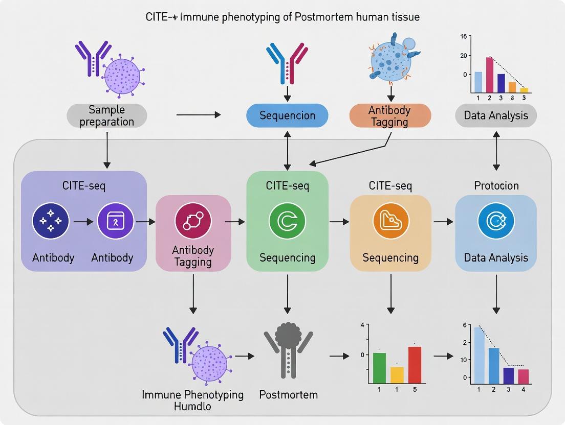
CITE-seq for Postmortem Human Tissue: A Complete Guide to Immune Profiling in Complex Samples
This comprehensive guide explores the application of CITE-seq (Cellular Indexing of Transcriptomes and Epitopes by Sequencing) for high-dimensional immune phenotyping of postmortem human tissue.
CITE-seq for Postmortem Human Tissue: A Complete Guide to Immune Profiling in Complex Samples
Abstract
This comprehensive guide explores the application of CITE-seq (Cellular Indexing of Transcriptomes and Epitopes by Sequencing) for high-dimensional immune phenotyping of postmortem human tissue. Targeted at researchers, scientists, and drug development professionals, it details the unique challenges and solutions for working with fixed, frozen, or archived samples. The article covers foundational principles, a step-by-step optimized protocol from tissue dissociation to data analysis, critical troubleshooting for sample degradation and autofluorescence, and validation strategies comparing CITE-seq to flow cytometry and spatial transcriptomics. It provides actionable insights for unlocking the immune atlas of human disease from precious biobank specimens, advancing biomarker discovery and therapeutic development.
Why Postmortem Tissue? Unlocking the Immune Atlas of Human Disease with CITE-seq
Application Notes and Protocols Thesis Context: Optimization of CITE-seq for immune phenotyping in postmortem human tissue, enabling deep profiling of disease states and therapeutic targets.
1. Core Principles and Quantitative Advantages CITE-seq (Cellular Indexing of Transcriptomes and Epitopes by Sequencing) couples oligo-tagged antibodies to single-cell RNA sequencing, allowing for the simultaneous quantification of surface protein abundance and transcriptional profiles within the same cell. This integration resolves key limitations of single-modality assays, particularly for immune cells where protein expression (e.g., CD markers, checkpoint receptors) often does not correlate directly with mRNA levels.
Table 1: Comparison of Single-Cell Multiomics Modalities for Immune Phenotyping
| Modality | Measured Features | Throughput (Cells) | Key Advantage for Postmortem Tissue | Primary Limitation |
|---|---|---|---|---|
| CITE-seq | mRNA + ~500 surface proteins | 5,000 - 100,000+ | Direct protein measurement on intact cells; critical for immunophenotyping with degraded RNA. | Limited to surface/secreted proteins; antibody panel cost. |
| REAP-seq | mRNA + surface proteins | Similar to CITE-seq | Comparable to CITE-seq. | Less commonly used; smaller commercial antibody panels. |
| scRNA-seq alone | mRNA (whole transcriptome) | 10,000 - 1,000,000+ | Unbiased gene discovery. | Missing key protein-level phenotypic data. |
| Flow/Mass Cytometry | 20-50 proteins | 1,000,000+ | High protein multiplexing; live cell sorting. | Limited mRNA data; higher input cell requirements. |
2. Detailed Protocol for Postmortem Human Lymphoid Tissue Critical Pre-Protocol Note: Postmortem tissues present challenges including RNA degradation, increased autofluorescence, and potential antigen degradation. Rapid processing or cryopreservation is essential.
Protocol 2.1: Nuclei Isolation and Antibody Staining for Frozen Tissue Objective: To generate a single-nucleus suspension labeled with TotalSeq antibodies for CITE-seq from snap-frozen human spleen or lymph node. Materials:
- Frozen tissue section (≈30 mg).
- Dounce homogenizer.
- Nuclei Extraction Buffer: 10 mM Tris-HCl (pH 7.4), 146 mM NaCl, 1 mM CaCl2, 21 mM MgCl2, 0.05% BSA, 0.2% Nonidet P-40 Substitute.
- Nuclei Wash Buffer: 1x PBS, 1% BSA, 0.2 U/µl RNase Inhibitor.
- TotalSeq Antibody Panel (Human, e.g., CD45, CD3, CD19, CD11b, CD16, HLA-DR, PD-1).
- Fc Receptor Blocking Solution (Human TruStain FcX).
Procedure:
- Homogenization: Place frozen tissue in 2 mL ice-cold Nuclei Extraction Buffer in a Dounce. Homogenize with 10-15 strokes of the loose pestle, then 10-15 strokes of the tight pestle.
- Filtration & Centrifugation: Filter through a 40 µm flow-through cell strainer. Centrifuge at 500 rcf for 5 min at 4°C.
- Wash: Gently resuspend pellet in 2 mL Nuclei Wash Buffer. Centrifuge at 500 rcf for 5 min at 4°C.
- Blocking & Staining: Resuspend pellet in 100 µl of Fc Block + Wash Buffer. Incubate 10 min on ice. Add pre-titrated TotalSeq antibody cocktail. Incubate for 30 min on ice, protected from light.
- Final Wash: Add 1 mL Wash Buffer, centrifuge at 500 rcf for 5 min at 4°C. Repeat. Resuspend in appropriate buffer for your chosen single-cell platform (e.g., 10x Genomics).
Protocol 2.2: Single-Cell Library Preparation and Sequencing Objective: To generate sequencing-ready libraries from antibody-labeled nuclei/cells. Procedure:
- Single-Cell Partitioning: Load the stained suspension into a 10x Chromium Chip (3' v3.1 or 5' v2). Follow manufacturer's instructions to generate gel bead-in-emulsions (GEMs).
- cDNA Synthesis & Amplification: Perform reverse transcription within GEMs. Break emulsions, recover cDNA, and amplify via PCR.
- Library Construction:
- Gene Expression Library: Fragment amplified cDNA, add adaptors, and index via a standard 10x protocol.
- Antibody-Derived Tag (ADT) Library: Isolate antibody-derived cDNA amplicons via a separate PCR using a universal primer set specific for the TotalSeq backbone. Index with a unique sample index.
- Sequencing: Pool libraries. Recommended sequencing depths:
- Gene Expression: ≥ 20,000 reads/cell.
- ADT Library: ≥ 5,000 reads/cell.
- Use a dual-indexed sequencing configuration (e.g., Illumina NovaSeq).
3. Data Analysis Workflow
Diagram Title: CITE-seq Data Analysis Pipeline
Diagram Title: CITE-seq Experimental Workflow
4. The Scientist's Toolkit: Key Research Reagent Solutions
Table 2: Essential Materials for CITE-seq on Postmortem Tissue
| Item | Supplier Examples | Function & Critical Notes |
|---|---|---|
| TotalSeq Antibodies | BioLegend, Bio-Rad | Oligo-tagged antibodies. Pre-titrate on matched tissue. Use hashed antibodies for sample multiplexing. |
| Chromium Controller & Kits | 10x Genomics | Single-cell partitioning and library prep. The 5' kit is optimized for protein detection. |
| RNase Inhibitor | Takara, Lucigen | Essential for preserving RNA integrity during nuclei extraction from postmortem tissue. |
| Fc Receptor Block | BioLegend, Miltenyi | Reduces non-specific antibody binding, crucial for clean ADT signal. |
| Viability Stain | BioLegend (Zombie dyes) | Distinguish live/dead cells in fresh preparations. Less critical for nuclei. |
| Cell Hashtag Antibodies | BioLegend (TotalSeq-C) | Enables sample multiplexing, reduces batch effects, and lowers cost. |
| Single-Cell Analysis Software | Seurat (R), Scanpy (Python) | Primary tools for integrated RNA+protein analysis, including WNN (Weighted Nearest Neighbors). |
The Critical Value of Postmortem Human Tissue in Biomedical Research
Postmortem human tissue (PMT) is an indispensable resource for validating and contextualizing findings from in vitro and animal models, particularly in immune system research. Within the thesis framework focusing on CITE-seq (Cellular Indexing of Transcriptomes and Epitopes by Sequencing) protocol development for immune phenotyping, PMT provides the critical "ground truth." It enables the characterization of the human immune landscape in its native tissue architecture and disease state, which is impossible to fully replicate in models. Utilizing PMT with CITE-seq allows for the simultaneous measurement of surface protein expression and transcriptomic data from single cells, offering an unprecedentedly detailed view of immune cell identity, activation state, and functional potential in health and disease. This application note details protocols and considerations for integrating PMT into such a research pipeline.
Table 1: Key Research Applications of Postmortem Tissue in Immunology
| Application | Key Insight Gained | Representative PMT Source | CITE-seq Advantage |
|---|---|---|---|
| Tumor Microenvironment (TME) Profiling | Spatial organization of exhausted T cells, tumor-associated macrophages, myeloid-derived suppressor cells. | Brain (GBM), lung, melanoma, colon. | Links surface immune checkpoint markers (PD-1, CTLA-4) to transcriptional programs. |
| Neuroinflammation | Microglia and astrocyte activation states in Alzheimer's, Parkinson's, multiple sclerosis. | Brain (various regions), spinal cord. | Identifies protein surface markers (TMEM119, CD11b) with simultaneous disease-associated gene expression. |
| Autoimmune Disease | Tertiary lymphoid structure formation, plasma cell and memory B cell niches. | Synovium (RA), gut (Crohn's), skin (psoriasis). | Phenotypes B cell maturation (CD19, CD27, CD38) alongside antibody class-switch transcripts. |
| Infectious Disease | Tissue-resident memory T cell (Trm) persistence and localization post-infection/vaccination. | Lung, lymph nodes, liver, gut. | Defines Trm via CD69/CD103 protein co-expression and residency gene signatures. |
| Baseline Immune Atlas | Defining normal immune cell frequency and phenotype across all human tissues. | Spleen, lymph node, bone marrow, non-diseased tissues. | Creates a multi-modal reference for detecting disease-specific deviations. |
Table 2: Critical Quantitative Factors for PMT CITE-seq Studies
| Parameter | Typical Target/Impact Range | Protocol Consideration |
|---|---|---|
| Postmortem Interval (PMI) | ≤24 hours (optimal); viability decreases significantly >48h. | Shorter PMI correlates with higher cell viability and RNA integrity number (RIN). |
| Cell Viability (Pre-enrichment) | 40-80% is common; >70% is ideal for CITE-seq. | Vital dyes (DAPI, 7-AAD) for flow cytometry; use of viability antibody tags in CITE-seq. |
| RNA Integrity Number (RIN) | >7.0 for robust transcriptomics; 5.0-7.0 may be acceptable with UMIs. | Assessed via Bioanalyzer/TapeStation; informs cDNA amplification cycles. |
| Antibody-Derived Tag (ADT) Signal | Can be more robust than RNA in sub-optimal PMI samples. | Normalize ADT counts using isotype controls or background from negative cells. |
| Estimated Cell Yield | Highly tissue-dependent: 1x10^6 to 1x10^7 cells/gram tissue. | Influences library complexity and need for sample multiplexing. |
Detailed Protocol: CITE-seq on Postmortem Lymph Node Tissue
Protocol 3.1: Tissue Dissociation & Single-Cell Suspension Preparation
Objective: Generate a viable, single-cell suspension suitable for barcoding and library construction. Reagents & Equipment: GentleMACS Octo Dissociator, RPMI 1640 medium, collagenase IV (1 mg/mL), DNase I (0.1 mg/mL), Fetal Bovine Serum (FBS), 70µm cell strainer, pre-cooled PBS.
- Tissue Collection & Transport: Collect tissue in cold (4°C) preservation medium (e.g., Hypothermosol) and process immediately or store at 4°C for <24h.
- Dissociation: Mince ~1cm^3 tissue with scalpel in 5mL of digestion medium (RPMI + collagenase IV + DNase I). Transfer to GentleMACS C-tube.
- Mechanical Dissociation: Run the "gentleMACS" program 37CmTDK_1 on the Octo Dissociator. Incubate for 30 minutes at 37°C with rotation.
- Termination: Add 10mL of cold RPMI + 10% FBS to stop digestion. Filter suspension through a 70µm strainer.
- Wash & Count: Centrifuge at 500g for 5 min at 4°C. Resuspend pellet in PBS + 0.04% BSA. Count cells using hemocytometer and assess viability (e.g., Trypan Blue).
Protocol 3.2: Viable Cell Enrichment & Antibody Staining
Objective: Enrich for live cells and label with CITE-seq antibody conjugates. Reagents & Equipment: Dead Cell Removal Kit (e.g., Miltenyi), Fc Receptor Blocking Solution, TotalSeq-B/C Antibody Panel (e.g., BioLegend), PBS + 0.04% BSA.
- Dead Cell Removal: Follow manufacturer's protocol for dead cell removal. This step is critical for PMT.
- Fc Block & Surface Staining: Resuspend ~1x10^6 cells in 100µL PBS/BSA. Add Fc block (10µL), incubate 10 min on ice.
- Antibody Tagging: Without washing, add pre-titrated TotalSeq-B antibody cocktail. Incubate for 30 min on ice in the dark.
- Wash: Wash cells 2x with 2mL PBS/BSA. Resuspend in PBS/BSA at ~1000 cells/µL. Keep on ice.
Protocol 3.3: Single-Cell Library Preparation & Sequencing
Objective: Generate barcoded cDNA and antibody-derived tag (ADT) libraries. Reagents & Equipment: 10X Genomics Chromium Controller & Single Cell 5' Kit, TotalSeq-B Add-on Kit, SPRIselect beads, Bioanalyzer.
- Gel Bead-in-Emulsion (GEM) Generation: Load cells, TotalSeq-B antibodies (if not already stained), and master mix onto a Chromium chip. Target recovery of 5,000-10,000 cells.
- Post-GEM-RT Cleanup & cDNA Amplification: Follow 10X protocol for reverse transcription, cDNA cleanup, and amplification (12-14 cycles recommended for PMT).
- Library Construction: Construct the gene expression library per 10X protocol. Construct the ADT library separately using the TotalSeq-B kit, which adds P5/P7 handles and sample indices via a PCR (14-16 cycles).
- Quality Control & Sequencing: Assess library size distribution (Bioanalyzer). Pool libraries proportionally. Sequence on Illumina platform: ~50,000 reads/cell for gene expression, ~5,000 reads/cell for ADT.
Experimental Workflow & Pathway Diagrams
Title: PMT CITE-seq Experimental Workflow
Title: CITE-seq Data Integration & Analysis Pathways
The Scientist's Toolkit: Research Reagent Solutions
Table 3: Essential Reagents for PMT CITE-seq Studies
| Reagent Category | Specific Product/Example | Critical Function |
|---|---|---|
| Tissue Preservation | Hypothermosol (BioLife Solutions) | Extends viable processing window by stabilizing pH and reducing cold shock. |
| Enzymatic Dissociation | Liberase TL Research Grade (Roche) | Gentle, tissue-specific enzyme blend for maximizing viable immune cell yield. |
| Dead Cell Removal | Dead Cell Removal Kit (Miltenyi Biotec) | Magnetic negative selection of viable cells; crucial for low-viability PMT samples. |
| Fc Receptor Block | Human TruStain FcX (BioLegend) | Blocks non-specific antibody binding, improving ADT signal-to-noise ratio. |
| CITE-seq Antibodies | TotalSeq-B/C Anti-Human Hashtags & Phenotyping Panels (BioLegend) | Oligo-tagged antibodies for multiplexing samples and surface protein detection. |
| Cell Barcoding | Chromium Single Cell 5' Kit (10x Genomics) | Standardized reagents for partitioning cells into GEMs and barcoding RNA/ADT. |
| Viability Assessment | 7-AAD Viability Staining Solution (BioLegend) | Flow cytometric discrimination of live/dead cells prior to loading on Chromium. |
| RNA Protection | RNAlater Stabilization Solution (Thermo Fisher) | Optional for tissue aliquots intended for bulk RNA-seq validation. |
| Library QC | High Sensitivity D1000 ScreenTape (Agilent) | Accurate sizing and quantification of final GEX and ADT libraries pre-pooling. |
Introduction CITE-seq (Cellular Indexing of Transcriptomes and Epitopes by Sequencing) enables simultaneous high-dimensional transcriptome and surface protein profiling at single-cell resolution. Its application to postmortem human tissue for immune phenotyping presents unique challenges: rapid RNA degradation due to the absence of perfusion and cold ischemia time, loss of conformational protein epitopes, and increased autofluorescence from lipofuscin and other age-related pigments. These factors directly impact data quality, requiring tailored protocols to ensure viability and specificity.
Application Notes & Data Summary
Table 1: Impact of Postmortem Interval (PMI) on Sample Quality Metrics
| Metric | PMI < 6 hrs (Ideal) | PMI 6-12 hrs (Moderate) | PMI 12-24 hrs (Degraded) | Mitigation Strategy |
|---|---|---|---|---|
| RNA Integrity Number (RIN) | 7.5 - 10 | 5.5 - 7.4 | 2.0 - 5.4 | Immediate tissue freezing or fixation |
| % Viable Cells (Flow) | 70-90% | 50-70% | 20-50% | Pre-processing enzymatic digestion at 4°C |
| Autofluorescence Index | Low | Moderate | High | Chemical quenching, spectral unmixing |
| Antigen Detection Score (MFI) | High | Reduced for sensitive epitopes | Low/Broad | Antigen retrieval, validated antibody clones |
Table 2: Reagent Solutions for Postmortem CITE-seq
| Reagent | Function & Rationale |
|---|---|
| RNase Inhibitors (e.g., RNasin Plus) | Suppresses endogenous RNase activity during tissue dissociation. |
| Cold-Active Protease (e.g., Liberase TL) | Efficient tissue digestion at 4°C, minimizing ambient RNA degradation. |
| Methanol Fixation Buffer | Preserves RNA and protein integrity simultaneously; compatible with CITE-seq. |
| Sudan Black B / TrueBlack Lipofuscin Autofluorescence Quencher | Reduces nonspecific signal by quenching lipofuscin and cellular autofluorescence. |
| Hashtag Oligonucleotide Antibodies (TotalSeq-A) | Multiplex samples to control for batch effects and identify doublets. |
| Cell Staining Buffer (BSA + Fc Block) | Reduces nonspecific antibody binding, critical for high protein background tissues. |
| Dead Cell Removal Microbeads | Enriches for live cells, improving sequencing library quality. |
Detailed Protocols
Protocol 1: Tissue Harvest & Nuclei Isolation for Degraded RNA Objective: Recover high-quality nuclei from tissue with extended PMI (>12 hrs) where cytoplasmic RNA is severely degraded.
- Snap-Freezing: Subdissect tissue region of interest on dry ice. Store at -80°C.
- Homogenization: In a pre-chilled Dounce homogenizer, add 1-2 mL of Lysis Buffer (10mM Tris-HCl, 146mM NaCl, 1mM CaCl2, 21mM MgCl2, 0.05% BSA, 0.2% Nonidet P-40, 1U/μL RNase inhibitor). Dounce 15-20 strokes on ice.
- Filtration & Washing: Filter through a 40μm flow-through strainer. Centrifuge nuclei at 500 x g for 5 min at 4°C. Gently resuspend pellet in 1 mL Wash Buffer (PBS + 1% BSA + 1U/μL RNase inhibitor).
- Debris Removal: Layer suspension over a 1.5 mL cushion of 29% iodixanol. Centrifuge at 13,000 x g for 20 min at 4°C. Collect nuclei band at interface.
- Count & Proceed: Count using AO/PI on a hemocytometer. Use immediately for CITE-seq antibody labeling (nuclear antigens) or 10x Genomics library prep.
Protocol 2: Protein Integrity & Autofluorescence Mitigation for Surface CITE-seq Objective: Preserve surface epitopes and quench autofluorescence prior to antibody staining.
- Cell Suspension Preparation: Generate single-cell suspension from fresh tissue using cold-active protease digestion. Filter through a 70μm strainer.
- Methanol Fixation (Optional): For long-term storage or epitope stabilization, resuspend pellet in ice-cold 80% Methanol drop-wise while vortexing. Incubate 10 min on ice. Wash 2x with Cell Staining Buffer.
- Autofluorescence Quenching: Resuspend cell pellet in 0.1% Sudan Black B (in 70% ethanol) or 1X TrueBlack in PBS. Incubate for 20-30 minutes at room temperature in the dark.
- Wash: Centrifuge at 300 x g for 5 min. Wash thoroughly 2x with 3 mL Cell Staining Buffer.
- Fc Blocking & Staining: Resuspend in Cell Staining Buffer with Human TruStain FcX (1:100). Incubate 10 min on ice. Add TotalSeq-A antibody cocktail directly. Proceed with standard CITE-seq protocol.
Protocol 3: Multiplexed Hashtagging for Batch Normalization Objective: Control for technical variation across multiple postmortem samples with varying quality.
- Sample Barcoding: Prior to pooling, label each individual cell suspension from different donors or PMIs with a unique TotalSeq-A Hashtag antibody (1:200 dilution in 50μL). Incubate 30 min on ice.
- Pooling: Wash all samples twice with Cell Staining Buffer. Combine all hashtagged samples into one single tube.
- Proceed with CITE-seq: Perform the remaining TotalSeq-A antibody staining for surface proteins (if not already multiplexed) on the pooled sample. This ensures identical staining conditions.
- Demultiplexing: After sequencing, use the hashtag antibody-derived tags (HTOs) to assign each cell to its original sample, enabling downstream batch correction.
Visualizations
Title: Experimental Workflow for Postmortem CITE-seq
Title: Challenges & Solutions Framework
This application note details the suitability of four key tissue types—brain, spleen, lymph node, and solid tumors—for immune cell phenotyping using CITE-seq (Cellular Indexing of Transcriptomes and Epitopes by Sequencing) in the context of postmortem human tissue research. A primary thesis in this field posits that systematic, multi-tissue CITE-seq analysis from postmortem donors can unlock unprecedented maps of the human immune system, revealing tissue-specific residency, trafficking, and functional states that are critical for understanding disease pathogenesis and developing novel immunotherapies. The unique challenges of postmortem tissue, including RNA degradation and variable antigen integrity, necessitate tailored protocols and a clear understanding of each tissue's inherent cellular composition and structural properties.
Tissue Characteristics & Suitability Analysis
The following table summarizes the key characteristics, advantages, and challenges of each tissue type for postmortem CITE-seq analysis.
Table 1: Tissue Suitability for Postmortem CITE-seq Immune Phenotyping
| Tissue | Primary Immune Context | Key Immune Cell Types | Advantages for CITE-seq | Major Challenges (Postmortem) |
|---|---|---|---|---|
| Brain | Immune-privileged, specialized niche. | Microglia, tissue-resident macrophages, limited T-cells. | Low baseline immune infiltrate simplifies focus on CNS-specific residents. Highly defined cellular states. | Rapid RNA degradation postmortem. Delicate cell types sensitive to isolation. Low cell yield. |
| Spleen | Secondary lymphoid organ; blood filter. | B-cells, T-cells, macrophages, dendritic cells, RBCs. | Extremely high immune cell density and diversity. Excellent for systemic immune profiling. | High red blood cell & platelet contamination. Prone to rapid autolysis. |
| Lymph Node | Secondary lymphoid organ; adaptive immunity hub. | Naïve/activated T & B cells, dendritic cells, follicular helper cells. | Ideal for studying antigen-specific responses and lymphocyte trafficking. Structured microenvironment. | Often fibrotic or diseased in donors. Requires careful dissection to isolate follicles. |
| Solid Tumor | Variably immunosuppressive microenvironment. | Tumor-infiltrating lymphocytes (TILs), myeloid-derived suppressor cells, TAMs. | Direct profiling of therapeutic target—the tumor immune microenvironment (TIME). | Extreme heterogeneity. High enzymatic digestion needed can damage surface epitopes. High ambient RNA. |
Core Protocol: Postmortem Tissue Processing for CITE-seq
This standardized protocol is adapted for the challenges of postmortem human tissue, with tissue-specific notes.
Protocol 3.1: Tissue Dissociation & Single-Cell Suspension Preparation Objective: To obtain a viable, single-cell suspension with preserved RNA and surface protein integrity from postmortem tissues. Key Reagent Solutions: See Section 5.
- Tissue Acquisition & Preservation: Record postmortem interval (PMI). Mince tissue (≤ 2-3 mm³ pieces) in cold preservation medium (e.g., Hypothermosol) on ice. Critical: Process brain tissue within <12h PMI; lymphoid tissues within <24h PMI.
- Enzymatic Dissociation: Use a gentle, titrated approach.
- Spleen/Lymph Node: Use a gentle collagenase/DNase I blend (e.g., 1 mg/mL Collagenase IV, 20 U/mL DNase I) in RPMI for 20-30 min at 37°C with gentle agitation.
- Solid Tumor: Use a multi-enzyme cocktail (e.g., Collagenase IV, Hyaluronidase, Dispase) for 45-60 min. Optimize time per tumor type.
- Brain: Use a neural tissue dissociation kit (papain-based) for 15-20 min. Avoid over-digestion.
- Mechanical Dissociation: Pass digested tissue through a 70 µm strainer. For brain and tumors, use a plunger. For spleen, lyse RBCs using ACK buffer (2 min on ice).
- Wash & Debris Removal: Wash cells 2x in PBS + 0.04% BSA. Use a dead cell removal kit or Percoll/density gradient centrifugation (for spleen/brain) to enrich live cells.
- Cell Counting & Viability Assessment: Count using trypan blue or AO/PI on an automated counter. Acceptable Postmortem Viability: >70% for lymphoid tissues, >50% for brain/tumors.
Protocol 3.2: CITE-seq Library Generation Objective: To barcode cellular transcripts and antibody-derived tags (ADTs) from the single-cell suspension.
- Cell Staining for ADTs: Wash cell pellet in PBS + 0.04% BSA. Stain with a pre-titrated, DNA-barcoded antibody cocktail (e.g., TotalSeq) for 30 min on ice. Include a viability marker (e.g., TotalSeq-C). Critical: Titrate antibodies extensively for each postmortem tissue type due to potential epitope degradation.
- Wash & Resuspension: Wash cells 3x thoroughly to remove unbound antibodies. Resuspend at target concentration (700-1200 cells/µL) for your platform (e.g., 10x Genomics).
- Single-Cell Partitioning & Library Prep: Follow manufacturer's protocol (e.g., 10x Genomics Chromium Next GEM). Generate separate cDNA and ADT libraries.
- cDNA Amplification: 12-14 cycles recommended for postmortem samples with potentially lower RNA quality.
- ADT Amplification: Use 12-18 PCR cycles, determined by antibody panel signal strength.
- Sequencing: Pool libraries. Sequence cDNA library deeply (~50,000 reads/cell) and ADT library modestly (~5,000 reads/cell) on an Illumina platform.
Data Analysis Workflow & Pathway Diagrams
Title: CITE-seq Data Analysis Workflow for Multi-tissue Studies
Title: Postmortem Tissue Challenges & CITE-seq Protocol Solutions
The Scientist's Toolkit: Key Research Reagent Solutions
Table 2: Essential Reagents for Postmortem Tissue CITE-seq
| Reagent / Kit | Primary Function | Tissue-Specific Application Note |
|---|---|---|
| Hypothermosol FRS | Hypothermic tissue preservation medium. Slows metabolism & autolysis. | Critical for all tissues. Use immediately upon collection to extend viable processing window. |
| Gentle MACS Dissociator | Standardized mechanical dissociation. | Provides reproducible agitation for spleen/LN/tumor enzymatic digestion. Use gentle programs for brain. |
| Liberase TL / TM | Research-grade enzyme blends for gentle tissue dissociation. | Preferred over crude collagenase for better epitope preservation, especially in tumors. |
| Papain-based Neural Dissociation Kit | Enzyme mix optimized for neural tissue. | Essential for brain. Yields viable microglia with intact surface markers. |
| TotalSeq Antibody Panels | DNA-barcoded antibodies for CITE-seq. | Must include lineage & activation markers. Require extensive titration on postmortem tissue. |
| LIVE/DEAD Fixable Viability Dyes | Distinguishes live/dead cells during staining. | Use near-IR dye for less spectral overlap. Critical for postmortem samples with high debris. |
| Dead Cell Removal Kit | Magnetic removal of apoptotic/necrotic cells. | Recommended for all tissues. Dramatically improves sequencing data quality from low-viability samples. |
| Chromium Next GEM Single Cell 5' Kit | Partitioning, RT, and library prep for 5' gene expression + ADT. | Standardized workflow. For postmortem samples, consider increasing cDNA PCR cycles. |
| Cell Ranger / Cite-seq-Count | Software pipelines for demultiplexing, alignment, and ADT counting. | Antibody Panel CSV file must be meticulously curated for correct ADT quantification. |
Ethical Considerations and Biobanking Best Practices for CITE-seq Studies
1. Introduction and Context within Postmortem Human Tissue Immune Phenotyping Research
Postmortem human tissue (PMHT) is an indispensable resource for validating immunological discoveries made in model systems and for understanding human-specific disease pathophysiology in situ. CITE-seq (Cellular Indexing of Transcriptomes and Epitopes by Sequencing), which simultaneously quantifies single-cell RNA expression and surface protein abundance, is a powerful tool for deep immune phenotyping of these tissues. However, the application of CITE-seq to PMHT introduces a nexus of unique ethical and practical challenges that must be addressed to ensure scientific rigor, reproducibility, and public trust. This document outlines critical ethical considerations and biobanking best practices, framing them within the workflow of a broader CITE-seq-based thesis on human immune system analysis in health and disease.
2. Core Ethical Considerations
2.1. Donor Consent and Governance The ethical foundation of PMHT research rests on informed, broad, and often tiered consent. Consent must explicitly cover high-dimensional genomic and proteomic data generation, data sharing in controlled-access databases (e.g., dbGaP, AnVIL), and potential for future unspecified research use.
2.2. Privacy and Data Security CITE-seq data is inherently identifiable. Robust de-identification protocols must be applied, and data must be classified according to relevant regulations (e.g., GDPR, HIPAA). A typical data security framework is summarized below:
Table 1: Data Security Tiers for CITE-seq Derived from PMHT
| Data Tier | Description | Access Control | Example |
|---|---|---|---|
| Tier 1: Raw Data | Unprocessed BCL or FASTQ files. | Strictest control, limited to primary processing team. | BCL files from sequencer. |
| Tier 2: Processed Data | Gene-cell (RNA) and antibody-cell (ADT) count matrices, cell metadata. | Controlled access via data use agreements (DUAs). | H5AD or Seurat objects. |
| Tier 3: Analyzed Data | Annotated clusters, differential expression results, visualizations. | Can often be published with manuscripts or in public repositories. | UMAP plots, marker gene lists. |
2.3. Return of Results and Incidental Findings Given the complexity of CITE-seq data, return of individual results to donor families is generally not feasible or appropriate. Policies must be clearly defined in the consent form and reviewed by an Institutional Review Board (IRB) or Ethics Committee.
2.4. Equity and Justice Biobanks must actively work to ensure donor populations are diverse and representative to avoid perpetuating health disparities in research outcomes.
3. Biobanking Best Practices for CITE-seq Quality
Pre-analytical variables are the greatest source of technical noise in PMHT CITE-seq. Standardized protocols are essential.
3.1. Tissue Procurement and Preservation Protocol
- Title: Rapid Procurement and Multi-Modal Preservation of PMHT for CITE-seq.
- Objective: To minimize postmortem interval (PMI) effects and preserve both RNA and epitope integrity.
- Materials: Sterile dissection tools, RNA-later stabilization solution, OCT compound, chilled PBS, isopentane (pre-chilled on dry ice), labeled cryovials, -80°C freezer.
- Procedure:
- Documentation: Record exact PMI, cause of death, and tissue location.
- Dissection: Rapidly dissect target tissue (e.g., lymph node, spleen parenchyma) on ice.
- Multi-Aliquot Preservation:
- Aliquot A (CITE-seq): Mince ~100 mg of tissue into 1-2 mm³ pieces in cold PBS. Immediately proceed to single-cell suspension protocol (3.2).
- Aliquot B (Nucleic Acids): Place ~50 mg in 1 ml RNA-later. Store at 4°C overnight, then transfer to -80°C.
- Aliquot C (Morphology): Embed ~50 mg in OCT, freeze in isopentane cooled on dry ice. Store at -80°C for spatial validation (e.g., CODEX/IF).
- Storage: Log all aliquots in the biobank inventory management system.
3.2. Single-Cell Suspension Protocol from PMHT for CITE-seq
- Title: Gentle Enzymatic and Mechanical Dissociation for Viable Immune Cell Recovery.
- Objective: To generate a high-viability, high-yield single-cell suspension suitable for CITE-seq library preparation.
- Materials: GentleMACS Dissociator (or similar), Tumor Dissociation Kit (human), DNase I, RPMI 1640 + 10% FBS, 70µm cell strainer, 40µm flow cytometry mesh, Dead Cell Removal Kit, Automated Cell Counter (e.g., with AO/PI staining).
- Procedure:
- Dissociation: Transfer tissue pieces (from 3.1, Aliquot A) to a C-tube containing enzyme mix. Run the "gentleMACS" program appropriate for soft tissue (e.g.,
m_spleen_01). - Incubation: Incubate the tube at 37°C for 15-20 minutes with gentle rotation.
- Termination: Add 10 ml of cold RPMI+10%FBS to stop digestion. Filter through a 70µm strainer.
- RBC Lysis: If erythrocyte contamination is high, perform ACK lysis for 2 minutes on ice.
- Debris Removal: Filter through a 40µm mesh. Optionally, use a dead cell removal kit.
- Assessment: Count cells and assess viability via AO/PI. Target: >70% viability for optimal CITE-seq performance.
- Staining: Proceed directly to antibody staining for CITE-seq (see 3.3).
- Dissociation: Transfer tissue pieces (from 3.1, Aliquot A) to a C-tube containing enzyme mix. Run the "gentleMACS" program appropriate for soft tissue (e.g.,
3.3. CITE-seq Antibody Staining and Library Preparation Protocol
- Title: Hashtag-Oligo and TotalSeq-B Antibody Staining for Multiplexed PMHT CITE-seq.
- Objective: To label cells from multiple donors/samples with unique barcodes (Hashtag Oligos - HTOs) and detect surface proteins (TotalSeq-B antibodies) prior to pooled library generation.
- Materials: Human TruStain FcX, TotalSeq-B antibody cocktail, CellPlex (Hashtag) antibody cocktail, Cell Staining Buffer, 0.04% BSA-PBS.
- Procedure:
- Fc Block: Resuspend up to 1x10⁶ cells in 50µl Cell Staining Buffer with 5µl Human TruStain FcX. Incubate on ice for 10 minutes.
- Hashtag Staining: Add a unique CellPlex HTO antibody (1:100 dilution) to each sample. Incubate on ice for 30 minutes.
- Surface Protein Staining: Without washing, add the pre-titrated TotalSeq-B antibody cocktail (e.g., 50-100 antibodies). Incubate on ice for 30 minutes.
- Washing: Wash cells twice with 1 ml of 0.04% BSA-PBS.
- Pooling: Combine all individually barcoded samples into one single-cell suspension.
- Library Preparation: Count cells and load onto the Chromium Controller (10X Genomics) per manufacturer's instructions for Gene Expression and Cell Surface Protein libraries. Key Modification: Use a reduced reverse transcription cycle number (e.g., 50% of standard) for ADT library amplification to mitigate amplification bias.
4. Visualization: CITE-seq Workflow from Biobank to Analysis
Title: PMHT CITE-seq Workflow with Critical QC Checkpoints
5. The Scientist's Toolkit: Essential Reagent Solutions
Table 2: Key Research Reagent Solutions for PMHT CITE-seq Studies
| Item | Function | Key Consideration for PMHT |
|---|---|---|
| RNA-later Stabilization Solution | Preserves RNA integrity in tissue aliquots by inhibiting RNases. | Critical for validating transcriptional profiles from CITE-seq suspensions against bulk tissue. |
| TruStain FcX (Fc Receptor Block) | Blocks non-specific binding of antibodies to Fc receptors on immune cells. | Essential for reducing background signal in protein detection, especially in myeloid-rich tissues. |
| TotalSeq-B Antibody Panels | Oligo-tagged antibodies for simultaneous protein detection with scRNA-seq. | Require extensive titration and validation on PMHT due to potential antigen degradation. |
| CellPlex (Hashtag Oligo) Kit | Allows multiplexing of up to 12 samples by labeling cells with sample-specific barcodes. | Maximizes throughput and reduces batch effects; crucial for cohort studies with limited cell yields per sample. |
| Dead Cell Removal Kit | Selectively removes non-viable cells via magnetic separation. | Improves sequencing library efficiency and data quality by reducing background from dead cells (common in PMHT). |
| GentleMACS Dissociator & Kits | Standardized mechanical and enzymatic tissue dissociation. | Provides reproducible cell yields; program selection is tissue-specific (e.g., brain vs. spleen). |
| Chromium Next GEM Chip Kits (10X) | Microfluidic partitioning for single-cell gel bead-in-emulsion (GEM) generation. | Standardized workflow; for PMHT, consider loading a higher cell concentration to account for lower viability. |
Step-by-Step: An Optimized CITE-seq Protocol for Fixed and Frozen Human Tissues
Within CITE-seq protocol postmortem human tissue immune phenotyping research, the initial tissue dissociation step is the critical bottleneck. The viability, yield, and transcriptional fidelity of isolated immune cells directly dictate the success of downstream single-cell multi-omic analysis. Postmortem tissues present unique challenges, including increased hypoxia, onset of apoptosis, and release of endogenous nucleases and proteases. This application note details optimized dissociation strategies to maximize viable immune cell recovery from complex solid tissues for advanced immunophenotyping.
Quantitative Comparison of Dissociation Methods
The following table summarizes key performance metrics for common dissociation strategies applied to lymphoid and non-lymphoid postmortem human tissues.
Table 1: Performance Metrics of Tissue Dissociation Methods for Immune Cell Yield
| Method | Principle | Avg. Viable Cell Yield (cells/g) | Avg. Viability (%) | Key Immune Cell Types Preserved | Relative Stress Signature |
|---|---|---|---|---|---|
| Mechanical Only | Homogenization, mincing | 0.5 - 2 x 10⁶ | 40-60% | Robust lymphocytes (T/B cells) | Very High |
| Enzymatic (Gentle) | Collagenase IV, DNase I, 37°C, <60 min | 3 - 8 x 10⁶ | 70-85% | Myeloid cells, T cells, B cells | Moderate |
| Enzymatic (Aggressive) | Multi-enzyme cocktails, 37°C, >90 min | 5 - 15 x 10⁶ | 50-75% | Tissue-resident macrophages, Tregs | High |
| Combined Mechanical & Enzymatic | Minced tissue + Gentle Enzymatic | 4 - 10 x 10⁶ | 75-90% | Broad spectrum (incl. fragile innate lymphoid cells) | Low |
| Commercial Multi-Step Kits | Optimized reagent sequences | 3 - 9 x 10⁶ | 80-88% | Consistent across tissue types | Low-Moderate |
Detailed Protocols
Protocol 1: Optimized Combined Dissociation for Lymphoid Tissue (Spleen, Lymph Node)
Objective: Maximize yield of intact lymphocyte subsets with minimal activation.
- Preparation: Work in cold (4°C) RPMI medium. Pre-chill all instruments.
- Tissue Mincing: Place tissue in a petri dish with 5 mL cold PBS-2% FBS. Using scalpels, mince tissue into <1 mm³ fragments.
- First Dissociation: Transfer fragments to a gentleMACS C Tube containing 5 mL of cold RPMI. Run program "mspleen01" on the gentleMACS Dissociator.
- Enzymatic Digestion: Filter supernatant through a 70µm strainer. Resuspend remaining fragments in 5 mL of pre-warmed Digestion Buffer (RPMI, 1 mg/mL Collagenase IV, 20 µg/mL DNase I).
- Incubation: Incubate at 37°C for 20 minutes with slow tilting rotation.
- Termination & Filtration: Add 10 mL of cold PBS-2% FBS-1mM EDTA to stop digestion. Pass the entire suspension through a 70µm strainer, followed by a 40µm strainer.
- Wash: Centrifuge at 400 x g for 5 min at 4°C. Resuspend pellet in 10 mL cold PBS-2% FBS. Count using trypan blue or an automated cell counter.
Protocol 2: Gentle Multi-Step Dissociation for Inflamed Non-Lymphoid Tissue (Lung, Colon)
Objective: Recover both stromal and infiltrating immune cells while minimizing cell death.
- Vascular Perfusion (if possible): Gently flush tissue with cold PBS via major vessels to remove circulating blood cells.
- Initial Processing: Mince tissue finely in cold PBS with 0.5% BSA.
- Sequential Enzymatic Steps:
- Step A (Disaggregation): Incubate fragments in 5 mL of Buffer A (RPMI, 1.5 mg/mL Collagenase D, 0.5 mg/mL Dispase II) for 30 min at 37°C, slow rotation.
- Step B (Clump Dissolution): Pellet fragments, resuspend in 5 mL of Buffer B (RPMI, 0.2 mg/mL Collagenase D, 20 µg/mL DNase I) for 10 min at 37°C.
- Mechanical Release: Use a 10 mL syringe plunger to gently press tissue against the strainer wall during filtration through a 100µm then 40µm strainer.
- Density Gradient Centrifugation: Layer cell suspension over Lymphoprep. Centrifuge at 800 x g for 20 min at 4°C, with brake off. Harvest the mononuclear cell layer at the interface.
- Wash & Count: Wash twice with PBS-0.5% BSA-1mM EDTA. Assess viability with AO/PI staining.
Visualization of Workflows and Pathways
Diagram 1: Postmortem Tissue Dissociation Decision Workflow
Diagram 2: Stress & Apoptosis Pathways in Postmortem Dissociation
The Scientist's Toolkit: Essential Research Reagents & Solutions
Table 2: Key Reagents for Postmortem Tissue Dissociation
| Reagent/Solution | Function in Protocol | Critical Note for Postmortem Tissue |
|---|---|---|
| Cold PBS + 2% FBS + 1mM EDTA | Wash & suspension buffer; EDTA inhibits adhesion and metaloproteases. | Pre-chill to 4°C; essential to slow metabolic decay. |
| Collagenase IV (or D) | Digests collagen in basement membranes to release cells. | Use purified, low-endotoxin grades. D is gentler than IV. |
| DNase I (RUO Grade) | Degrades extracellular DNA released by dead cells, reducing clumping. | Absolutely critical for postmortem tissue with high necrosis. |
| Dispase II | Neutral protease cleaving fibronectin and collagen IV; good for epithelial tissues. | Helps maintain cell surface protein integrity for CITE-seq. |
| RBC Lysis Buffer | Removes contaminating red blood cells after digestion. | Use after digestion/filtration to avoid lysing fragile immune cells. |
| Lymphoprep or Percoll | Density gradient medium for enriching mononuclear cells. | Clears debris and dead cells, improving viability for sorting. |
| Cell Staining Buffer (CSB) | PBS-based with BSA/EDTA for antibody staining post-isolation. | Use for CITE-seq antibody cocktail staining; prevents Fc-mediated binding. |
| Viability Dye (e.g., Zombie NIR) | Distinguishes live/dead cells for flow sorting pre-CITE-seq. | Imperative for excluding dead cells which cause high background. |
| Rnasin Plus/RNase Inhibitor | Inhibits RNases during dissociation. | Consider adding to digestion mix for transcriptome preservation. |
Antibody Panel Design and Conjugation for Complex Human Immune Phenotyping
Application Notes
This protocol details the design, validation, and conjugation of TotalSeq antibodies for CITE-seq (Cellular Indexing of Transcriptomes and Epitopes by Sequencing) applications within postmortem human tissue research. The integration of high-parameter protein and transcriptome measurement is critical for dissecting the complex immune landscape in tissues, such as brain, lung, and gut, obtained from postmortem donors. This work supports a broader thesis aiming to map immune dysfunction in neurological and inflammatory diseases using a multimodal single-cell approach.
Panel Design Strategy for Postmortem Tissue
Designing an antibody panel for degraded or fixed postmortem tissue requires careful consideration of epitope stability and antigen accessibility. Panels should prioritize antibodies known to withstand mild fixation and target epitopes resilient to postmortem degradation. A typical panel for comprehensive immune phenotyping includes markers for:
- Major Lineages: CD45, CD3, CD19, CD14, CD56.
- T Cell Subsets: CD4, CD8, CD25, CD127, CD45RA, CD45RO, PD-1.
- Myeloid Cells: CD11c, CD123, CD16, CD163, HLA-DR.
- Activation & Functional Markers: CD69, CD38, ICOS, CTLA-4.
- Tissue-Resident Markers: CD103, CD49a, CXCR6.
Table 1: Example 30-Marker Antibody Panel for Postmortem Tissue Immune Profiling
| Target | Clone | Isotope | Function in Panel | Validation Notes for Tissue |
|---|---|---|---|---|
| CD45 | HI30 | [89Y] | Leukocyte common antigen | Robust signal in fixed tissue |
| CD3 | UCHT1 | [141Pr] | Pan T-cell marker | Epitope stable post-fixation |
| CD19 | HIB19 | [142Nd] | B cells | Validated for CITE-seq on nuclei |
| CD14 | M5E2 | [143Nd] | Monocytes/ Macrophages | |
| CD4 | RPA-T4 | [144Nd] | Helper T cells | |
| CD8 | SK1 | [145Nd] | Cytotoxic T cells | |
| CD56 | NCAM16.2 | [146Nd] | NK cells | |
| CD45RA | HI100 | [147Sm] | Naïve T cells | |
| CD45RO | UCHL1 | [148Nd] | Memory T cells | |
| HLA-DR | L243 | [149Sm] | Antigen presentation | May require antigen retrieval |
| CD11c | Bu15 | [150Nd] | Dendritic cells, macrophages | |
| CD16 | 3G8 | [151Eu] | FcγRIII, neutrophils, NK cells | |
| CD127 | A019D5 | [152Sm] | IL-7Rα, T cell subsets | |
| CD25 | BC96 | [153Eu] | IL-2Rα, Tregs, activation | |
| PD-1 | EH12.2H7 | [154Sm] | Exhaustion marker | Critical for tissue contexts |
| CD69 | FN50 | [155Gd] | Early activation marker | |
| CD103 | Ber-ACT8 | [156Gd] | Tissue-resident T cells | Key for tissue studies |
| CD335 (NKp46) | 9E2 | [158Gd] | Natural cytotoxicity receptor | |
| CTLA-4 | L3D10 | [159Tb] | Immune checkpoint | Intracellular target |
| CD278 (ICOS) | C398.4A | [160Gd] | Co-stimulatory molecule | |
| CD183 (CXCR3) | G025H7 | [161Dy] | Th1-associated chemokine receptor | |
| CD185 (CXCR5) | J252D4 | [162Dy] | Tfh-associated receptor | |
| CD196 (CCR6) | G034E3 | [163Dy] | Th17-associated receptor | |
| CD194 (CCR4) | L291H4 | [164Dy] | Th2-associated receptor | |
| CD197 (CCR7) | G043H7 | [165Ho] | Lymphoid homing receptor | |
| CD27 | O323 | [166Er] | Memory B & T cell marker | |
| CD28 | CD28.2 | [167Er] | T cell co-stimulation | |
| CD39 | A1 | [168Er] | Immunoregulatory ectoenzyme | Important in tissue tolerance |
| CD73 | AD2 | [169Tm] | Immunoregulatory ectoenzyme | |
| CD161 | HP-3G10 | [170Er] | NK, MAIT, and Th17 cells |
Protocols
Protocol 1: Antibody Conjugation to TotalSeq Oligonucleotides
This protocol is adapted for conjugating commercially available purified antibodies to TotalSeq hashtag or feature barcode oligonucleotides.
Materials:
- Purified antibody (carrier-free, 0.5-1.0 mg/mL in PBS).
- TotalSeq oligonucleotide conjugation kit (e.g., from BioLegend, containing modified oligos, conjugation buffer, quenching reagent).
- PD-10 desalting columns or 100 kDa molecular weight cutoff centrifugal filters.
- UV-Vis spectrophotometer (Nanodrop).
Method:
- Antibody Preparation: Transfer 50-100 µg of antibody to a low-protein-binding microcentrifuge tube. Ensure the antibody is in a conjugation-compatible buffer (PBS, pH 7.2-7.4). Remove amines (e.g., Tris, glycine, azide) using a desalting column if present.
- Reduction: Add a 10-20 molar excess of TCEP (tris(2-carboxyethyl)phosphine) to the antibody. Incubate at 37°C for 30-60 minutes to reduce inter-chain disulfide bonds.
- Conjugation: Add a 5-10x molar excess of maleimide-modified TotalSeq oligonucleotide to the reduced antibody mixture. Incubate in the dark at room temperature for 2 hours.
- Quenching: Add a 50x molar excess of cysteine or the provided quenching reagent to stop the reaction. Incubate for 15 minutes.
- Purification: Purify the antibody-oligo conjugate from free oligonucleotides using a 100 kDa centrifugal filter. Wash 3-4 times with PBS + 0.05% BSA + 0.02% sodium azide (Staining Buffer).
- Quantification & Validation: Measure the concentration at A280 (antibody) and A260 (oligo). Calculate the oligo-to-antibody ratio (OAR). An optimal OAR is 0.5-2. Target 1-2 oligos per antibody for best staining performance. Validate conjugation by running a sample on an SDS-PAGE gel with nucleic acid staining.
- Storage: Aliquot and store at 4°C. Avoid freeze-thaw cycles.
Protocol 2: CITE-seq Staining of Single-Cell Suspensions from Postmortem Tissue
This protocol details the staining of single-cell/nuclei suspensions prepared from enzymatically digested or mechanically dissociated postmortem tissue.
Materials:
- Single-cell/nuclei suspension from postmortem tissue (viability >70% recommended).
- Conjugated TotalSeq antibody panel (Table 1), titrated and pooled.
- Fc Receptor Blocking Solution (Human TruStain FcX).
- Cell Staining Buffer (PBS + 0.5% BSA + 2mM EDTA).
- LIVE/DEAD Fixable Viability Dye (e.g., Zombie NIR).
- Fixation/Permeabilization Buffer (if intracellular targets are included).
- Magnetic separator and sorting buffers (for cell hashing).
Method:
- Cell Preparation: Count cells and aliquot up to 1x10^6 cells per sample into a V-bottom plate. Pellet at 300-400 x g for 5 minutes. Aspirate supernatant.
- Viability Staining & Fc Block: Resuspend cell pellet in 50 µL of Cell Staining Buffer containing a 1:1000 dilution of viability dye. Incubate for 15 minutes at 4°C in the dark. Wash with 150 µL buffer. Pellet and aspirate. Resuspend in 20 µL of Fc Block solution. Incubate for 10 minutes on ice.
- Surface Antibody Staining: Without washing, add the pre-titrated, pooled TotalSeq antibody cocktail directly to the cells. The final staining volume should be 50-100 µL. Mix gently and incubate for 30 minutes on ice in the dark.
- Wash: Add 150 µL of Cell Staining Buffer. Pellet cells at 300-400 x g for 5 min. Aspirate supernatant carefully. Repeat wash step twice.
- (Optional) Intracellular Staining: If panel includes intracellular targets (e.g., CTLA-4), fix and permeabilize cells using a commercial kit (e.g., Foxp3/Transcription Factor Staining Buffer Set). Stain with conjugated intracellular antibodies for 30-60 minutes on ice. Wash twice with Permeabilization Buffer, then once with Cell Staining Buffer.
- Final Resuspension: Resuspend the stained cell pellet in an appropriate volume of Cell Staining Buffer for loading onto a single-cell partitioning device (e.g., 10X Genomics Chromium). Filter through a 35 µm cell strainer cap immediately before loading.
Protocol 3: Titration and Validation of Conjugated Antibodies
Method:
- Prepare a test sample of cells (e.g., PBMCs or dissociated tissue cells).
- Serially dilute the conjugated antibody (e.g., 1:50, 1:100, 1:200, 1:500) in Cell Staining Buffer.
- Stain 5x10^4 cells with each dilution alongside an unstained control and a control stained with a standard fluorescently-labeled antibody for the same target.
- Process samples for CITE-seq library preparation on a small scale or analyze by flow cytometry if the oligo is also conjugated to a fluorophore.
- Determine the optimal staining dilution as the concentration that provides the best separation from background (highest signal-to-noise ratio) without causing aggregation or non-specific binding. Re-titrate after pooling antibodies.
Visualizations
Title: CITE-seq Antibody Panel Workflow for Tissue
Title: CITE-seq Antibody Binding & Library Construction
The Scientist's Toolkit
Table 2: Essential Research Reagent Solutions for CITE-seq on Postmortem Tissue
| Item | Function & Rationale |
|---|---|
| TotalSeq Antibodies | Pre-conjugated or custom conjugation-ready antibodies for simultaneous detection of surface proteins alongside mRNA. |
| Chromium Next GEM Chip Kits (10X Genomics) | Microfluidic chips for partitioning single cells into Gel Bead-In-Emulsions (GEMs) for barcoding. |
| Human TruStain FcX | Monoclonal antibody to block Fc receptors, critical for reducing non-specific antibody binding in tissue-derived cells. |
| Zombie NIR Fixable Viability Kit | Amine-reactive fluorescent dye for identifying dead cells in fixed samples, essential for postmortem tissue analysis. |
| Liberase TL Research Grade | Enzyme blend for gentle tissue dissociation, preserving surface epitopes critical for antibody staining. |
| Foxp3/Transcription Factor Staining Buffer Set | For intracellular staining of targets like cytokines or transcription factors after surface staining. |
| Cell Staining Buffer (BSA/EDTA) | Optimized buffer for antibody dilutions and washes to maintain cell viability and minimize background. |
| SPRIselect Beads | For clean-up and size selection of cDNA and antibody-derived tag (ADT) libraries post-amplification. |
| Cell Hashing Antibodies (TotalSeq-C) | For sample multiplexing, allowing pooling of multiple postmortem samples to reduce batch effects and cost. |
| Nuclei Isolation Kits (for frozen tissue) | For extracting nuclei from frozen or difficult-to-dissociate postmortem tissue when cytoplasm is degraded. |
Within the framework of a thesis on CITE-seq-based immune phenotyping of postmortem human tissue, achieving clean, specific, and reproducible antibody-derived tag (ADT) signals is paramount. Postmortem tissues present unique challenges, including increased autofluorescence, higher levels of non-specific antibody binding, and potential antigen degradation. This application note details a systematic approach to optimize sample staining—focusing on blocking, antibody titration, and wash stringency—to maximize signal-to-noise ratio in CITE-seq experiments for drug discovery and immunological research.
Key Challenges in Postmortem Tissue CITE-seq
- Increased Non-specific Binding: Higher levels of endogenous proteins and debris.
- Autofluorescence: Compromises fluorescent viability staining and can interfere with some detection methods.
- Antigen Integrity: Variable postmortem intervals can affect epitope availability.
- Sample Heterogeneity: Immune cell populations may be in varying states of activation or apoptosis.
Optimization Protocols
Protocol 1: Fc Receptor Blocking & Non-specific Binding Reduction
Objective: Minimize non-specific antibody binding to Fc receptors and other cellular components. Materials: Human TruStain FcX (Fc Receptor Blocking Solution), PBS, 1% BSA in PBS, 0.1M EDTA, Human IgG, Zombie NIR Fixable Viability Kit. Method:
- Prepare a single-cell suspension from postmortem human lymphoid tissue (e.g., spleen, lymph node) using a validated gentle mechanical and enzymatic dissociation protocol.
- Wash cells twice with cold PBS + 1% BSA.
- Primary Block: Resuspend cell pellet (1x10⁶ cells) in 100µL of PBS + 1% BSA containing Human TruStain FcX (1:100 dilution). Incubate on ice for 10 minutes.
- Secondary Block (Optional, for high background): Without washing, add 10µL of purified human IgG (1mg/mL final) and incubate on ice for an additional 10 minutes.
- Add Zombie NIR viability dye (1:1000) directly to the blocking mixture. Incubate for 15 minutes in the dark at RT.
- Wash cells with 2mL of cold PBS + 1% BSA. Centrifuge at 300 x g for 5 minutes. Decant supernatant.
Protocol 2: Antibody Titration for TotalSeq-B/CITE-seq Antibodies
Objective: Determine the optimal antibody concentration that maximizes the separation index (SI) between positive and negative populations. Materials: Panel of TotalSeq-B antibodies (e.g., CD45, CD3, CD19, CD11c), Cell Staining Buffer (CSB, PBS + 0.5% BSA + 2mM EDTA), 1.5mL microcentrifuge tubes. Method:
- Prepare 5 aliquots of 1x10⁵ cells (from Protocol 1, post-viability stain) per antibody to be titrated.
- Prepare a 2X serial dilution series of each TotalSeq-B antibody in CSB (e.g., 1:25, 1:50, 1:100, 1:200, 1:400). Include a "staining buffer only" negative control.
- Resuspend each cell aliquot in 50µL of the diluted antibody. Incubate for 30 minutes on ice in the dark.
- Wash cells twice with 1mL of CSB. Centrifuge at 300 x g for 5 minutes.
- Resuspend cells in CSB + 1% formaldehyde (or proceed directly to hashing/library preparation if compatible). Acquire data on a flow cytometer or proceed to CITE-seq library prep and sequencing.
- Analysis: Calculate the Separation Index (SI) = (MFIpositive - MFInegative) / (2 * SDnegative) for each dilution. Plot SI vs. antibody dilution. The optimal dilution is the one yielding the highest SI before the MFIpositive plateaus.
Protocol 3: High-Stringency Wash Optimization
Objective: Reduce background by optimizing wash buffer composition and volume. Materials: PBS, CSB, 0.05% Tween-20 in PBS (PBS-T), 0.5M EDTA. Method:
- After antibody staining (using an optimal dilution from Protocol 2), split cells into three wash condition groups (n=1x10⁵ cells/group).
- Group A (Standard): Wash 2x with 1mL CSB.
- Group B (Increased Volume/Stringency): Wash 3x with 2mL CSB.
- Group C (Detergent Wash): Wash 2x with 1mL PBS-T, followed by 1x wash with 1mL CSB.
- For all groups, centrifuge at 300 x g for 5 minutes at 4°C. Carefully aspirate supernatant.
- Resuspend and analyze. Compare the MFI of negative populations and the SI across groups.
Table 1: Titration Results for Common Immune Markers in Postmortem Spleen
| TotalSeq-B Antibody | Optimal Dilution (Lot #XYZ) | Separation Index (SI) at Optimum | MFI Negative (Background) |
|---|---|---|---|
| CD45 (Pan-Leukocyte) | 1:100 | 42.5 | 155 |
| CD3 (T Cells) | 1:150 | 38.2 | 142 |
| CD19 (B Cells) | 1:200 | 35.7 | 138 |
| CD11c (Myeloid/DCs) | 1:75 | 25.1 | 210* |
*Higher background noted for CD11c, necessitating stringent blocking.
Table 2: Impact of Wash Stringency on Signal-to-Noise Ratio
| Wash Condition (Post-Staining) | Background MFI (Neg. Pop.) | CD45 SI | Cell Loss (%) | Recommendation |
|---|---|---|---|---|
| Standard (2x CSB, 1mL) | 160 | 40.1 | <5% | Baseline |
| High Volume (3x CSB, 2mL) | 145 | 43.5 | 8% | For high background samples |
| Detergent (2x PBS-T, 1x CSB) | 118 | 45.2 | 12% | Optimal for postmortem tissue |
Visualized Workflows and Pathways
Optimized CITE-seq Staining Workflow for Postmortem Tissue
Problem-Solution Framework for Staining Background
The Scientist's Toolkit: Essential Reagents & Materials
| Item | Function in Postmortem CITE-seq | Key Consideration |
|---|---|---|
| Human TruStain FcX | Blocks Fcγ receptors on human immune cells, reducing non-specific antibody binding. | Critical for postmortem tissue due to exposed Fc receptors on activated/apoptotic cells. |
| Purified Human IgG | Provides excess, non-specific immunoglobulin to saturate low-affinity Fc interactions and other non-specific sites. | Use as a secondary block for stubborn background. |
| Zombie NIR Fixable Viability Kit | Distinguishes live from dead cells. The NIR fluorophore is outside typical autofluorescence spectra. | Essential for postmortem tissue; allows gating out of dead cells which bind antibodies non-specifically. |
| TotalSeq-B Antibodies | Oligo-tagged antibodies for CITE-seq. Bind surface proteins and are later converted to sequencing libraries. | Must be titrated for every new tissue type and lot number. |
| Cell Staining Buffer (CSB) | Preserves cell viability and prevents clumping during staining and washes. The BSA acts a carrier protein. | Standard wash buffer. |
| PBS with 0.05% Tween-20 (PBS-T) | Mild detergent wash buffer. Disrupts hydrophobic and charge-based non-specific interactions. | Key for final wash to reduce background without damaging epitopes. |
| Magnetic Separation Racks | For bead-based washes during library preparation. Ensures minimal cell loss post-staining. | Use wide-bore/low-retention tips when handling fragile postmortem cells. |
Cell Hashing with Hashtag Antibodies for Sample Multiplexing and Doublet Detection
This application note details the integration of Cell Hashing within a broader CITE-seq protocol for high-parameter immune phenotyping of postmortem human tissue. A primary challenge in such studies is the technical variability introduced when processing samples individually, which confounds biological interpretation. Furthermore, the limited cellular yield from rare tissue samples can hinder robust analysis. Cell Hashing enables the multiplexing of up to 12 or more samples in a single CITE-seq run by labeling cells from each donor or condition with a unique, sample-specific hashtag antibody (HTO). This approach minimizes batch effects, reduces reagent costs, and increases throughput. Crucially, post-sequencing computational demultiplexing allows for the confident assignment of single cells to their sample of origin and the identification of inter-sample doublets—a significant source of artifact in single-cell data from complex, dissociated tissues. This protocol is therefore essential for scalable, rigorous immune atlas construction from postmortem human tissue specimens.
Application Notes & Key Data
Principles and Advantages
Cell Hashing utilizes oligonucleotide-conjugated antibodies that bind ubiquitously expressed surface proteins (e.g., CD298, CD45). Each sample is labeled with a distinct Hashtag Oligo (HTO) before pooling. During the CITE-seq workflow, HTOs are captured alongside cellular mRNAs and surface protein-derived Antibody-Derived Tags (ADTs). Bioinformatic deconvolution separates the single-cell data by original sample.
The following table summarizes key performance metrics from published Cell Hashing experiments relevant to tissue immunophenotyping.
Table 1: Cell Hashing Performance Metrics
| Metric | Typical Performance Range | Implications for Postmortem Tissue Studies |
|---|---|---|
| Sample Multiplexing Capacity | 2 - 12+ samples per lane/run | Enables pooling of control/disease pairs or multiple donors, controlling for run-to-run variability. |
| Cell Recovery Rate per Sample | >90% (post-demultiplexing) | Maximizes data yield from precious, limited tissue samples. |
| Doublet Detection Rate | Identification of 1-10% of total cells as inter-sample doublets | Critical for data quality; doublet rates increase with cells loaded and samples multiplexed. |
| Signal-to-Noise (HTO) | High (Clear separation of positive/negative distributions) | Allows for confident sample assignment using algorithms like Seurat's HTODemux or MULTIseqDemux. |
| Cross-Reactivity / Background | <1% misassignment rate with optimized titration | Ensures sample identity integrity for downstream differential analysis. |
| Cost Savings | ~60-80% reduction in library prep reagents | Makes large-scale cohort studies financially feasible. |
Detailed Protocol
Hashtag Antibody Staining of Postmortem Tissue Cell Suspensions
Goal: Label single-cell suspensions from individual postmortem tissue samples with unique HTOs.
Materials (Research Reagent Solutions):
- Single-cell suspensions from dissociated human postmortem tissue (e.g., spleen, lymph node, brain).
- Cell Hashing Antibodies (HTOs): TotalSeq-C or similar (e.g., BioLegend). Function: Binds ubiquitous surface antigen; contains a sample-specific DNA barcode.
- Cell Staining Buffer: PBS + 0.5% BSA + 2mM EDTA. Function: Maintains cell viability, reduces non-specific binding.
- Human TruStain FcX (Fc Receptor Blocking Solution). Function: Blocks non-specific antibody binding via Fc receptors.
- Viability Dye (optional): e.g., DAPI or Propidium Iodide. Function: Discriminates live/dead cells.
- Cell Strainer (40µm). Function: Ensures single-cell suspension prior to pooling.
- Centrifuge and Refrigerated (4°C) Microfuge.
Procedure:
- Prepare Cells: Generate a high-viability single-cell suspension from each postmortem tissue sample using a validated dissociation protocol. Count cells and determine viability.
- Aliquot: Aliquot up to 1x10⁶ cells per sample into individual 1.5mL microcentrifuge tubes. Pellet cells (400 x g, 5 min, 4°C).
- Block and Stain: Resuspend each pellet in 100µL of cold Cell Staining Buffer containing Human TruStain FcX (1:100 dilution). Add the predetermined optimal concentration of the unique HTO antibody to each tube (typically 0.5-2µg per 1x10⁶ cells; titration is essential).
- Incubate: Mix well and incubate for 30 minutes on a rotator at 4°C, protected from light.
- Wash: Add 1mL of cold Cell Staining Buffer to each tube. Pellet cells (400 x g, 5 min, 4°C). Aspirate supernatant carefully. Repeat wash step twice more (total of 3 washes).
- Resuspend: After the final wash, resuspend each stained sample in a known volume (e.g., 200µL) of cold Cell Staining Buffer. Count cells again.
- Pool Samples: Combine equal numbers of cells from each uniquely HTO-labeled sample into a single, new tube to create the multiplexed pool. Pass the pooled sample through a 40µm cell strainer.
- Proceed to CITE-seq: The pooled, hashed sample is now ready for the standard CITE-seq protocol, including staining with a panel of TotalSeq-B conjugated antibodies for surface protein detection (ADTs), followed by single-cell library preparation (separate mRNA, ADT, and HTO libraries).
Bioinformatic Demultiplexing and Doublet Detection
Goal: Assign cells to original samples and identify doublets using HTO count matrices.
Tools: Seurat R package, MULTIseqDemux R script, or Cell Ranger multi pipeline.
Procedure using Seurat:
- Data Import: Create a Seurat object from the mRNA gene expression matrix. Load the HTO count matrix (a cells x HTOs matrix).
- Quality Control: Add the HTO matrix as an independent assay (
"HTO") in the Seurat object. - Normalization: Normalize HTO data with a centered log-ratio (CLR) transformation:
NormalizeData(object, assay = "HTO", normalization.method = "CLR"). - Demultiplex: Use
HTODemux()to perform positive/negative classification for each HTO per cell. This function:- Fits a Gaussian mixture model for each HTO.
- Classifies cells as "Positive" or "Negative" for each hashtag.
- Assigns each cell a single-sample identity (singlet) if positive for exactly one HTO.
- Classifies cells as "Doublet" if positive for more than one HTO.
- Classifies cells as "Negative" if not confidently positive for any HTO.
- Visualize & Filter: Visualize results using
RidgePlot()orHTOHeatmap(). Remove "Negative" and "Doublet" cells from downstream integrated analysis. The resulting singlet assignments enable sample-aware, batch-corrected analysis of the multiplexed CITE-seq data.
Visualizations
Cell Hashing & CITE-seq Integrated Workflow
Cell Hashing Workflow for CITE-seq
Bioinformatic Demultiplexing Logic
HTO Demultiplexing Classification Logic
The Scientist's Toolkit
Table 2: Essential Research Reagents & Materials for Cell Hashing
| Item | Function / Role in Protocol | Example Product / Note |
|---|---|---|
| TotalSeq-C Hashtag Antibodies | Sample-specific labeling. Binds ubiquitous antigen (e.g., CD298) and carries a unique DNA barcode (HTO). | BioLegend TotalSeq-C Human Universal Hashtag antibodies (e.g., Hashtag 1-12). |
| TotalSeq-B Antibody Panel | Immunophenotyping. Conjugated to a different DNA barcode (ADT) for surface protein detection via CITE-seq. | Custom or pre-designed panels for human immunology (BioLegend). |
| Single-Cell RNA-seq Kit w/ Feature Barcoding | Library preparation. Enables capture of mRNA, ADTs, and HTOs in parallel. | 10x Genomics Chromium Single Cell 5' v2 with Feature Barcoding kit. |
| Cell Staining Buffer (BSA/EDTA) | Staining medium. Reduces cell clumping and non-specific antibody binding during HTO/ADT staining. | Home-made (PBS/0.5% BSA/2mM EDTA) or commercial (BioLegend Cat. No. 420201). |
| Human Fc Receptor Blocking Solution | Reduces background. Blocks non-specific, Fc-mediated antibody binding to immune cells. | Human TruStain FcX (BioLegend). Critical for tissue-derived cells. |
| Viability Dye | Live/Dead discrimination. Allows exclusion of dead cells which cause high background. | DAPI, Propidium Iodide, or Live/Dead Fixable stains compatible with fixation. |
| Cell Strainer (40µm) | Clump removal. Ensures a true single-cell suspension prior to loading on chip. | Pluristrainer (PluriSelect) or similar. |
| Demultiplexing Software | Data analysis. Classifies cells into singlets, doublets, and negatives based on HTO counts. | Seurat R package (HTODemux), MULTIseqDemux, or Cell Ranger multi. |
This document provides detailed application notes and protocols for single-cell RNA and protein sequencing (CITE-seq) within the context of a broader thesis focusing on immune phenotyping of postmortem human tissues. The integration of cellular indexing of transcriptomes and epitopes by sequencing (CITE-seq) enables the simultaneous quantification of transcriptomic and surface protein expression from thousands of single cells, which is crucial for comprehensive immune profiling in complex tissue environments like the postmortem human brain or lymph nodes. This protocol covers library preparation, sequencing, and the standard computational pipeline using Cell Ranger and Seurat.
Key Research Reagent Solutions
The following table lists essential reagents and materials for CITE-seq experiments on postmortem human tissue.
Table 1: Essential Research Reagents for Postmortem Tissue CITE-seq
| Reagent/Material | Function in Protocol |
|---|---|
| Viability Dye (e.g., Zombie NIR) | Distinguishes live cells from dead cells in postmortem samples, which often have high background mortality. |
| Human Fc Receptor Blocking Reagent | Reduces non-specific antibody binding, critical for accurate surface protein detection. |
| TotalSeq-C Antibody Panel | Oligo-tagged antibodies for measuring surface protein abundance via sequencing. |
| Chromium Next GEM Chip G | Part of the 10x Genomics platform for single-cell partitioning and GEM (Gel Bead-in-emulsion) generation. |
| Chromium Single Cell 5' Library & Gel Bead Kit v2 | Contains reagents for reverse transcription, cDNA amplification, and 5' gene expression library construction. |
| Chromium Single Cell 5' Feature Barcode Kit | Enables the capture of antibody-derived tags (ADTs) for CITE-seq analysis. |
| SPRIselect Beads | For size selection and clean-up of cDNA and final libraries. |
| Dual Index Kit TT Set A | Provides unique dual indices for multiplexed sequencing of multiple samples. |
| RNase Inhibitor | Preserves RNA integrity during tissue dissociation and library prep. |
| GentleMACS Dissociator | For mechanical dissociation of tough postmortem tissues into single-cell suspensions. |
Detailed Experimental Protocols
Postmortem Tissue Processing & Single-Cell Suspension
Objective: To generate a high-viability, single-cell suspension from frozen or freshly collected postmortem human tissue (e.g., brain, spleen) for CITE-seq.
- Tissue Dissociation: Thaw frozen tissue block in ice-cold, RNAse-free PBS. Mince tissue into ~1 mm³ pieces using a sterile scalpel.
- Enzymatic Digestion: Transfer pieces to a GentleMACS C Tube containing an appropriate enzyme mix (e.g., collagenase IV/DNase I in PBS). Run the "brain01" or "spleen01" program on the GentleMACS Dissociator.
- Quenching & Filtration: Quench digestion with cold PBS + 10% FBS. Filter the suspension through a 70 µm followed by a 40 µm Flowmi cell strainer.
- RBC Lysis & Wash: If red blood cells are present, resuspend pellet in 2 mL of ACK Lysing Buffer for 2 minutes on ice. Quench with 10 mL PBS+FBS.
- Viability Staining & Count: Resuspend cells in PBS. Incubate an aliquot with a viability dye (Zombie NIR, 1:1000) for 15 minutes in the dark. Count live cells using an automated cell counter or hemocytometer. Target viability >80% is ideal; >70% may be acceptable with careful bioinformatics filtering.
- Centrifugation: Pellet cells at 300-500 rcf for 5 min at 4°C. Resuspend in PBS + 0.04% BSA at a target concentration of 700-1200 cells/µL.
Cell Surface Staining with TotalSeq Antibodies
Objective: To label cell surface proteins with oligonucleotide-conjugated antibodies for subsequent sequencing.
- Blocking: Resuspend up to 1x10⁶ cells in 100 µL of FACS buffer (PBS + 2% FBS) containing Human TruStain FcX (1:100). Incubate for 10 minutes on ice.
- Antibody Staining: Add the pre-titrated TotalSeq-C antibody cocktail directly to the cells. Typical Volume: 2-5 µL of each antibody per 100 µL staining reaction. Incubate for 30 minutes on ice in the dark.
- Wash: Add 2 mL of cold FACS buffer. Pellet cells at 300 rcf for 5 min at 4°C. Repeat wash step twice to remove unbound antibodies.
- Resuspension: After the final wash, resuspend the stained cell pellet in the appropriate volume of PBS + 0.04% BSA to achieve 700-1200 cells/µL. Keep on ice until loading onto the Chromium Chip.
10x Genomics Library Preparation
Objective: To generate barcoded single-cell RNA-seq (GEX) and Antibody-Derived Tag (ADT) libraries.
- Single-Cell Partitioning: Load the cell suspension, Master Mix, and Partitioning Oil onto a Chromium Next GEM Chip G. Use the Chromium Controller to generate single-cell GEMs. Target Recovery: 10,000 cells.
- Reverse Transcription & cDNA Amplification: Perform RT and cDNA amplification in a thermal cycler according to the Chromium Single Cell 5' Reagent Kit v2 protocol. Amplify for 12-14 cycles.
- cDNA Clean-up: Purify amplified cDNA using SPRIselect beads.
- Library Construction – GEX & ADT:
- Gene Expression (GEX) Library: Fragment the cDNA, attach sample indexes via end repair, A-tailing, adapter ligation, and PCR amplification (10-14 cycles) using the Dual Index Kit.
- Feature Barcode (ADT) Library: Amplify the antibody-derived tags from a separate aliquot of the cDNA product using a specific primer set and PCR (13-17 cycles).
- Library QC & Quantification: Assess library quality and concentration using a Bioanalyzer (Agilent) or Fragment Analyzer and qPCR (Kapa Biosystems). Expected Profiles:
- GEX Library: Broad peak ~400-500 bp.
- ADT Library: Sharp peak ~150-250 bp.
Sequencing
Objective: To sequence libraries to an appropriate depth for robust gene and protein detection. Table 2: Recommended Sequencing Parameters for CITE-seq
| Library Type | Recommended Platform | Read Length (Cycle) | Recommended Depth per Cell | Purpose |
|---|---|---|---|---|
| Gene Expression (GEX) | Illumina NovaSeq 6000 | Read 1: 28, i7: 10, i5: 10, Read 2: 90 | 20,000 - 50,000 Reads | Transcriptome coverage |
| Feature Barcode (ADT) | Illumina NovaSeq 6000 | Read 1: 28, i7: 10, i5: 10, Read 2: 30 | 5,000 - 20,000 Reads | Antibody tag counting |
Computational Pipeline
Primary Analysis with Cell Ranger
Objective: To demultiplex raw sequencing data, perform alignment, barcode counting, and generate feature-barcode matrices.
- Setup: Create a
fastqdirectory with sequencing output and areferencedirectory with the pre-built human reference (GRCh38) and the antibody feature reference CSV file. - Run
cellranger multi: This is the primary command for integrated analysis of GEX and ADT data from a single sample.
- Output: The primary output is the
outsfolder containing the filtered feature-barcode matrices (raw_feature_bc_matrix.h5), web summary files, and cloupe files for visualization in Loupe Browser.
Secondary Analysis with Seurat in R
Objective: To perform quality control, normalization, integration, clustering, and joint analysis of multimodal CITE-seq data. Protocol:
- Load Data & Create Seurat Object:
Quality Control & Filtering:
Normalization & Scaling:
Dimensionality Reduction & Clustering (on RNA assay):
Multimodal Visualization & Analysis:
Diagrams of Workflows and Relationships
Diagram 1: CITE-seq Experimental & Computational Workflow
Diagram 2: Seurat R Analysis Pipeline Steps
Solving Common Pitfalls: Degradation, Autofluorescence, and Low Cell Viability
Mitigating Effects of Postmortem Interval (PMI) on RNA Quality and Cell Surface Epitopes
Application Notes
Postmortem human tissues are an invaluable resource for immune phenotyping studies, particularly when paired with high-parameter technologies like CITE-seq (Cellular Indexing of Transcriptomes and Epitopes by Sequencing). However, the postmortem interval (PMI)—the time between death and tissue preservation—induces significant degradation of RNA and protein epitopes, confounding data interpretation. Successful CITE-seq profiling in this context requires deliberate strategies to mitigate PMI effects. Key principles include rapid tissue procurement, immediate stabilization, and the use of degradation-resistant assays. The following protocols and data summaries are framed within a thesis on adapting CITE-seq for robust immune cell profiling in postmortem human lymphoid tissue.
Quantitative Impact of PMI on Sample Quality
Table 1: Effect of PMI on Key Quality Metrics in Human Spleen Tissue
| PMI (Hours) | RIN (RNA Integrity Number) | % Viable Cells (Flow Cytometry) | Median ADT (Antibody-Derived Tag) Counts per Cell | % of Surface Epitopes Detectable vs. Fresh Control |
|---|---|---|---|---|
| 0-2 (Control) | 8.5 ± 0.4 | 92 ± 3 | 12,450 ± 1,200 | 100% |
| 6-8 | 6.1 ± 0.8 | 75 ± 7 | 8,330 ± 950 | 82% ± 6% |
| 12-24 | 4.3 ± 0.9 | 45 ± 12 | 3,150 ± 1,100 | 58% ± 11% |
| 24-48 | 2.8 ± 0.7 | 18 ± 8 | 950 ± 450 | 32% ± 9% |
Table 2: Efficacy of Stabilization Reagents on PMI-Extended Samples (24h PMI)
| Stabilization Method | RIN Post-Stabilization | % Viable Cells Post-Stabilization | ADT Library Complexity (Unique Tags) |
|---|---|---|---|
| Immediate Snap-Freeze (Control) | 2.9 ± 0.6 | 20 ± 6 | Low |
| RNAlater (4°C immersion) | 5.8 ± 0.5 | 52 ± 10 | Medium |
| Commercial Tissue Stabilizer (with protease inhibitors) | 6.2 ± 0.4 | 65 ± 8 | High |
| Perfusion with Fixative (e.g., 1% PFA) | 4.1 ± 0.7* | 88 ± 5 | Medium-High |
RNA from fixed tissue requires specialized extraction kits. *Viability assays not applicable post-fixation; value represents intact nuclei yield.
Protocols
Protocol 1: Rapid Procurement and Stabilization of Postmortem Lymphoid Tissue for CITE-seq
Objective: To minimize degradation during tissue collection. Materials: Sterile dissection tools, pre-cooled containers, RNAlater or commercial nucleic acid/protein stabilizer (e.g., Allprotect Tissue Reagent), labels, liquid nitrogen. Procedure:
- Documentation: Record precise time of death and time of tissue collection to calculate PMI.
- Dissection: Perform dissection promptly. For spleen/lymph node, remove a 5-10 mm³ section.
- Immediate Processing: Divide tissue into three aliquots:
- A: Snap-freeze. Submerge directly in liquid nitrogen for 30 sec, store at -80°C for RNA/protein.
- B: Chemical Stabilization. Immerse tissue in 5x volume of stabilizer (e.g., RNAlater) at 4°C for 24h, then store at -80°C.
- C: Single-Cell Suspension. Place tissue in cold PBS for immediate dissociation (see Protocol 2).
- Storage: All stored samples must be clearly labeled with PMI and stabilization method.
Protocol 2: Degradation-Resistant Single-Cell Suspension Preparation from PMI-Extended Tissue
Objective: To generate viable single-cell suspensions preserving surface epitopes. Materials: GentleMACS Dissociator, cold RPMI medium, collagenase IV (1 mg/mL), DNase I (0.1 mg/mL), protease inhibitor cocktail (PIC), FBS, cell strainers (70µm, 40µm), RBC lysis buffer, viability dye (e.g., Zombie NIR), autofluorescence quenching kit. Procedure:
- Dissociation: Mince tissue with scalpels in cold RPMI + PIC. Transfer to GentleMACS C-tube with 5 mL RPMI containing collagenase IV, DNase I, and PIC.
- Run program: Use "mspleen01" or equivalent gentle program on the Dissociator at 37°C for 15-20 min.
- Filter and Wash: Pass suspension through 70µm then 40µm strainers. Wash with cold RPMI + 10% FBS + PIC.
- RBC Lysis: If needed, resuspend pellet in 2 mL RBC lysis buffer for 5 min on ice. Quench with excess medium.
- Viability Staining and Quenching: Resuspend in PBS with viability dye. Incubate 10 min in dark. Wash. Apply autofluorescence quenching reagent per kit instructions.
- Count: Use automated cell counter with trypan blue. Proceed to CITE-seq only if viability >50% post-PMI.
Protocol 3: Modified CITE-seq Protocol for Postmortem Tissue-Derived Cells
Objective: To maximize ADT (antibody-derived tag) and GEX (gene expression) library quality from degraded samples. Materials: TotalSeq-C antibody cocktail, FC blocker, hashing antibodies (if multiplexing), Chromium Next GEM Single Cell 5' Kit v2, SPRIselect beads, thermocycler. Key Modifications:
- Enhanced Blocking: Incubate cells with FC blocker for 20 min on ice before adding TotalSeq antibody cocktail. This reduces non-specific ADT binding common in postmortem tissue.
- Increased Antibody Concentration: Use a 1.5x concentration of TotalSeq antibodies to compensate for potential epitope degradation. Incubate for 30 min on ice.
- Reduced Cell Input: Given lower viability, target 1.5x the desired cell recovery (e.g., input 15,000 cells to recover 10,000).
- Doublet Removal: Aggressively remove doublets via hashing (with cell hashing antibodies) and bioinformatic tools (e.g.,
HashtagTools). - ADT Library Amplification: Add 2 extra PCR cycles during ADT library prep to compensate for lower tag counts.
- Sequencing Depth: Plan for deeper sequencing (≥ 50,000 reads/cell) to capture lower-quality libraries adequately.
The Scientist's Toolkit
Table 3: Essential Research Reagent Solutions for PMI Mitigation in CITE-seq
| Item | Function & Rationale |
|---|---|
| RNAlater / Allprotect Tissue Reagent | Chemical stabilizer that rapidly penetrates tissue to inhibit RNases and proteases, slowing degradation during PMI. |
| Protease Inhibitor Cocktail (PIC) | Added to all dissociation and wash buffers to halt proteolytic degradation of cell surface epitopes. |
| Collagenase IV (Low Activity) | Gentle enzyme for tissue dissociation that minimizes damage to cell surface proteins compared to other collagenases. |
| TotalSeq-C Antibodies with Barcoded Oligos | Antibodies conjugated to unique DNA barcodes for simultaneous surface protein detection via sequencing; robust despite some epitope loss. |
| FC Receptor Blocking Reagent | Crucial for blocking non-specific antibody binding, which is increased in postmortem tissue due to exposed Fc receptors. |
| Viability Dye (Zombie NIR, Fixable) | Distinguishes intact, permeable cells from dead cells for downstream analysis and gating. |
| Autofluorescence Quenching Kit | Reduces background autofluorescence common in stressed/degrading cells, improving ADT signal clarity. |
| Chromium Next GEM Chip K (Single Cell 5') | Enables partitioning of single cells for GEX and ADT library generation; the "K" series allows for lower viability inputs. |
| SPRIselect Beads | For precise size selection and cleanup of cDNA and ADT libraries, critical for removing primer dimers. |
| Cell Hashing Antibodies (TotalSeq-C) | Enables sample multiplexing, allowing pooling of low-viability samples to reduce batch effects and costs. |
Diagrams
Title: Postmortem Tissue CITE-seq Workflow
Title: PMI Degradation Pathways and Mitigation Strategies
Within the context of developing a robust CITE-seq (Cellular Indexing of Transcriptomes and Epitopes by Sequencing) protocol for postmortem human tissue immune phenotyping, addressing intrinsic autofluorescence is paramount. Fixed tissues, particularly from human autopsy samples, exhibit significant autofluorescence due to lipofuscin, red blood cells, and collagen cross-links, which can obscure antibody-derived fluorescent signals in downstream flow cytometry or imaging-based protein detection. This application note details chemical quenching treatments and computational correction strategies to enhance data fidelity.
Chemical Treatment Strategies for Autofluorescence Reduction
Chemical treatments aim to reduce the autofluorescence signal at the source before antibody staining.
The following table compares the efficacy, mechanism, and compatibility of common quenching agents.
Table 1: Comparison of Autofluorescence Quenching Reagents
| Reagent | Primary Mechanism | Typical Incubation | Key Advantages | Considerations for Postmortem/CITE-seq |
|---|---|---|---|---|
| Sudan Black B | Binds to lipofuscin and lipids, masking fluorescence. | 0.1% in 70% EtOH, 30 min, RT. | Highly effective on lipofuscin in aged/human tissues. Non-covalent. | May require optimization for tissue permeability. Compatible with most epitopes. |
| TrueBlack Lipofuscin Autofluorescence Quencher | Proprietary, high-affinity non-covalent quencher. | 1:20 in PBS or 70% EtOH, 30 sec - 2 min, RT. | Fast, specific, works in aqueous or alcoholic buffers. | Cost. Effective for formalin-fixed paraffin-embedded (FFPE) and frozen sections. |
| Sodium Borohydride (NaBH4) | Reduces Schiff bases and aldehyde-induced fluorescence. | 0.1% in PBS, 30 min, on ice. | Reduces aldehyde-fixation induced fluorescence. Inexpensive. | Can damage some epitopes. Requires cold temperature and fresh preparation. |
| Ammonium Sulfate (NH4)2SO4 + Triton X-100 | Charge-based quenching, particularly of eosinophil/RBC fluorescence. | Incubate tissue in PBS with 1% TX-100, then 5-10 min in sat. (NH4)2SO4. | Targets specific emission spectra. Good for red/blue autofluorescence. | May be less effective on broad-spectrum lipofuscin. |
| Trypan Blue | Quenches extracellular and surface autofluorescence. | 0.05% in PBS, 10 min, RT. | Simple, inexpensive. | Only effective on extracellular emission; not for intracellular lipofuscin. |
Detailed Protocol: Integrated Sudan Black B Quenching for Fixed Tissue CITE-seq Samples
Objective: To suppress lipofuscin autofluorescence in fixed, permeabilized single-cell suspensions from postmortem human lymphoid tissue prior to antibody staining for CITE-seq.
Materials (Research Reagent Solutions Toolkit):
- Tissue Dissociation Reagents: Collagenase IV, DNase I.
- Fixation/Permeabilization: 4% Paraformaldehyde (PFA), 0.1% Triton X-100 in PBS.
- Quenching Solution: 0.1% (w/v) Sudan Black B in 70% molecular grade ethanol. Filter through a 0.22 µm filter before use. Caution: Stains plastics.
- Staining Buffer: PBS with 0.5% BSA and 0.02% sodium azide.
- Antibody Cocktail: TotalSeq-C antibodies (BioLegend) or equivalent hashtag/panel antibodies for CITE-seq.
Procedure:
- Single-Cell Suspension Preparation: Generate a single-cell suspension from postmortem human spleen/LN using a gentle mechanical and enzymatic (Collagenase IV + DNase I) dissociation protocol. Filter through a 70 µm strainer.
- Fixation and Permeabilization: Fix cells in 4% PFA for 20 minutes at room temperature (RT). Quench fixation with 100mM Glycine in PBS for 5 min. Wash twice with staining buffer. Permeabilize cells with 0.1% Triton X-100 in PBS for 10 min on ice. Wash twice.
- Sudan Black B Quenching: Resuspend the fixed, permeabilized cell pellet in 1 mL of 0.1% Sudan Black B solution. Incubate for 30 minutes at RT in the dark with gentle agitation.
- Washing: Centrifuge cells at 500 x g for 5 min. Carefully aspirate the dark blue supernatant. Wash cells 3-4 times with large volumes (5 mL) of staining buffer until the supernatant is clear.
- Antibody Staining: Proceed with standard CITE-seq antibody staining protocol. Incubate cells with the TotalSeq antibody cocktail (e.g., CD45, CD3, CD20, etc.) for 30 min on ice. Wash thoroughly.
- Downstream Processing: After final wash, proceed to cell hashing, viability staining, and library preparation per standard CITE-seq protocols (e.g., 10x Genomics).
Data Correction Strategies
When chemical quenching is insufficient or alters epitope integrity, computational subtraction is required.
Spectral Unmixing and Compensation
For data acquired on spectral flow cytometers or imaging platforms, use single-stain controls and autofluorescence controls (unstained, fixed tissue) to create a spectral library. Software (e.g., SpectroFlo, inForm) can then mathematically unmix and subtract the autofluorescence signal from each channel.
Computational Subtraction in Sequencing Data
For CITE-seq, the Antibody-Derived Tag (ADT) count matrix can be corrected.
Table 2: Computational Correction Methods for ADT Data
| Method | Principle | Implementation | Reference (Example) |
|---|---|---|---|
| dsb (Denoised and Scaled by Background) | Uses isotype controls and empty droplet background to model and remove technical and ambient noise. | R package dsb. Normalizes ADT counts using background protein levels. |
Mulè et al., Nat Biotechnol, 2022 |
| CLR (Centered Log-Ratio) Transformation with Background Subtraction | Standard CITE-seq normalization, often improved by subtracting the mean signal from negative cells or a "autofluorescence gate". | Seurat function NormalizeData(..., normalization.method = 'CLR', margin = 2). Manually define negative population. |
Stoeckius et al., Nat Methods, 2017 |
| Regression-based Methods | Regress out signal correlated with autofluorescence markers or aggregate metrics (e.g., total ADT counts in non-immune channels). | Include a "nuisance variable" in the ScaleData function in Seurat. |
Detailed Protocol: dsb Normalization for Postmortem Tissue ADT Data
Objective: To normalize ADT counts and subtract ambient noise and nonspecific binding common in complex postmortem tissue suspensions.
Procedure:
- Create Background Matrix: Isolate empty droplets (cell-free barcodes) and droplets containing only damaged/dead cells (if identified) from your Cell Ranger output. Their ADT counts form the
backgroundmatrix. - Create Isotype Control Matrix: If isotype control antibodies were included in the panel, isolate counts for these channels across all cells.
- Run dsb:
- Integrate with Seurat: Add the
dsb_normmatrix to your Seurat object as a new assay and use it for clustering and visualization.
Signaling Pathways and Workflows
Title: Autofluorescence Combat Workflow for CITE-seq
Title: Chemical Quenching Mechanisms Map
The Scientist's Toolkit: Research Reagent Solutions
Table 3: Essential Reagents for Autofluorescence Management in Fixed Tissue CITE-seq
| Item | Function & Rationale | Example/Supplier |
|---|---|---|
| Sudan Black B | Lipophilic dye that non-covalently binds to and masks autofluorescent lipofuscin granules. Critical for aged/human tissues. | Sigma-Aldrich, Cat# 199664 |
| TrueBlack Lipofuscin Autofluorescence Quencher | Ready-to-use, rapid quencher for lipofuscin. Effective in aqueous buffers, preserving tissue morphology and many epitopes. | Biotium, Cat# 23007 |
| TotalSeq-C Antibody Panel | Antibody-oligonucleotide conjugates designed for CITE-seq. Allow simultaneous protein and RNA measurement from single cells. | BioLegend |
| Collagenase IV | Gentle enzyme for dissociating postmortem tissues while preserving cell surface epitopes for subsequent staining. | Worthington, Cat# CLS-4 |
| DNase I | Reduces cell clumping during and after tissue dissociation by digesting free DNA released from dead cells. | STEMCELL Tech, Cat# 07900 |
| dsb R Package | Key computational tool for normalizing ADT data by modeling and removing technical noise and ambient background. | CRAN / GitHub (https://github.com/NNKennedy/dsb) |
| Sodium Borohydride (NaBH4) | Reduces fluorescent Schiff bases formed during aldehyde fixation. Simple, low-cost treatment for fixation-induced fluorescence. | Sigma-Aldrich, Cat# 452882 |
| Cell Strainers (70 µm) | Essential for generating single-cell suspensions free of aggregates that can clog microfluidic devices in CITE-seq workflows. | Falcon, Cat# 352350 |
Application Notes for Postmortem Human Tissue CITE-seq
This document provides application notes and protocols for the dissociation of postmortem human lymphoid tissue for downstream CITE-seq immune phenotyping. The choice between enzymatic digestion and mechanical dissociation is critical for optimizing cell viability, recovery, and the fidelity of surface epitope preservation.
Key Considerations:
- Tissue Heterogeneity: Postmortem tissues present increased autofluorescence, variable RNA integrity (RIN), and potential for cold ischemia time artifacts.
- Epitope Integrity: Enzymatic cocktails, particularly collagenase-based, can cleave surface proteins, affecting TotalSeq antibody-derived tag (ADT) detection in CITE-seq.
- Viability vs. Yield: Mechanical methods preserve epitopes but can result in lower yields and higher debris from damaged cells.
Table 1: Comparison of Dissociation Methods for Spleen/Lymph Node
| Metric | Enzymatic Digestion (Mild) | Gentle Mechanical Dissociation |
|---|---|---|
| Average Cell Viability | 75-85% | 85-95% |
| Total Cell Yield per Gram | 1.5-3.0 x 10^7 | 0.8-1.5 x 10^7 |
| CD45+ Leukocyte Recovery | High | Moderate-High |
| Epitope Damage Risk (e.g., CD62L) | Moderate-High | Low |
| Processing Time | 60-90 mins | 20-40 mins |
| Debris Content | Moderate | Low-Moderate |
| Recommended for CITE-seq | With validation & epitope rescue | Preferred, if yield sufficient |
Table 2: Impact on CITE-seq Data Quality
| Parameter | Enzymatic Digestion Effect | Mechanical Dissociation Effect |
|---|---|---|
| ADT Library Complexity | Potentially reduced for sensitive epitopes | Generally preserved |
| Doublet Rate | Slightly increased | Standard |
| Mitochondrial Read % | Often increased | Lower |
| Cell Type Bias | Potential loss of fragile subsets (e.g., plasma cells) | Better preservation of diverse subsets |
| Data Integration Ease | May require batch correction | More straightforward |
Detailed Protocols
Protocol A: Gentle Enzymatic Digestion for Dense Tissues
Objective: To dissociate firm, fibrous postmortem tissues where mechanical methods alone yield insufficient cells.
Reagents & Materials:
- Cold PBS (Ca2+/Mg2+-free)
- Gentle Dissociation Cocktail: 1 mg/mL Collagenase P, 0.1 mg/mL DNase I in RPMI-1640.
- Fetal Bovine Serum (FBS)
- Cell Strainers (70µm, 100µm)
- Refrigerated centrifuge
Procedure:
- Tissue Preparation: Mince 1g of tissue on a petri dish with scalpels into ~2mm³ fragments in 5mL of cold PBS.
- Digestion: Transfer fragments to 15mL tube. Add 10mL of pre-warmed (37°C) Gentle Dissociation Cocktail.
- Incubate: Place tube in a shaking incubator (or orbital shaker in 37°C warm room) at 200 rpm for 25 minutes.
- Neutralization: Add 2mL of cold FBS to inhibit enzyme activity.
- Mechanical Agitation: Pipet the suspension up and down 10x with a 10mL serological pipette.
- Filtration: Filter through a 100µm strainer, followed by a 70µm strainer into a 50mL tube containing 10mL cold PBS + 2% FBS.
- Wash: Centrifuge at 400 x g for 5 min at 4°C. Resuspend pellet in 10mL cold PBS. Repeat wash.
- Count & Proceed: Resuspend in appropriate buffer for dead cell removal and CITE-seq antibody staining.
Protocol B: Gentle Mechanical Dissociation (Preferred)
Objective: To maximize viability and surface epitope integrity for CITE-seq.
Reagents & Materials:
- Cold PBS + 0.04% BSA (Ca2+/Mg2+-free)
- GentleMACS Octo Dissociator with heaters (or similar) and C Tubes.
- GentleMACS Program "mspleen01"
- DNase I (1 mg/mL stock)
- Cell Strainers (70µm, 100µm)
Procedure:
- Tissue Preparation: Place 1g of tissue in a GentleMACS C Tube with 5mL of cold PBS/0.04% BSA.
- Programmatic Dissociation: Attach the C Tube to the GentleMACS Octo Dissociator and run the pre-programmed "mspleen01" cycle (approx. 1 min).
- Incubation & DNase Treatment: Incubate the tube for 5 minutes on ice. Add 50µL of DNase I stock (final ~10 µg/mL) to reduce clumping.
- Repeat Mechanical Step: Run the "mspleen01" program a second time.
- Filtration: Immediately filter the homogenate through a 100µm strainer placed on a 50mL tube. Rinse with 10mL cold PBS/BSA.
- Secondary Filtration: Pass the cell suspension through a pre-wet 70µm strainer.
- Wash & Count: Centrifuge at 300 x g for 5 min at 4°C. Resuspend in 10mL cold PBS/BSA. Count using an automated cell counter with acridine orange/propidium iodide staining.
Visualizations
Diagram 1: CITE-seq Workflow Post-Dissociation
Diagram 2: Decision Logic for Dissociation Method
The Scientist's Toolkit: Essential Reagents & Materials
| Item | Function in Protocol | Key Consideration for Postmortem CITE-seq |
|---|---|---|
| Collagenase P (Low Activity) | Degrades collagen network in tissues. | Use low concentration/short time; known to cleave CD62L, CD8β. |
| DNase I | Degrades extracellular DNA from dead cells, reducing clumping. | Essential for postmortem tissue with high levels of cell death. |
| RPMI-1640 (No Phenol Red) | Base medium for digestion cocktail. | Prevents phenol red interference with fluorescence/sequencing. |
| Fetal Bovine Serum (FBS) | Enzyme neutralization; cell protection. | Use to quench enzymes; source can affect background in assays. |
| PBS (Ca2+/Mg2+-free) | Washing and suspension buffer. | Prevents cell clumping and unwanted enzymatic activity. |
| GentleMACS System | Standardized mechanical dissociation. | Provides reproducible agitation, critical for protocol consistency. |
| 70µm & 100µm Cell Strainers | Removal of tissue aggregates and debris. | Pre-clog strainers with PBS/BSA for better recovery of fragile cells. |
| Dead Cell Removal Kit | Removes non-viable cells pre-staining. | Critical for postmortem samples to lower background in ADT/RNA data. |
| TotalSeq Antibodies | Oligo-tagged antibodies for CITE-seq. | Titrate carefully; epitope damage from digestion may affect binding. |
| RNAse Inhibitor | Preserves RNA during processing. | Mandatory additive in all buffers post-dissociation for RNA integrity. |
Addressing Background Noise and Non-Specific Antibody Binding
In the context of a broader thesis on CITE-seq (Cellular Indexing of Transcriptomes and Epitopes by Sequencing) protocol development for postmortem human tissue immune phenotyping research, managing background noise and non-specific antibody binding is paramount. Postmortem tissues present unique challenges, including increased autofluorescence, higher protease activity, and elevated levels of cellular debris and endogenous immunoglobulins. These factors exacerbate non-specific signal, compromising the resolution of true antibody-derived tagging (ADT) data. This application note details current methodologies and protocols to mitigate these issues, ensuring high-fidelity surface protein data crucial for drug development and translational immunology research.
Quantitative assessment of noise sources from recent literature is summarized below.
Table 1: Primary Contributors to Background in Postmortem Tissue Staining
| Noise Source | Impact on CITE-seq ADT | Typical Increase vs. Fresh Tissue | Proposed Mitigation |
|---|---|---|---|
| Cellular Autofluorescence | High background across all channels, mimics antibody signal. | 2-3 fold increase in MFI. | Photobleaching, use of quenching agents (e.g., TrueBlack, Sudan Black B). |
| Endogenous Ig Binding (Fc receptors) | Non-specific binding of antibody conjugates. | Highly variable; can obscure low-abundance epitopes. | Fc Receptor blocking (human, mouse, rat sera, or commercial blockers). |
| Protein Degradation & Exposed Hydrophobic Regions | Increased hydrophobic interactions with antibody aggregates. | Not quantified; qualitative increase in "sticky" cells. | Use of ultra-purified, pre-cleared antibodies; inclusion of carrier proteins (BSA). |
| Cellular Debris & Free Nucleic Acids | Non-specific adsorption of antibody-oligo conjugates. | Significant increase in event count in "debris" gates. | Enhanced filtration (e.g., 40µm strainer, density gradient centrifugation). |
| Antibody Aggregate Formation | Binding to multiple cell types non-specifically. | Major cause of high background in ~5-15% of lots. | High-speed centrifugation of antibody cocktail pre-incubation (18,000g for 15 min). |
Detailed Protocols
Protocol 3.1: Comprehensive Cell Preparation and Blocking for Postmortem Tissue
Objective: To minimize non-specific binding and reduce autofluorescence prior to CITE-seq antibody staining.
Materials:
- Single-cell suspension from postmortem human tissue (e.g., brain, lung).
- Staining Buffer: PBS + 0.5% BSA + 2mM EDTA.
- Fc Receptor Blocking Solution: Human TruStain FcX (BioLegend) or equivalent.
- Autofluorescence Quencher: TrueBlack Lipofuscin Autofluorescence Quencher (Biotium) or 0.1% Sudan Black B in 70% ethanol.
- DNAse I (optional, for reducing clumps).
- 40µm Flowmi cell strainers.
Method:
- Debris Removal: Pass the single-cell suspension through a pre-wet 40µm strainer. Centrifuge at 300g for 5 min at 4°C.
- DNase Treatment (Optional): Resuspend pellet in 1 mL of PBS with 50 U/mL DNAse I. Incubate for 10 min at RT. Quench with 2 mL of staining buffer.
- Fc Receptor Blocking: Resuspend cell pellet (up to 1x10⁷ cells) in 100 µL of staining buffer containing a 1:50 dilution of Human TruStain FcX. Incubate for 15 minutes on ice.
- Autofluorescence Quenching (Pre-staining): For TrueBlack, follow manufacturer's instructions for live cells. For Sudan Black B, after blocking, resuspend cells in 0.1% Sudan Black B solution. Incubate for 20 minutes on ice in the dark. Wash 2x with 2 mL staining buffer.
- Proceed to CITE-seq antibody labeling (Protocol 3.2).
Protocol 3.2: Optimized CITE-seq Antibody Staining with Noise Reduction
Objective: To achieve specific antibody-oligo conjugate binding with minimal background.
Materials:
- TotalSeq-B/C antibodies (BioLegend) or other commercially conjugated oligo-antibodies.
- Antibody Stabilizer: PBS with 0.5% BSA and 0.02% Sodium Azide (for storage).
- Wash Buffer: PBS + 0.1% BSA + 0.02% Sodium Azide.
- Benchtop centrifuge capable of 18,000g.
Method:
- Antibody Cocktail Pre-Clearing: Prepare the master mix of TotalSeq antibodies in staining buffer at 2x the final desired concentration. Centrifuge the cocktail at 18,000g for 15 minutes at 4°C. Immediately after the spin, carefully pipette the supernatant into a new tube, avoiding the pellet (potential aggregates).
- Cell Staining: Resuspend the blocked cells from Protocol 3.1 in the pre-cleared, 2x antibody cocktail. Mix equal volumes of cell suspension and antibody cocktail for the final 1x concentration. Incubate for 30 minutes on ice in the dark.
- Stringent Washes: Wash cells 3 times with 2 mL of wash buffer. For the final wash, consider filtering through a 40µm strainer into the capture tube to remove any aggregates formed during staining.
- Post-staining Quenching (Alternative): If autofluorescence remains high, a post-staining application of TrueBlack (following its live-cell protocol) can be applied after the final wash. This step must be validated, as it may affect some antibody conjugates.
- Proceed to cell counting, viability assessment, and library preparation per standard CITE-seq protocols.
Visualization of Workflows and Concepts
Diagram 1: Postmortem CITE-seq Noise Mitigation Workflow
Title: Workflow for Reducing Noise in Postmortem CITE-seq
Title: Mapping Noise Sources to Specific Solutions
The Scientist's Toolkit: Essential Reagent Solutions
Table 2: Key Research Reagents for Noise Reduction in CITE-seq
| Reagent / Material | Primary Function | Key Consideration for Postmortem Tissue |
|---|---|---|
| Human TruStain FcX (BioLegend) | Blocks human Fc receptors to prevent non-specific antibody binding. | Essential. Use at higher concentration or longer incubation for postmortem tissue with presumed high FcR expression. |
| TrueBlack Lipofuscin Autofluorescence Quencher (Biotium) | Quenches broad-spectrum autofluorescence from lipofuscin and other pigments. | Highly effective for aged and postmortem tissues (e.g., brain). Compatible with live-cell staining. |
| Sudan Black B | Low-cost alternative to quench autofluorescence, particularly in the green/red spectrum. | Requires ethanol-based staining; must be thoroughly washed out and viability monitored. |
| UltraPure BSA (0.5-1%) | Carrier protein to block non-specific hydrophobic and electrostatic interactions. | Use nuclease-free, IgG-free grade to avoid introducing new contaminants. |
| Sodium Azide (0.02-0.05%) | Preservative that inhibits bacterial growth and can modulate some cell surface interactions. | Caution: Toxic. Do not use if cells are to be cultured. Compatible with downstream sequencing. |
| High-Protein-Binding Filters (e.g., Pall AcroPrep) | For pre-filtering antibody cocktails to remove aggregates. | Pre-wet with BSA-containing buffer to prevent non-specific antibody loss. |
| DNAse I (Rapidase Grade) | Reduces cell clumping caused by free DNA from dead/dying cells. | Critical for tissues with high necrosis. Use in a Ca²⁺/Mg²⁺-free buffer to prevent cell activation. |
| Viability Dyes (e.g., PI, 7-AAD, DAPI) | Distinguish live from dead cells for gating; dead cells are primary source of noise. | Use a membrane-impermeant dye. DAPI can be used if not conflicting with oligo sequences. |
Within a thesis on CITE-seq protocol for postmortem human tissue immune phenotyping, defining success is paramount. Postmortem tissue presents unique challenges: variable autolysis times, RNA degradation, and antigen integrity loss. A successful run balances high-quality single-cell transcriptome data with robust surface protein detection. This note details the quantitative metrics, protocols, and reagents essential for validating postmortem CITE-seq experiments.
Core Quality Control Metrics
A successful postmortem CITE-seq experiment must satisfy dual-modality QC thresholds. Key metrics, derived from recent literature and best practices, are summarized below.
Table 1: Mandatory QC Metrics for Postmortem CITE-seq Data
| Metric Category | Specific Metric | Target Threshold (Viable Nuclei) | Failure Indicator | Primary Cause in Postmortem Tissue |
|---|---|---|---|---|
| Library & Sequencing | Median Genes per Nucleus | > 1,000 | < 500 | RNA degradation, poor nuclear isolation |
| Total RNA Reads Aligned to Genome | > 80% | < 60% | Excessive ambient RNA, degradation | |
| Fraction of Reads in Cells | > 60% | < 40% | High debris, poor cell calling | |
| Sequencing Saturation | > 50% | < 30% | Insufficient sequencing depth | |
| CITE-seq Antibody Data | Total Antibody-Derived Tags (ADTs) per Nucleus | > 5,000 | < 1,000 | Poor antibody conjugation/staining, antigen decay |
| Background (Negative Control) ADT Count | < 100 (median) | > 500 (median) | Non-specific binding, high debris | |
| Signal-to-Noise Ratio (Key vs. Iso. Ctrl) | > 5 | < 2 | Antibody aggregation, low antigen availability | |
| Doublet & Viability | Doublet Rate (Inferred) | < 10% | > 20% | Overloading, clumped nuclei |
| Mitochondrial RNA Fraction | < 20%* | > 30%* | Apoptotic/necrotic cells, tissue autolysis |
Note: A higher mtRNA threshold may be acceptable for postmortem tissue but should be consistent across compared samples.
Detailed Experimental Protocol: Postmortem Tissue CITE-seq
Protocol: Nuclei Isolation and Staining from Frozen Postmortem Human Brain Tissue
Adapted from current methodologies for immune phenotyping in neurological studies.
I. Materials & Reagents
- Frozen tissue section (≤ 1 cm³).
- Dounce homogenizer (loose & tight pestles).
- Nuclei EZ Lysis Buffer (Sigma, NUC101) or similar.
- PBS + 1% BSA + 0.2 U/µl RNase Inhibitor.
- Fc Receptor Block (Human TruStain FcX).
- Totally Tagged (TT) CITE-seq Antibody Panel (BioLegend): Pre-conjugated antibodies against CD45, CD11b, CD3, HLA-DR, etc., plus isotype controls.
- Cell Staining Buffer (BioLegend, 420201).
- Sucrose cushion (30% sucrose in PBS).
- 40 µm Flowmi cell strainers.
II. Procedure
- Tissue Homogenization: On ice, mince 1cm³ tissue with scalpel. Transfer to Dounce with 2 ml cold Lysis Buffer. Homogenize with 15 strokes of loose pestle, then 15 strokes of tight pestle. Incubate on ice 5 min.
- Nuclei Purification: Filter homogenate through 40 µm strainer. Layer filtrate over 1 ml sucrose cushion. Centrifuge at 850g for 10 min at 4°C. Discard supernatant.
- Wash: Gently resuspend pellet in 2 ml PBS+1%BSA+RNase Inhibitor. Centrifuge at 500g for 5 min at 4°C.
- Fc Block & Stain: Resuspend pellet in 100 µl Cell Staining Buffer. Add 5 µl Fc Block. Incubate 10 min on ice.
- Antibody Incubation: Add pre-titrated TT CITE-seq antibody cocktail. Incubate for 30 min on ice, protected from light.
- Final Wash: Wash nuclei twice with 2 ml Cell Staining Buffer, centrifuging at 500g for 5 min at 4°C.
- Resuspension & Counting: Resuspend in PBS+0.04%BSA+RNase Inhibitor. Count with trypan blue or AO/PI on a hemocytometer. Proceed to 10x Genomics library preparation (Chromium Next GEM Single Cell 5' v3).
Protocol: In-silico QC and Data Alignment Workflow
Diagram Title: Postmortem CITE-seq Computational QC Workflow
Critical Signaling Pathways in Immune Cell Viability & Activation
Understanding pathways affected postmortem is crucial for interpreting data.
Diagram Title: Key Postmortem Cell Death & Immune Pathways
The Scientist's Toolkit: Essential Research Reagents & Materials
Table 2: Key Reagent Solutions for Postmortem CITE-seq
| Reagent/Material | Supplier Examples | Function in Postmortem Context |
|---|---|---|
| RNase Inhibitor | Takara, Lucigen | Critical for preserving degraded RNA during nuclei isolation and staining. |
| Nuclei Isolation Kit (EZ Lysis) | Sigma-Aldrich | Gentle, consistent lysis of tough postmortem tissue to release nuclei. |
| TotalSeq/Totally Tagged Antibodies | BioLegend, BioRad | Pre-conjugated, validated antibodies reduce steps and variability in protein detection. |
| Human Fc Receptor Blocking Solution | BioLegend | Vital for reducing non-specific antibody binding common in postmortem tissue. |
| Sucrose Solution (OptiPrep) | Sigma-Aldrich, Cosmo Bio | Creates density cushion for cleaner nuclei isolation, removing myelin/debris. |
| Chromium Next GEM Chip K | 10x Genomics | Optimal for lower expected nuclei recovery from postmortem samples. |
| Doublet Removal Kit (Enzyme-based) | 10x Genomics (Multiome ATAC) | Physical doublet removal option complementary to in-silico tools. |
| Ambient RNA Removal Tool (SoupX) | Open Source (R) | Computational correction for high ambient RNA typical in dissociated tissue. |
Benchmarking CITE-seq: Validation Against Flow Cytometry and Spatial Platforms
This application note details a critical validation workflow within a broader thesis on CITE-seq (Cellular Indexing of Transcriptomes and Epitopes by Sequencing) protocol development for postmortem human tissue immune phenotyping. Accurately defining immune cell identities in complex tissues like brain, liver, or tumor microenvironments is paramount. While transcriptomics is powerful, protein expression remains the gold standard for cell classification. This protocol describes a systematic approach to correlate Antibody-Derived Tag (ADT) counts from oligonucleotide-conjugated antibodies with mRNA expression data to rigorously validate canonical and novel immune cell subsets identified in CITE-seq experiments.
Key Experimental Protocol: Integrated CITE-seq Data Processing & Correlation Analysis
Objective: To process paired ADT and mRNA data from a single-cell suspension, perform multimodal clustering, and calculate correlation metrics to validate protein-mRNA correspondence for key immune markers.
Materials:
- Postmortem human tissue single-cell suspension (e.g., prefrontal cortex, spleen).
- Pre-titrated TotalSeq-C antibody panel (Human Immunology V3.0, 50+ antibodies).
- 10x Genomics Chromium Next GEM Single Cell 5' Kit v2.
- Chromium Controller.
- High-throughput sequencer (NovaSeq, NextSeq).
- Computational resources (Linux server, >=32GB RAM).
Procedure:
Library Preparation & Sequencing: Perform CITE-seq using the 10x Genomics 5' v2 kit according to the manufacturer's instructions, with the addition of the TotalSeq-C antibody cocktail during the cell staining step. Generate paired-end reads for Gene Expression (Read 1: cDNA; i7 index: sample index; i5 index: TruSeq Read 2: ADT and HTO).
Bioinformatic Processing:
- Demultiplexing & Alignment: Use
Cell Ranger(v7.0+) with the--feature-refflag specifying the antibody barcode CSV file to align mRNA to GRCh38 and count ADT features. - Doublet Removal & Normalization: Load the feature-barcode matrices into
Seurat(v5.0.0). Remove doublets usingDoubletFinder. Normalize mRNA data usingSCTransform. Normalize ADT data using centered log ratio (CLR) transformation across cells. - Multimodal Integration & Clustering: Use
Seurat's weighted nearest neighbor (WNN) analysis to integrate mRNA and ADT modalities. Perform graph-based clustering on the WNN graph.
- Demultiplexing & Alignment: Use
Correlation Analysis:
- Isolate raw counts for key marker pairs (e.g., CD3E mRNA vs. CD3 ADT; CD19 mRNA vs. CD19 ADT).
- For each cell population (cluster), calculate the Spearman's rank correlation coefficient (ρ) between the log-normalized expression values of the mRNA and its corresponding protein ADT.
- Perform this correlation across all cells as a global measure and per cluster to identify discordant populations.
Data Presentation
Table 1: Protein-mRNA Correlation for Canonical Immune Markers in Postmortem Human Spleen
| Cell Cluster (WNN) | Marker Pair (ADT-mRNA) | Spearman's ρ (Global) | Spearman's ρ (Within-Cluster) | P-value (Within) | Validation Outcome |
|---|---|---|---|---|---|
| CD4+ T Cells | CD4 - CD4 | 0.78 | 0.85 | <2.2e-16 | Strong Concordance |
| CD8+ T Cells | CD8a - CD8A | 0.72 | 0.91 | <2.2e-16 | Strong Concordance |
| B Cells | CD19 - CD19 | 0.81 | 0.88 | <2.2e-16 | Strong Concordance |
| Monocytes | CD14 - CD14 | 0.69 | 0.79 | 1.5e-10 | Strong Concordance |
| NK Cells | CD56 - NCAM1 | 0.45 | 0.62 | 3.7e-05 | Moderate Concordance |
| Putative Microglia (Brain) | CD11b - ITGAM | 0.51 | 0.71 | 8.9e-09 | Concordant |
| Regulatory T Cells | CD25 - IL2RA | 0.38 | 0.41 | 0.003 | Low Concordance* |
*Low correlation suggests post-transcriptional regulation or technical artifact; necessitates flow validation.
Table 2: Essential Research Reagent Solutions
| Item | Function | Example/Product Code |
|---|---|---|
| TotalSeq-C Antibodies | Oligo-conjugated antibodies for simultaneous protein detection in scRNA-seq. | BioLegend TotalSeq-C Human Universal Cocktail |
| Cell Staining Buffer | Buffer for antibody dilution and washing to minimize non-specific binding. | BioLegend Cell Staining Buffer (Cat #420201) |
| Human Fc Receptor Blocking Solution | Reduces nonspecific antibody binding via Fc receptors. | BioLegend Human TruStain FcX |
| Viability Dye | Distinguishes live from dead cells prior to library prep. | Thermo Fisher LIVE/DEAD Fixable Near-IR |
| Single Cell 5' Gel Beads | Contains barcoded oligonucleotides for capturing mRNA and ADT. | 10x Genomics Chromium Next GEM Single Cell 5' Kit v2 |
| Feature Barcode CSV File | Links antibody barcode sequences to target protein names for Cell Ranger. | Custom-generated per panel. |
Visualizations
Title: CITE-seq Workflow for Protein-mRNA Correlation
Title: Logic of Protein-mRNA Correlation Validation
1. Introduction in Thesis Context Within a thesis focused on optimizing CITE-seq for postmortem human tissue immune phenotyping, a rigorous comparison with conventional flow cytometry is essential. This analysis validates the multimodal single-cell RNA sequencing (scRNA-seq) data against the established gold standard, ensuring the reliability of protein expression measurements from often degraded or challenging tissue sources. These application notes provide a protocol for running matched samples on both platforms to enable direct, quantitative comparison.
2. Experimental Protocols
2.1. Protocol A: CITE-seq for Postmortem Lymph Node Tissue
- Tissue Dissociation: Mechanically dissociate ~1 cm³ of fresh or cryopreserved postmortem lymph node tissue using a gentleMACS Dissociator in cold RPMI with collagenase IV (1 mg/mL) and DNase I (20 µg/mL) for 30 mins at 37°C. Pass through a 70-µm filter.
- Cell Viability Enrichment: Use a dead cell removal kit. Centrifuge at 300 x g for 5 mins.
- Cell Surface Staining (CITE-seq Antibody-Derived Tags - ADTs): Resuspend up to 1x10⁶ viable cells in 100 µL of PBS + 0.04% BSA. Add a pre-titrated, human validated TotalSeq antibody cocktail. Incubate for 30 mins on ice in the dark. Wash twice with 2 mL of PBS+BSA.
- Cell Hashing (Optional for Multiplexing): If multiplexing samples, stain cells with a unique TotalSeq hashtag antibody per sample during the surface stain step.
- Cell Counting & Viability: Count using an automated cell counter with acridine orange/propidium iodide.
- Library Preparation: Load targeted cell number (e.g., 10,000 cells) onto the 10x Genomics Chromium Controller using the Single Cell 5' or 3' v3.1/v3.2 kit, following the manufacturer's protocol. This generates separate cDNA libraries for gene expression and antibody-derived tags (ADTs).
- Sequencing: Pool libraries and sequence on an Illumina platform. Recommended sequencing depth: ~20,000 reads/cell for gene expression, ~5,000 reads/cell for ADTs.
2.2. Protocol B: Conventional Flow Cytometry on Matched Aliquot
- Sample Preparation: From the same dissociated cell suspension used in 2.1, aliquot 0.5-1x10⁶ cells per tube.
- Fc Receptor Blocking: Incubate cells in human Fc block (e.g., TruStain FcX) for 10 mins on ice.
- Surface Staining: Add a pre-optimized flow cytometry antibody cocktail targeting the same protein markers as the CITE-seq panel. Incubate 30 mins on ice in the dark. Wash twice with FACS buffer (PBS + 2% FBS).
- Viability Staining: Resuspend in a viability dye (e.g., Zombie NIR) for 15 mins on ice. Wash.
- Fixation: Fix cells in 2% paraformaldehyde for 20 mins at 4°C. Wash and resuspend in FACS buffer.
- Data Acquisition: Acquire data on a high-parameter flow cytometer (e.g., 3-laser, 10-color or greater) within 24 hours. Collect at least 50,000 events per sample.
- Compensation & Controls: Use single-stain controls on compensation beads or cells for all fluorochromes.
3. Data Analysis & Comparative Metrics Key quantitative outputs from both platforms are compared below.
Table 1: Platform Characteristics & Outputs
| Metric | CITE-seq | Conventional Flow Cytometry |
|---|---|---|
| Cells Profiled (Typical) | 5,000 - 20,000 per lane | 50,000 - 1,000,000+ per tube |
| Multiplexing Capacity (Proteins) | 100+ (limited by antibody pool) | 10-40 (limited by fluorochrome spillover) |
| Parallel Measurement | Surface protein + Transcriptome + (optional) VDJ/ATAC | Surface protein (+ intracellular protein) |
| Throughput (Samples/Day) | Low-Medium (Library prep: 2-3 days) | High (Acquisition: 10s-100s samples) |
| Cell Type Resolution | High (Unsupervised clustering via transcriptome + protein) | Medium (Gating on pre-defined protein combinations) |
| Cost per Cell | High | Low |
Table 2: Representative Data from Matched Postmortem Tissue Analysis
| Measurement | CITE-seq Result (Mean ± SD) | Flow Cytometry Result (Mean ± SD) | Correlation (Spearman r) |
|---|---|---|---|
| % CD45+ Leukocytes | 78.5% ± 6.2% | 81.3% ± 5.8% | 0.94 |
| % CD3+ T Cells | 52.1% ± 8.7% | 55.4% ± 7.9% | 0.91 |
| CD4:CD8 Ratio | 2.8 ± 0.9 | 3.1 ± 1.1 | 0.87 |
| % CD19+ B Cells | 15.3% ± 4.5% | 13.8% ± 3.9% | 0.89 |
| Median Protein Expression (MFI/ADT) | Varies by marker | Varies by marker | 0.75 - 0.95 |
4. The Scientist's Toolkit: Key Research Reagent Solutions
| Item | Function in Postmortem Tissue Analysis |
|---|---|
| gentleMACS Dissociator | Standardized mechanical dissociation of fibrous human tissue. |
| TotalSeq Antibodies | Oligo-tagged antibodies for CITE-seq; require validation for postmortem antigen preservation. |
| Cell Hashing Antibodies (TotalSeq-H) | Enables sample multiplexing, reducing batch effects and costs. |
| 10x Genomics Chromium Kit | Microfluidic platform for single-cell gel bead-in-emulsion (GEM) generation. |
| Dead Cell Removal Kit | Critical for enriching viable cells from degraded postmortem samples. |
| Human TruStain FcX | Blocks non-specific antibody binding to Fc receptors on immune cells. |
| Zombie NIR Viability Dye | Fixed-cell compatible dye to exclude dead cells in flow cytometry. |
| UltraComp eBeads | Compensation beads for flow cytometry panel setup and calibration. |
5. Visualization Diagrams
Matched Sample Analysis Workflow
CITE-seq to Flow Cytometry Validation Logic
Integrating with Spatial Transcriptomics (e.g., Visium, CODEX) for Contextual Validation.
Application Notes
Within the thesis on CITE-seq-based immune phenotyping of postmortem human tissue, a critical gap is the lack of spatial context. CITE-seq provides high-dimensional protein and gene expression data from dissociated cells but loses the native tissue architecture. Integrating spatial transcriptomics (ST) enables the contextual validation of CITE-seq-derived immune cell phenotypes and their localization within disease-relevant tissue niches (e.g., tumor microenvironments, tertiary lymphoid structures, inflamed parenchyma). This integration validates population identities and reveals spatially-resolved cell-cell communication networks.
Key Quantitative Comparisons of Spatial Platforms
Table 1: Comparison of Featured Spatial Transcriptomics Platforms for Contextual Validation
| Platform | Spatial Resolution | Molecular Capture | Throughput (Areas per run) | Best For Validating: |
|---|---|---|---|---|
| 10x Genomics Visium | 55-µm spots (~1-10 cells) | Whole Transcriptome (or Targeted), Protein (IF) | 1-4 slides (1-8 capture areas) | Regional gene expression signatures, immune niche mapping, correlating phenotype with histology. |
| Akoya CODEX/Phenocycler | Single-cell (~1 µm) | Multiplexed Protein (40-100+ markers) | 1-4 slides | High-plex protein-based cell typing, precise spatial neighborhood analysis, and receptor-ligand co-localization. |
| Nanostring GeoMx Digital Spatial Profiler | User-defined ROI (single cell to >600µm) | Whole Transcriptome, Targeted RNA, Protein | 1-11 slides (theoretically unlimited ROIs) | Profiling specific tissue morphologies or rare cell clusters identified in CITE-seq. |
Experimental Protocols
Protocol 1: Sequential CITE-seq and Visium Analysis from Adjacent Tissue Sections
This protocol validates CITE-seq clusters and maps them to tissue compartments.
- Tissue Preparation: Obtain a fresh-frozen human postmortem tissue block (e.g., lymph node, brain, tumor). Section at optimal cutting temperature (OCT) compound.
- Sectioning: Cut sequential 5-10 µm cryosections. For CITE-seq: collect sections in a tube containing cold digestion buffer (e.g., RPMI + 0.2% BSA + enzymatic cocktail). For Visium: mount directly onto Visium slides and fix immediately in pre-chilled methanol.
- Parallel Processing:
- CITE-seq Arm: Follow standard postmortem tissue CITE-seq protocol (tissue digestion, live cell enrichment, antibody staining with hashtags and TotalSeq-C antibodies, pooling, and library preparation).
- Visium Arm: Follow Visium Spatial Gene Expression protocol (fixation, H&E staining & imaging, permeabilization optimization, cDNA synthesis, and library preparation).
- Data Integration & Validation:
- Process CITE-seq data to identify immune cell clusters and define differential gene expression (DEG) signatures.
- Process Visium data, align with H&E image, and perform spot deconvolution (using tools like
Cell2location,SpatialDWLS, orRCTD) with the CITE-seq-derived clusters as a reference. - Visually validate the spatial localization of predicted cell types in histologically defined regions.
Protocol 2: CODEX Multiplexed Imaging for Protein-based Spatial Validation
This protocol provides high-plex, single-cell protein spatial mapping to confirm protein phenotypes from CITE-seq.
- Antibody Conjugation: Conjugate antibodies targeting key immune markers (CD45, CD3, CD20, CD68, CD4, CD8, etc.) and tissue structural markers (Collagen IV, Pan-CK, GFAP) to Akoya-specific DNA barcodes (CODEX) using the manufacturer’s conjugation kit.
- Tissue Staining: Use a formalin-fixed, paraffin-embedded (FFPE) section adjacent to the tissue used for CITE-seq.
- Deparaffinize, rehydrate, and perform antigen retrieval.
- Incubate with the conjugated antibody cocktail overnight at 4°C.
- CODEX Imaging Cycle:
- Mount the tissue on the CODEX instrument.
- Perform iterative cycles of (a) fluorescent reporter hybridization, (b) imaging, and (c) dye inactivation. Repeat for all markers (e.g., 40+ cycles).
- Data Analysis & Correlation:
- Generate a single-cell, multiplexed protein expression matrix with X-Y coordinates.
- Perform dimensionality reduction and clustering; correlate clusters with CITE-seq-defined immune phenotypes.
- Quantify cell-cell adjacency and neighborhood composition to define spatial immune structures.
Visualization
Title: Workflow for Integrating CITE-seq with Spatial Transcriptomics
The Scientist's Toolkit: Research Reagent Solutions
Table 2: Essential Reagents for Integrated Spatial Validation Experiments
| Item | Function |
|---|---|
| 10x Genomics Visium Spatial Gene Expression Slide & Reagents | Provides the substrate and chemistry for capturing spatially barcoded whole transcriptome data from tissue sections. |
| Akoya CODEX Antibody Conjugation Kit & Imaging Buffers | Enables custom conjugation of antibodies to oligonucleotide barcodes and supplies buffers for cyclical imaging. |
| TotalSeq-C Antibody Cocktail (Human) | Pre-conjugated antibodies for CITE-seq that can also be validated in CODEX with custom conjugates. |
| Multi-tissue Control Slides (e.g., Tissue Microarrays) | Positive controls for optimizing staining and imaging protocols across platforms. |
| Nuclease-free Water and RNAse Inhibitors | Critical for all steps involving RNA to maintain transcript integrity, especially in postmortem tissue. |
| Validated Primary Antibodies for FFPE/cryo (Unconjugated) | For custom conjugation to CODEX barcodes or use in supplementary immunofluorescence (IF) on Visium. |
| Cell Hashing Antibodies (TotalSeq-C) | Allows multiplexing of samples in CITE-seq, correlating more conditions to spatial slides. |
| Live/Dead Fixable Viability Dyes | Essential for postmortem tissue CITE-seq to exclude dead cells and improve data quality. |
Application Notes
The integration of CITE-seq (Cellular Indexing of Transcriptomes and Epitopes by Sequencing) with formalin-fixed, paraffin-embedded (FFPE) tissue analysis represents a transformative approach in retrospective oncology research. This case study details a validated pipeline for performing single-cell immune phenotyping on archived FFPE blocks, directly supporting a broader thesis on postmortem human tissue immune mapping. The protocol enables the concurrent measurement of whole transcriptome and over 100 surface protein markers from the same single-cell suspension derived from decade-old FFPE tumor blocks, unlocking deep, multidimensional analysis of preserved tumor microenvironments (TMEs).
Key quantitative outcomes from a representative study using triple-negative breast cancer (TNBC) blocks (5-10 years old) are summarized below:
Table 1: Key Metrics from FFPE-Derived CITE-seq Analysis of TNBC Block
| Metric | Result | Notes |
|---|---|---|
| Viable Nuclei Yield | 3,500 - 8,000 nuclei/mg tissue | Post-decrosslinking and digestion |
| Cell Ranger Recovery | 65-75% | Median genes per cell: 1,200-1,800 |
| Protein Detection (CITE-seq) | 85-110 antibodies | After hashtag demultiplexing |
| Median Protein UMIs/Cell | 950 - 1,500 | For a 130-antibody panel |
| Major Cell Populations Identified | T cells (CD3+), Myeloid (CD11b+), B cells (CD19+), Stromal | Confirmed by protein & RNA |
Table 2: Differential Features Identified in Archived vs. Fresh TME
| Feature | Archived FFPE Analysis | Typical Fresh Tissue Analysis |
|---|---|---|
| RNA Transcript Length | Primarily 3'-biased, shorter fragments | Full-length or 5' biased possible |
| Autolysis Effect | Minimized by fixation | Critical confounder in postmortem studies |
| Antigen Retrieval | Required (heat-induced, enzymatic) | Not required |
| Data Integration Potential | High for retrospective cohorts | Limited to prospective collection |
Protocols
Protocol 1: Nuclei Isolation from Archived FFPE Tissue Blocks Objective: To extract intact, protein-accessible nuclei from FFPE tissue sections for CITE-seq.
- Sectioning: Cut 4-6 x 50 μm curls or 8-10 x 10 μm slides from the FFPE block into a 1.5 mL LoBind tube.
- Deparaffinization: Add 1 mL xylene, vortex, incubate 10 min at RT. Centrifuge 2 min at 16,000 rcf. Discard supernatant. Repeat once.
- Rehydration: Wash with 1 mL of graded ethanol series (100%, 100%, 95%, 70%, 50%), centrifuging and discarding supernatant each time.
- Decrosslinking & Digestion: Resuspend pellet in 1 mL of digestion buffer (20 mM Tris-HCl pH 8, 2 mM CaCl2, 1% SDS, 500 μg/mL Proteinase K). Incubate 1 hour at 56°C with agitation (1000 rpm).
- Quenching & Washing: Add 1 mL of 1X PBS with 0.04% BSA and 0.2 U/μL RNase inhibitor. Centrifuge 5 min at 500 rcf. Wash pellet twice with 1 mL of wash buffer (1X PBS, 0.04% BSA, 0.2 U/μL RNase inhibitor).
- Mechanical Disruption: Resuspend pellet in 1 mL wash buffer. Pass through a 21G needle 10-15 times, then through a 40 μm flow cytometry strainer. Centrifuge and resuspend in final wash buffer for counting.
Protocol 2: CITE-seq Antibody Conjugation & Staining for FFPE-Derived Nuclei Objective: To label nuclei with hashtag oligo (HTO)- and feature barcode (ABO)-conjugated antibodies for multiplexed protein detection.
- Antibody Conjugation: Conjugate TotalSeq-C antibodies (BioLegend) using manufacturer’s protocol. Validate titration on control FFPE nuclei.
- Antigen Retrieval (in suspension): Incubate nuclei in 100 μL retrieval buffer (10 mM Tris, 1 mM EDTA, 0.05% Tween-20, pH 9.0) for 10 min at 75°C. Immediately cool on ice.
- Antibody Staining: Block with Human TruStain FcX (1:50) in wash buffer for 10 min on ice. Add pre-titrated, pooled CITE-seq antibody cocktail (including HTOs for sample multiplexing). Incubate for 30 min on a rotator at 4°C.
- Washing: Wash nuclei three times with 1 mL of wash buffer, centrifuging at 500 rcf for 5 min between washes.
- Resuspension: Resuspend stained nuclei in wash buffer at 1,000 nuclei/μL. Count and assess viability with acridine orange/propidium iodide.
Protocol 3: Single-Cell Library Preparation & Sequencing Objective: To generate Gene Expression (GEX), Antibody-Derived Tag (ADT), and HTO libraries using the 10x Genomics Chromium Next GEM Single Cell 5' Kit v3.
- Single-Cell Partitioning: Combine stained nuclei (aiming for 10,000 recovery) with Master Mix and Feature Barcode reagents. Load into a Chromium Chip G.
- Gel Bead-In-Emulsions (GEMs) Generation & RT: GEM generation and reverse transcription performed per manufacturer’s protocol. The template switch oligonucleotide captures poly-A RNA, while antibody-derived oligonucleotides are also reverse-transcribed.
- Post-RT Cleanup & cDNA Amplification: Break GEMs, recover cDNA, and perform PCR amplification (12 cycles).
- Library Construction: Fractionate cDNA for GEX library construction. Separate ADT/HTO libraries via a second PCR (14 cycles) using a Feature Barcode-specific primer set and sample index kits.
- Sequencing: Pool libraries at recommended ratios (e.g., GEX: 85%, ADT: 10%, HTO: 5%). Sequence on an Illumina NovaSeq 6000 (GEX: Read1-28, i7-10, i5-10, Read2-90; ADT/HTO: Read1-28, i7-10, Read2-90).
Visualizations
Title: FFPE to CITE-seq Single-Cell Analysis Workflow
Title: Thesis Context for FFPE CITE-seq Protocol
The Scientist's Toolkit
Table 3: Key Research Reagent Solutions for FFPE CITE-seq
| Item | Function & Rationale |
|---|---|
| Proteinase K | Enzymatically reverses formalin crosslinks to release nucleic acids and recover protein epitopes. |
| TotalSeq-C Antibody Panels | Pre-conjugated oligonucleotide-labeled antibodies for simultaneous protein detection with 10x Genomics platforms. |
| Cell Multiplexing Oligos (HTOs) | Sample-specific hashtag antibodies enable sample pooling, reducing batch effects and costs. |
| 10x Genomics Chromium Next GEM Single Cell 5' Kit | Provides reagents for GEM generation, RT, and library construction compatible with feature barcoding. |
| RNase Inhibitor | Critical for preserving already-fragmented RNA from FFPE-derived nuclei throughout isolation and staining. |
| TruStain FcX (Fc Receptor Blocking) | Reduces nonspecific antibody binding to myeloid and other Fc receptor-expressing cells in the TME. |
| Magnetic Cell Separation Kits (e.g., CD45+) | Optional for pre-enrichment of immune cells from complex FFPE tissue digests. |
Reproducibility and Robustness Across Different Laboratories and Biobanks
Abstract: Achieving reproducible CITE-seq immune phenotyping from postmortem human tissue is a critical challenge. This application note details standardized protocols and quality control (QC) checkpoints designed to mitigate variability arising from tissue acquisition, nucleus isolation, library preparation, and data analysis across independent laboratories and biobanks.
Postmortem human tissues are invaluable for studying the immune landscape in health and disease. However, variables such as postmortem interval (PMI), agonal state, tissue procurement protocols, and downstream processing introduce significant noise. When coupled with the technical complexity of CITE-seq (Cellular Indexing of Transcriptomes and Epitopes by Sequencing), these factors can severely impact the robustness of findings across studies. This document outlines a harmonized workflow to ensure that immune cell phenotypes and transcriptomes measured via CITE-seq are reliable and comparable, regardless of the source biobank or processing laboratory.
The major sources of non-biological variability are summarized below with proposed mitigation strategies.
Table 1: Major Variability Sources and Standardization Controls
| Variable Factor | Impact on CITE-seq Data | Proposed Standardization Control |
|---|---|---|
| Postmortem Interval (PMI) | RNA degradation, protein epitope stability. | Stratify samples by PMI (<24h optimal). Use RNA Integrity Number (RIN) and antibody signal controls. |
| Tissue Dissociation / Nuclei Isolation | Cell/nuclei yield, viability, and subtype-specific loss. | Adopt a single, validated protocol for each tissue type (e.g., brain vs. spleen). Include spike-in cells (e.g., 10x Genomics HTSD) for yield QC. |
| Antibody Conjugation & Staining | Non-specific binding, batch effects, signal dropout. | Use validated, commercially available TotalSeq-B antibodies. Implement antibody titration and staining normalization with cell hashing (e.g., TotalSeq-H). |
| Sequencing Depth & Platform | Gene/feature detection sensitivity. | Target a minimum of 50,000 reads per cell. Use the same sequencing platform (e.g., NovaSeq X) and kit version across labs. |
| Bioinformatic Analysis | Clustering artifacts, population definitions. | Use a standardized pipeline (e.g., Cell Ranger > Seurat) with fixed parameters. Implement mutual nearest neighbors (MNN) batch correction. |
Core Experimental Protocols
Protocol: Standardized Nuclei Isolation from Snap-Frozen Postmortem Tissue
Objective: To isolate high-quality, intact nuclei from frozen human tissue (e.g., brain, lymph node) for CITE-seq. Reagents: Dounce homogenizer, Nuclei EZ Lysis Buffer (Sigma NUC-101), RNase inhibitor, BSA, PBS. Procedure:
- Tome Weighing: In a pre-chilled petri dish on dry ice, weigh 20-50mg of frozen tissue.
- Dounce Homogenization: Quickly transfer tissue to a chilled Dounce homogenizer containing 2mL of ice-cold Lysis Buffer + RNase inhibitor. Homogenize with 10-15 strokes of the loose pestle (A), then 10-15 strokes of the tight pestle (B).
- Filtration & Washing: Filter homogenate through a 40μm flowmi cell strainer into a 15mL tube. Incubate on ice for 5 minutes.
- Centrifugation: Centrifuge at 500g for 5 minutes at 4°C. Carefully decant supernatant.
- Resuspension & Counting: Resuspend pellet in 1mL of Wash Buffer (PBS + 1% BSA + RNase inhibitor). Count nuclei using an automated cell counter (e.g., Countess 3) with acridine orange/propidium iodide staining. Target viability >85%.
- QC Checkpoint: Assess nuclei integrity under a microscope. Aliquots can be taken for RNA extraction and RIN analysis (target RIN >7.0).
Protocol: Hashed Antibody Staining and Multiplexing
Objective: To label nuclei with hashtag antibodies (for sample multiplexing) and protein-specific TotalSeq-B antibodies (for surface phenotyping) while controlling for batch effects. Reagents: TotalSeq-B Antibody Cocktail (custom), TotalSeq-H Hashtag Antibodies (BioLegend), Fc Receptor Blocker. Procedure:
- Fc Blocking: Resuspend up to 1x10^6 nuclei in 100μL of PBS + 1% BSA + Fc blocker. Incubate for 10 minutes on ice.
- Hashtag Staining: Add a unique TotalSeq-H hashtag antibody (1:100 dilution) to each sample tube. Incubate for 30 minutes on ice.
- Wash: Add 1mL Wash Buffer, centrifuge at 500g for 5 min at 4°C. Decant supernatant.
- Pooling: Pool all hashtag-stained samples into one tube.
- Surface Protein Staining: Add pre-titrated TotalSeq-B antibody cocktail to the pooled sample. Incubate for 30 minutes on ice.
- Final Wash: Wash twice with 1mL Wash Buffer. Resuspend in PBS + 0.04% BSA at target concentration for 10x Genomics loading.
Data Analysis & Integration Workflow
A standardized bioinformatics pipeline is crucial for robust cross-lab comparisons.
(Diagram 1: CITE-seq Cross-Study Data Processing Pipeline)
The Scientist's Toolkit: Essential Research Reagent Solutions
Table 2: Key Reagents and Materials for Reproducible Postmortem CITE-seq
| Item | Function & Rationale | Example Product/Catalog |
|---|---|---|
| Nuclei Isolation Buffer | Gentle lysis of cytoplasm while preserving nuclear membrane and chromatin integrity. Critical for postmortem tissue. | Nuclei EZ Lysis Buffer (Sigma NUC-101) |
| RNase Inhibitor | Prevents degradation of often-fragile postmortem RNA during isolation. | Protector RNase Inhibitor (Roche 3335402001) |
| Validated TotalSeq-B Antibodies | Pre-conjugated, lot-controlled antibodies ensure consistent protein detection across batches and labs. | BioLegend TotalSeq-B Custom Cocktail |
| Cell Hashing Antibodies (TotalSeq-H) | Enables sample multiplexing, reduces batch effects, and improves doublet detection. | BioLegend TotalSeq-H Hashtag Antibodies |
| EQ Barcode Beads | Provides stable, synthetic mRNA spikes for normalization of sequencing depth and inter-run calibration. | 10x Genomics Single Cell 3' HT Spike-in Kit |
| Viability Stain | Accurate discrimination of intact nuclei from debris for quality gating. | Acridine Orange / Propidium Iodide (Thermo Fisher A35617) |
| Bench-top Centrifuge with Temp Control | Consistent, cold centrifugation is vital for nuclei pelleting and washing steps. | Eppendorf 5430 R (refrigerated) |
| Automated Cell Counter | Provides accurate, reproducible counts and viability metrics for loading optimization. | Countess 3 Automated Cell Counter (Thermo Fisher) |
Conclusion
CITE-seq represents a transformative tool for deep immune profiling of postmortem human tissues, turning archived biobank samples into high-dimensional molecular datasets. By understanding the foundational challenges, implementing a robust and optimized protocol, proactively troubleshooting degradation and signal issues, and rigorously validating findings against orthogonal methods, researchers can reliably map the human immune landscape in health and disease. This approach unlocks unprecedented opportunities for retrospective studies, biomarker discovery in rare diseases, and understanding tissue-specific immunity at scale. Future directions will focus on integrating CITE-seq with spatial multi-omics, improving analysis of formalin-fixed paraffin-embedded (FFPE) tissues, and establishing standardized biobanking protocols to maximize the utility of these irreplaceable human samples for next-generation therapeutics.