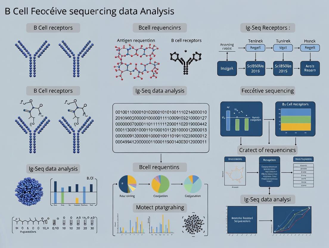
Decoding Immune Intelligence: A Comprehensive Guide to B Cell Repertoire Sequencing (Ig-Seq) Data Analysis
This article provides a detailed, end-to-end guide for researchers, scientists, and drug development professionals conducting B cell receptor repertoire sequencing (Ig-Seq) analysis.
Decoding Immune Intelligence: A Comprehensive Guide to B Cell Repertoire Sequencing (Ig-Seq) Data Analysis
Abstract
This article provides a detailed, end-to-end guide for researchers, scientists, and drug development professionals conducting B cell receptor repertoire sequencing (Ig-Seq) analysis. It begins with foundational concepts of adaptive immunity and the structure of immunoglobulins, explaining the biological significance of repertoire diversity. It then transitions to a practical, step-by-step walkthrough of the modern Ig-Seq analysis pipeline, from raw read processing and error correction to clonotype assignment, lineage tracing, and diversity quantification. The guide addresses common technical challenges, offering solutions for batch effects, contamination, and data normalization. Finally, it compares and validates different analytical tools and metrics, enabling robust interpretation for applications in vaccine development, autoimmunity, cancer immunology, and therapeutic antibody discovery. This resource synthesizes current methodologies to empower precise and reproducible immune repertoire research.
The Blueprint of Immunity: Understanding B Cells and the Power of Ig-Seq
Adaptive immunity provides vertebrates with a highly specific and memory-capable defense system. At its core are lymphocytes, with B cells playing the indispensable role of antibody production. This whitepaper provides a technical foundation for understanding B cell biology, explicitly framed within the context of B cell receptor (BCR) repertoire sequencing (Ig-Seq) data analysis research.
Core Principles of B Cell-Mediated Adaptive Immunity
B cells originate from hematopoietic stem cells in the bone marrow, where they undergo V(D)J recombination to generate a diverse primary BCR repertoire. Upon encountering a cognate antigen, B cells are activated, typically with T cell help, initiating a cascade of events: clonal expansion, somatic hypermutation (SHM), class switch recombination (CSR), and differentiation into antibody-secreting plasma cells or memory B cells.
Key Quantitative Metrics of B Cell Diversity: Table 1: Key Metrics in Primary B Cell Repertoire Generation
| Metric | Approximate Value | Biological Significance |
|---|---|---|
| Human Heavy Chain Gene Segments | ~44 V, ~23 D, 6 J | Raw genetic material for recombination. |
| Theoretical Combinatorial Diversity | ~10^12 | Diversity from V(D)J combination and junctional flexibility. |
| Estimated Actual Pre-immune Diversity | ~10^8 - 10^10 | Diversity after negative selection in bone marrow. |
| Somatic Hypermutation Rate | ~10^-3 per base per generation | Introduces point mutations in antigen-binding regions. |
B Cell Receptor Signaling and Activation Pathway
The BCR is a multi-protein complex composed of a membrane-bound immunoglobulin (mIg) non-covalently associated with a heterodimer of Igα (CD79a) and Igβ (CD79b). Antigen binding triggers a phosphorylation cascade.
Diagram 1: Core BCR signaling cascade leading to activation.
Ig-Seq Experimental Workflow for B Cell Repertoire Analysis
Ig-Seq enables high-throughput characterization of the BCR repertoire, providing insights into clonal dynamics, SHM, and isotype distribution.
Detailed Protocol: Library Preparation for Bulk BCR Sequencing
- Sample Input: Isolated PBMCs, sorted B cell subsets, or tissue biopsies.
- RNA/DNA Extraction: Use TRIzol (for RNA) or column-based kits (for gDNA). For RNA, perform reverse transcription using random hexamers or oligo-dT and a reverse transcriptase with high processivity.
- Target Amplification:
- For RNA/cDNA: Multiplex PCR is standard. Use multiple forward primers targeting V gene leader sequences and reverse primers targeting constant region genes (e.g., Cμ, Cγ, Cα). Critical: Use a high-fidelity polymerase to minimize PCR errors. Cycle number should be minimized (~20-25 cycles) to reduce bias.
- For gDNA: Similar multiplex PCR, but primers target V and J gene segments.
- Library Construction: Add sequencing adapters and sample indices via a second, limited-cycle PCR. Purify products using double-sided magnetic bead clean-up (e.g., 0.6x / 0.8x ratio).
- Quality Control & Quantification: Analyze fragment size distribution (Bioanalyzer/TapeStation) and quantify via qPCR or fluorometry.
- Sequencing: Pool libraries and sequence on platforms like Illumina MiSeq/NextSeq (paired-end 2x300bp is common for full-length V(D)J).
Diagram 2: End-to-end workflow for BCR repertoire sequencing.
The Scientist's Toolkit: Research Reagent Solutions
Table 2: Essential Reagents for B Cell & Ig-Seq Research
| Item | Function/Application | Example/Note |
|---|---|---|
| B Cell Isolation Kits | Negative or positive selection of human/mouse B cells from heterogeneous cell populations. | Magnetic-activated cell sorting (MACS) kits (e.g., Pan-B Cell Isolation Kit). |
| B Cell Stimulation Cocktails | Polyclonal activation of B cells in vitro for functional assays. | Combinations of anti-IgM/IgG F(ab')2, CD40L, CpG ODN, and cytokines (IL-2, IL-4, IL-21). |
| High-Fidelity Polymerase | Critical for accurate amplification of BCR genes with minimal PCR errors during library prep. | Enzymes like Q5 (NEB) or KAPA HiFi. |
| Multiplex V-Gene Primers | Sets of primers designed to amplify the vast majority of functional V genes with minimal bias. | Commercial primer sets (e.g., from iRepertoire) or carefully validated in-house mixes. |
| UMI (Unique Molecular Identifier) Adapters | Short random nucleotide tags added during cDNA synthesis to enable bioinformatic correction of PCR and sequencing errors. | Essential for accurate clonal quantification and mutation analysis. |
| Single-Cell Partitioning System | For linking heavy and light chain pairs from individual B cells. | Platforms like 10x Genomics Chromium, or microwell-based systems. |
| Flow Cytometry Antibodies | Phenotyping B cell subsets (Naive, Memory, Plasma), analyzing activation status, and sorting. | Anti-CD19, CD20, CD27, CD38, IgD, IgM, IgG, IgA. |
Key Data Analysis Metrics in Ig-Seq Research
Ig-Seq data analysis transforms raw sequences into biological insights, central to thesis research in this field.
Table 3: Core Analytical Outputs from Ig-Seq Data
| Analytical Goal | Key Metrics & Outputs | Significance for Research |
|---|---|---|
| Repertoire Diversity | Shannon Entropy, Clonality Index (1 - Pielou's evenness), Rarefaction Curves. | Quantifies repertoire breadth. Changes indicate immune activation, aging, or pathology. |
| Clonal Analysis | Clone Size Distribution, Largest Clone Frequency, Clonal Expansion Index. | Identifies antigen-driven responses. Tracks specific clones over time or between compartments. |
| Somatic Hypermutation | Mutation Frequency per clone, Mutation Hotspots (R/S ratios in CDRs vs. FWRs). | Measures affinity maturation. Aberrant patterns can indicate dysregulation (e.g., in autoimmunity). |
| Isotype/Class Switching | Isotype Distribution (IgM, IgG, IgA, etc.), Class Switch Recombination Events. | Induces effector function. Profiles humoral immune response quality (e.g., IgG1 vs. IgA). |
| Lineage Tree Reconstruction | Tree Topology, Branching Depth, Ancestral Sequence Inference. | Visualizes clonal evolution and intraclonal diversity, inferring antigen selection pressure. |
This whitepaper provides a technical examination of the genetic mechanisms underpinning antibody diversity, framed within the context of B cell receptor repertoire (Ig-Seq) data analysis. Understanding V(D)J recombination, somatic hypermutation (SHM), and class switch recombination (CSR) is paramount for interpreting high-throughput sequencing data in research and therapeutic discovery, from tracking clonal lineages to identifying vaccine-elicited responses.
V(D)J Recombination: Constructing the Primary Repertoire
V(D)J recombination is the site-specific genetic rearrangement that assembles variable (V), diversity (D), and joining (J) gene segments to create the coding sequence for the variable domains of immunoglobulin heavy (IgH) and light (IgL) chains. This process occurs in progenitor and precursor B cells in the bone marrow, generating a naive B cell repertoire with an estimated theoretical diversity of ~10^13 unique receptors.
Molecular Mechanism
The recombination is directed by recombination signal sequences (RSSs) flanking each V, D, and J gene segment. An RSS consists of a heptamer, a spacer (12 or 23 base pairs), and a nonamer. The "12/23 rule" ensures joining only between segments with RSSs of different spacer lengths.
The recombination is catalyzed by the RAG complex (RAG1 and RAG2). The key steps are:
- Synapsis: RAG complex binds to one RSS.
- Cleavage: Introduces a double-strand break between the coding segment and the RSS, generating hairpin-sealed coding ends and blunt signal ends.
- Hairpin Opening and Processing: The Artemis:DNA-PKcs complex opens hairpins, and exonucleases and terminal deoxynucleotidyl transferase (TdT) add or remove nucleotides, creating junctional diversity.
- Ligation: Non-homologous end joining (NHEJ) factors (Ku70/Ku80, XRCC4, DNA Ligase IV) ligate the processed coding ends.
Table 1: Key Quantitative Metrics of V(D)J Recombination
| Metric | Human IgH Locus | Human Igκ Locus | Contribution to Diversity |
|---|---|---|---|
| Functional Gene Segments | ~45 V, ~23 D, 6 J | ~35 V, 5 J | Combinatorial diversity |
| Theoretical Combinatorial Combinations | ~45 * 23 * 6 = ~6,210 | ~35 * 5 = 175 | ~1.1 x 10^6 VH:VL pairs |
| Junctional Diversity (N/P-additions) | Average 15-20 nt added per V-D-J junction | Average 5-10 nt added per V-J junction | Expands diversity by ~10^13 |
| Estimated Naive Repertoire Size | ~10^8 - 10^10 unique clonotypes in human periphery |
Experimental Protocol: Targeted Locus Amplification for V(D)J Arrangement
Purpose: To determine the complete V(D)J rearrangement status of an immunoglobulin or T cell receptor locus from a single cell or limited input.
Key Steps:
- Cell Lysis & DNA Isolation: Single B cells are lysed, and genomic DNA is released.
- Restriction Digest: Use of a frequent cutter (e.g., MseI) to fragment genomic DNA, leaving the locus of interest in large fragments.
- Ligation of Cassette Linkers: Specific double-stranded linkers are ligated to the digested DNA ends.
- Targeted PCR: Nested PCR is performed using one primer specific to the ligated linker and another primer specific to a known, conserved region within the locus (e.g., within the J-C intron or constant region).
- Sequencing & Analysis: PCR products are sequenced via Sanger or NGS. Sequences are aligned to the germline reference to identify the specific V, D, J segments used and the exact nucleotide sequence of the junctions.
Somatic Hypermutation (SHM): Fine-Tuning Affinity
Following antigen encounter, activated B cells proliferate within germinal centers and undergo SHM. This introduces point mutations into the rearranged V(D)J regions at a rate of ~10^-3 mutations per base pair per generation, approximately one million times higher than the spontaneous mutation rate.
Molecular Mechanism
SHM is initiated by Activation-Induced Cytidine Deaminase (AID), which deaminates deoxycytidine (dC) to deoxyuracil (dU) in single-stranded DNA, primarily within transcribed variable regions. The resulting dU:dG mismatch is then processed by error-prone repair pathways:
- Base Excision Repair (BER): Uracil-DNA glycosylase removes the uracil, creating an abasic site. Error-prone polymerases (e.g., Pol η) replicate across the lesion, potentially introducing mutations.
- Mismatch Repair (MMR): The MutSα complex (MSH2-MSH6) recognizes the mismatch. Exonuclease 1 excises a stretch of DNA, and error-prone polymerases fill the gap, leading to mutations clustered around the original site.
Mutations occur in "hotspots" defined by the AID target motif (WRRC, where W = A/T, R = purine). The outcome is affinity maturation, where B cells with mutations that confer higher affinity for antigen receive survival signals.
Table 2: SHM Characteristics and Analysis Metrics in Ig-Seq
| Parameter | Typical Value / Description | Significance in Repertoire Analysis |
|---|---|---|
| Mutation Rate | ~1 x 10^-3 / bp / generation | Drives affinity maturation. |
| Target Motif | WRRC (A/T A/G A/G C) | Explains biased mutation patterns. |
| Mutation Spectrum | Predominantly transitions (C→T, G→A) | Signature of AID activity. |
| Clonal Tree Analysis | Reconstruction of lineage from shared mutations | Tracks evolution of antigen-specific response. |
| Replacement/Silent (R/S) Ratio | Ratio of mutations in CDRs vs. FRs | Positive selection indicated by R/S > 2.9 in CDRs. |
Experimental Protocol:In VitroSHM Assay
Purpose: To measure the activity and specificity of AID or to screen for compounds that modulate SHM.
Key Steps:
- Reporter Construct: A cell line (e.g., Ramos Burkitt's lymphoma or engineered CH12F3) is used, or a non-B cell line (e.g., HEK293) is transfected with a plasmid containing an AID-sensitive reporter gene (e.g., a GFP gene rendered non-functional by a stop codon within an AID hotspot).
- AID Expression: The cells are engineered to express AID constitutively or upon induction (e.g., via a tet-on system).
- Mutation Induction & Selection: Cells are cultured for several days to allow SHM. If using a selectable reporter (e.g., GFP reversion or antibiotic resistance), cells that have acquired a reverting mutation are selected by FACS or drug treatment.
- Analysis: Mutation frequency is calculated as (number of revertant colonies / total number of viable cells). For deeper analysis, genomic DNA is extracted from the population, the reporter locus is amplified by PCR, and products are sequenced to characterize the spectrum and distribution of mutations.
Class Switch Recombination (CSR): Changing Effector Function
CSR alters the immunoglobulin isotype (e.g., from IgM/IgD to IgG, IgE, IgA) while retaining the antigen-specific variable region. This changes the antibody's effector functions (complement activation, placental transfer, mucosal secretion).
Molecular Mechanism
CSR is also initiated by AID, but targets switch (S) regions located upstream of each constant (CH) region (except Cδ). S regions are G-rich, repetitive, and transcriptionally active.
- Germline Transcription: Cytokines (e.g., IL-4, TGF-β, IFN-γ) induce transcription through target S regions (e.g., Sμ to Sγ1), making DNA accessible.
- AID Targeting: AID deaminates dCs within the transcribed S regions of both the donor (e.g., Sμ) and acceptor (e.g., Sγ1) regions.
- DSB Formation: The dU lesions are processed by BER/MMR into double-strand breaks (DSBs).
- Ligation: The DSBs in the donor and acceptor S regions are joined via a form of NHEJ (alternative end-joining, alt-EJ, involving microhomology) that results in the deletion of the intervening DNA loop.
Table 3: Cytokine Regulation of CSR
| Cytokine | Primary Induced Isotype(s) | Key Signaling Transcription Factor |
|---|---|---|
| IL-4 | IgG1, IgE | STAT6 |
| IFN-γ | IgG3, IgG2a (mouse) | STAT1, T-bet |
| TGF-β | IgG2b (mouse), IgA | Smad proteins |
| BAFF/APRIL | IgA (in conjunction with TGF-β) | NF-κB |
Experimental Protocol:In VitroCSR Assay
Purpose: To measure the efficiency of CSR in B cells in response to specific stimuli.
Key Steps:
- B Cell Isolation: Naive murine splenic B cells or human peripheral blood B cells are purified using negative selection magnetic bead kits.
- Stimulation: Cells are cultured with CSR-inducing stimuli:
- For IgM to IgG1/IgE: Anti-CD40 antibody (to mimic T cell help) + IL-4.
- For IgM to IgA: Anti-CD40 + TGF-β + IL-4/BAFF.
- LPS can be used as a T-independent stimulant for mouse B cells.
- Culture: Cells are cultured for 4-5 days to allow proliferation and switching.
- Analysis:
- Flow Cytometry: Surface staining for IgM, IgD, and the switched isotype of interest (e.g., IgG1, IgE, IgA). Frequency of switched (IgM- IgX+) cells is determined.
- Molecular Analysis (PCR/Southern Blot): Genomic DNA is analyzed for the presence of deletional switch junctions (e.g., using primers spanning Sμ and the target S region) or the absence of intervening constant regions.
The Scientist's Toolkit: Research Reagent Solutions
| Reagent / Material | Function & Application |
|---|---|
| Anti-CD40 Antibody | Agonistic antibody used in vitro to provide essential T cell-like co-stimulation for B cell activation, proliferation, and CSR induction. |
| Cytokines (IL-4, IFN-γ, TGF-β) | Recombinant proteins used to direct specific CSR pathways in cultured B cells by activating STAT and other signaling pathways. |
| AID Inhibitors (e.g., small molecules, HMBCA) | Chemical compounds used to specifically inhibit AID enzymatic activity in functional studies to dissect its role in SHM and CSR. |
| Uracil-DNA Glycosylase Inhibitor (UGI) | Protein inhibitor that blocks the base excision repair pathway of SHM, used to study the alternative MMR pathway and to trap uracils for sequencing methods. |
| 5'-Bromo-2'-deoxyuridine (BrdU) | Thymidine analog incorporated into DNA during replication. Used to label proliferating germinal center B cells undergoing SHM/CSR for flow cytometry or microscopy. |
| Switch-Specific PCR Primers | Oligonucleotide primers designed to anneal within Sμ and downstream S regions (e.g., Sγ1, Sε, Sα) to amplify and sequence switch junction fragments for CSR analysis. |
| Single-Cell BCR Amplification Kits | Commercial kits (e.g., from 10x Genomics, Takara Bio) for reverse transcription and multiplex PCR to amplify paired heavy and light chain V(D)J transcripts from single B cells. |
| RAG1/2 Recombinant Complex | Purified enzyme complex used in in vitro biochemical assays to study the kinetics and specificity of V(D)J cleavage on defined RSS substrates. |
Essential Visualizations for Pathway and Workflow Clarity
Title: V(D)J Recombination Core Steps
Title: Somatic Hypermutation Mechanism
Title: Class Switch Recombination Workflow
Title: Core Ig-Seq Data Analysis Pipeline
What is the B Cell Receptor Repertoire? Defining Clonotypes, Diversity, and Richness.
Within the broader thesis of B cell receptor repertoire sequencing (Ig-Seq) data analysis research, the B cell receptor (BCR) repertoire represents the total collection of immunoglobulins (Igs) expressed by all B cells in an individual at a given time. It is a functional readout of the adaptive immune system's capacity to recognize antigens. Ig-Seq research aims to decode this repertoire to understand immune responses in health, disease, and following therapeutic interventions, providing critical insights for vaccine development, oncology, and autoimmune disease diagnostics.
Core Definitions and Framework
Clonotype: The set of B cells descended from a single, common naïve progenitor, sharing an identical rearranged V(D)J nucleotide sequence for their BCR. Clonotype definition is the cornerstone of repertoire analysis.
Diversity: A statistical measure describing both the number of unique clonotypes present and the evenness of their frequency distribution within a repertoire. A highly diverse repertoire has many unique clonotypes at relatively equal frequencies.
Richness: The total number of distinct clonotypes present in a sample. It is a component of diversity but does not account for clonal frequency distribution.
Quantitative Metrics for Diversity and Richness
The following table summarizes common metrics used to quantify BCR repertoire properties.
| Metric | Formula / Description | Interpretation | Application in Ig-Seq |
|---|---|---|---|
| Clonal Richness | S = Number of distinct clonotypes. | Raw count of unique sequences. Simple but ignores abundance. | Initial sample comparison. |
| Shannon Index (H') | H' = -Σ(pi * ln(pi)); p_i = clonotype frequency. | Combines richness and evenness. Increases with more clonotypes and more even distribution. | General diversity assessment in immune monitoring. |
| Simpson Index (D) | D = Σ(p_i²). | Probability that two randomly selected sequences belong to the same clonotype. Emphasizes dominant clones. | Identifying clonal expansions (e.g., in leukemia). |
| Pielou's Evenness (J') | J' = H' / ln(S). | Measures how evenly frequencies are distributed (0 to 1). | Distinguishing if low diversity is due to few clones or dominance. |
| Chao1 Estimator | Ŝ = S_obs + (F1² / (2*F2)); F1=singletons, F2=doubletons. | Estimates true richness, correcting for unseen rare clonotypes. | Accounting for sequencing depth limitations. |
Detailed Experimental Protocol: BCR Repertoire Sequencing (Ig-Seq)
This protocol outlines a standard bulk RNA-based approach for BCR heavy-chain (IGH) repertoire sequencing.
1. Sample Preparation & RNA Isolation
- Source: Peripheral blood mononuclear cells (PBMCs), sorted B cells, or tissue biopsies.
- Lysis: Use TRIzol or RLT buffer with β-mercaptoethanol.
- RNA Extraction: Perform using silica-membrane columns (e.g., RNeasy Mini Kit, Qiagen). Include DNase I treatment.
- Quality Control: Assess RNA Integrity Number (RIN) >7.0 using Bioanalyzer.
2. cDNA Synthesis and Target Amplification
- Primer Strategy: Use multiplexed reverse transcription (RT) primers targeting the constant region (C region) of IGH transcripts (e.g., for IgG, IgA, IgM, IgD, IgE).
- RT Reaction: 1μg total RNA, gene-specific primers, and a reverse transcriptase (e.g., SuperScript IV).
- Primary PCR: Amplify the V(D)J region using a multiplex pool of V-gene forward primers and a C-gene reverse primer. Use a high-fidelity polymerase (e.g., KAPA HiFi) for 18-25 cycles.
- Indexing PCR (Nextera XT): Add sample-specific dual indices and sequencing adapters (Illumina) in a limited-cycle (8-10 cycles) PCR.
3. Library Purification and Quantification
- Size Selection: Use double-sided magnetic bead cleanup (e.g., AMPure XP beads) to remove primer dimers and large contaminants (~300-600 bp target).
- Quantification: Use fluorometric assays (Qubit dsDNA HS Assay).
- Quality Check: Assess library fragment size distribution via Bioanalyzer (High Sensitivity DNA chip).
4. High-Throughput Sequencing
- Platform: Illumina MiSeq (for exploratory) or NextSeq/NovaSeq (for deep repertoire).
- Configuration: 2x300 bp paired-end sequencing is standard for full V(D)J coverage.
- Depth: Aim for 5x10⁵ to 5x10⁶ reads per sample for robust diversity estimates.
5. Bioinformatic Analysis Pipeline
- Pre-processing: Merge paired-end reads (FLASH), quality filter (Trimmomatic).
- Alignment & Annotation: Map reads to IMGT reference V, D, J, and C genes (IgBLAST, MiXCR).
- Clonotype Definition: Cluster sequences with identical V and J gene calls and identical CDR3 nucleotide sequences.
- Diversity Analysis: Calculate richness, Shannon, Simpson indices using tools like Alakazam or immunarch.
- Visualization: Generate clonal abundance plots, phylogenetic trees, and diversity curve rarefaction plots.
Visualizations
Ig-Seq Experimental Workflow
Bioinformatic Clonotype Analysis Pipeline
Clonotype Definition Logic
The Scientist's Toolkit: Research Reagent Solutions
| Item | Function in BCR Repertoire Research |
|---|---|
| RNeasy Mini/Micro Kit (Qiagen) | Silica-membrane-based purification of high-quality total RNA from cell lysates, critical for accurate cDNA synthesis. |
| SuperScript IV Reverse Transcriptase (Thermo Fisher) | High-temperature, high-fidelity reverse transcriptase for generating full-length cDNA from BCR mRNA transcripts. |
| Multiplex IGH V-gene Primers | Pre-designed pools of primers targeting the leader or framework 1 regions of human/mouse IGHV genes for unbiased amplification. |
| KAPA HiFi HotStart ReadyMix (Roche) | High-fidelity DNA polymerase for low-error amplification of V(D)J regions during library construction, minimizing PCR artifacts. |
| AMPure XP Beads (Beckman Coulter) | Magnetic beads for size-selective purification of PCR products, removing primers, dimers, and non-specific products. |
| Illumina Nextera XT DNA Library Prep Kit | Facilitates rapid, simultaneous fragmentation and indexing of amplicons for Illumina sequencing. |
| MiXCR / IgBLAST Software | Essential bioinformatics tools for aligning sequence reads to immunoglobulin gene databases and performing clonotype assignment. |
| Anti-CD19/CD20 Magnetic Beads (e.g., Miltenyi) | For positive selection of B cells from PBMC samples to increase repertoire sequencing sensitivity and specificity. |
1. Introduction This whitepaper details the evolution of B cell receptor (BCR) repertoire analysis, charting its progression from low-resolution bulk techniques to single-cell, next-generation sequencing (NGS) paradigms. Framed within a broader thesis on Ig-Seq data analysis research, this guide underscores how technological leaps have enabled unprecedented insights into adaptive immune responses, with direct applications in vaccine development, autoimmune disease profiling, and therapeutic antibody discovery.
2. Historical & Methodological Progression The quantitative evolution of key metrics across technological generations is summarized in Table 1.
Table 1: Quantitative Comparison of Repertoire Sequencing Technologies
| Technology | Era | Readout | Throughput (Cells/Seq) | Key Resolvable Metric | Limitations |
|---|---|---|---|---|---|
| Spectratyping (CDR3-L) | 1990s | Fragment Length | Bulk Population (~10⁶) | CDR3 Length Distribution | No sequence identity; low multiplex. |
| Sanger Cloning | 2000s | Sequence | ~10² clones per run | Full V(D)J sequence for clones | Low depth, high cost, labor-intensive. |
| 1st-Gen NGS (454/Roche) | Mid-2000s | Sequence | 10⁴ - 10⁶ reads | Clonotype diversity & frequency | Short reads, high error rates in homopolymers. |
| 2nd-Gen NGS (Illumina) | 2010s | Sequence | 10⁷ - 10⁹ reads | High-resolution repertoire diversity | Paired-chain linkage lost in bulk methods. |
| Single-Cell NGS + 5'RACE | 2010s | Paired-chain Sequence | 10³ - 10⁵ cells | Native heavy & light chain pairing | Cell throughput limited by platform. |
| Single-Cell NGS + Barcoding | 2020s | Paired-chain + Transcriptome | 10⁴ - 10⁶ cells | Paired clonotype with cell phenotype (CITE-seq) | Complex data integration, higher cost. |
3. Detailed Experimental Protocols
3.1. Legacy Protocol: Spectratyping for CDR3 Length Analysis
- Principle: Amplify CDR3-encoding regions using V-family forward and constant region reverse primers, followed by capillary electrophoresis to profile amplicon lengths.
- Steps:
- RNA Isolation: Extract total RNA from PBMCs or lymphoid tissue.
- cDNA Synthesis: Reverse transcribe using constant region (Cγ, Cμ)-specific primers.
- Multiplex PCR: Perform separate PCR reactions for each VH family primer (e.g., VH1-VH7) with a fluorescently labeled constant region primer.
- Fragment Analysis: Pool PCR products and run on a capillary sequencer. Size distribution profiles (spectratypes) indicate repertoire skewing—a Gaussian distribution denotes polyclonality, while peaked distributions suggest clonal expansion.
3.2. Modern Protocol: High-Throughput Ig-Seq Library Preparation (5'RACE-based)
- Principle: Use 5' Rapid Amplification of cDNA Ends (RACE) with template switching to capture complete V(D)J transcripts without V-gene-specific primers, minimizing bias.
- Steps:
- Cell Lysis & Reverse Transcription: Lysate cells in a plate. Perform RT using a constant region (IgG/IgA/IgM)-specific primer and a template-switching oligo (TSO). A unique molecular identifier (UMI) is incorporated to correct for PCR errors and duplicates.
- cDNA Amplification: Amplify the full-length V(D)J-constant region cDNA using primers complementary to the TSO and constant region.
- Nested PCR for NGS: Perform a second, nested PCR to add platform-specific sequencing adapters (e.g., Illumina P5/P7) and sample indices (barcodes).
- Library QC & Sequencing: Purify, quantify, and size-select the library (typically ~500-700bp). Pool libraries for high-throughput sequencing on platforms like Illumina MiSeq/Novaseq (2x300bp paired-end).
3.3. Advanced Protocol: Single-Cell BCR-Seq with Feature Barcoding (CITE-seq)
- Principle: Partition single cells into droplets (e.g., 10x Genomics) where each cell's mRNA and surface proteins are barcoded with a unique cell identifier.
- Steps:
- Cell Preparation: Stain cells with antibody-derived tags (ADTs)—oligo-conjugated antibodies against surface markers (e.g., CD19, CD27).
- Gel Bead-in-Emulsion (GEM) Generation: Co-encapsulate single cells, lysis reagent, and gel beads in droplets. Each bead contains barcoded primers for mRNA capture and ADT capture.
- Inside-GEM RT: Lysed cells release mRNA and bound ADTs. Reverse transcription occurs, tagging each molecule with the cell's unique barcode and a UMI.
- Library Construction: Generate separate cDNA libraries for gene expression (from mRNA), BCR enriched V(D)J transcripts (via targeted PCR), and surface protein expression (from ADT-derived cDNA).
- Sequencing & Analysis: Sequence libraries and use computational tools (e.g., Cell Ranger V(D)J) to assemble contigs, identify paired heavy and light chains, and link clonotypes to cell phenotype.
4. Visualizing Key Workflows and Relationships
Title: Evolution from Bulk to Single-Cell BCR Analysis
Title: End-to-End Ig-Seq Workflow from Lab to Data
5. The Scientist's Toolkit: Key Research Reagent Solutions Table 2: Essential Reagents and Materials for Ig-Seq Experiments
| Item Name | Provider Examples | Function |
|---|---|---|
| SMARTer Human BCR Kit | Takara Bio | All-in-one kit for 5'RACE-based, bias-controlled amplification of human Ig transcripts from bulk RNA. |
| Chromium Next GEM Single Cell 5' Kit + Feature Barcode | 10x Genomics | Integrated reagent system for partitioning cells and barcoding mRNA & surface proteins (CITE-seq). |
| Human BCR Panel (Antibody-Oligo Conjugates) | BioLegend (TotalSeq) | Oligo-tagged antibodies for cell surface protein detection (e.g., CD19, CD20, CD27) in single-cell assays. |
| Unique Molecular Identifiers (UMI) | Integrated in kits (e.g., Illumina, 10x) | Short random nucleotide sequences added during RT to tag each original mRNA molecule, enabling accurate quantification and error correction. |
| PhiX Control v3 | Illumina | Sequencing control library for run quality monitoring, essential for low-diversity amplicon libraries like Ig-Seq. |
| SPRIselect Beads | Beckman Coulter | Magnetic beads for size selection and clean-up of NGS libraries, critical for removing primer dimers. |
| High-Fidelity DNA Polymerase | NEB (Q5), Thermo Fisher | Enzyme for low-error PCR amplification during library construction to minimize sequencing artifacts. |
Key Biological and Clinical Questions Addressable by Ig-Seq Analysis
Immunoglobulin or B-cell receptor sequencing (Ig-Seq) is a cornerstone of modern immunology research, enabling the high-resolution characterization of the adaptive immune repertoire. Within the context of a broader thesis on B cell repertoire sequencing data analysis, this whitepaper details the specific biological and clinical questions that can be addressed through rigorous Ig-Seq experimentation and computational interpretation.
Core Biological Questions
Repertoire Diversity and Clonal Architecture
A primary application of Ig-Seq is the quantitative assessment of the diversity of the B-cell repertoire, which is fundamental to understanding immune competence, response to antigenic challenge, and the detection of pathological skewing.
Key Quantitative Metrics: Table 1: Core Ig-Seq Diversity and Clonality Metrics
| Metric | Description | Typical Range (Peripheral Blood) | Biological Interpretation |
|---|---|---|---|
| Clonality Index | 1 - Pielou's evenness. 0=perfectly diverse, 1=monoclonal. | 0.01 - 0.15 (Healthy) | High clonality indicates antigen-driven expansion or malignancy. |
| Shannon Diversity | Entropy measure accounting for richness and evenness. | 8 - 12 (for ~50k sequences) | Higher values indicate a more diverse, balanced repertoire. |
| Unique Clones | Count of distinct nucleotide sequences. | 10^4 - 10^6 per sample | Lower counts may indicate immunosenescence or immunosuppression. |
| Gini Index | Inequality of clone size distribution. 0=perfect equality. | 0.7 - 0.9 (Healthy) | Higher index reflects greater dominance by large clones. |
Experimental Protocol: Bulk VDJ Sequencing for Diversity Analysis
- Sample Preparation: Isolate PBMCs or tissue-derived lymphocytes via density gradient centrifugation.
- Nucleic Acid Extraction: Extract total RNA (for expressed repertoire) or genomic DNA (for germline configuration) using column-based kits with DNase/RNase treatment as required.
- Library Preparation: Use multiplex PCR with primers targeting conserved framework regions of V genes and J genes. Incorporate unique molecular identifiers (UMIs) during reverse transcription or early PCR cycles to correct for amplification bias and sequencing errors.
- Sequencing: Perform high-throughput sequencing on platforms like Illumina MiSeq/Novaseq (2x300bp paired-end recommended for full-length VDJ).
- Bioinformatic Analysis: Process raw reads through a pipeline: UMI consensus calling, V(D)J alignment (using IMGT/HighV-QUEST, IgBLAST, or partis), clonotype clustering (≥97% nucleotide identity), and metric calculation with tools like Alakazam or immunarch.
Antigen-Driven Selection and Somatic Hypermutation (SHM)
Ig-Seq enables the study of adaptive immune maturation by quantifying and localizing SHM and analyzing selection pressures.
Key Quantitative Data: Table 2: Metrics for Somatic Hypermutation and Selection Analysis
| Metric | Calculation | Typical Value (Memory B Cells) | Interpretation |
|---|---|---|---|
| Mutation Frequency | # of mutations / length of productive V-region. | 2% - 8% | Higher frequency indicates greater antigen exposure and affinity maturation. |
| Replacement/Silent (R/S) Ratio | Ratio of amino acid-changing to silent mutations in Complementarity Determining Regions (CDRs) vs. Framework Regions (FRs). | CDR R/S > 2.9; FR R/S ~2.8 | CDR R/S > expected (~2.9) indicates positive selection. FR R/S < expected suggests negative selection. |
| Focusing Factor | Measures the concentration of mutations in CDRs. | >1 in antigen-selected repertoires | Values >1 indicate preferential targeting of mutations to CDRs. |
Experimental Protocol: Antigen-Specific B Cell Sorting and Ig-Seq
- Antigen Probe Design: Generate biotinylated recombinant antigen or use peptide-MHC tetramers.
- Cell Staining & Sorting: Label PBMCs with fluorescent antigen probes and antibodies for B cell markers (CD19, CD20). Use FACS to isolate antigen-binding (e.g., CD19+ Antigen+) and non-binding populations.
- Single-Cell or Bulk Sequencing: For high-resolution analysis, use single-cell Ig-Seq platforms (e.g., 10x Genomics VDJ solution). For deeper SHM analysis, bulk UMI-based sequencing from sorted populations is performed.
- Analysis: Align sequences, reconstruct clonal lineages, and calculate SHM and selection statistics using tools like Change-O and ShazaM. Perform phylogenetic tree construction (via dnaml or IgPhyML) to model clonal evolution.
Diagram 1: SHM and Selection in B Cell Maturation (76 chars)
Translational and Clinical Questions
Biomarker Discovery for Autoimmunity and Cancer
Clonal expansions and specific antibody signatures serve as diagnostic, prognostic, and minimal residual disease (MRD) biomarkers.
Key Quantitative Data: Table 3: Clinical Biomarkers Detectable by Ig-Seq
| Condition | Ig-Seq Biomarker | Detection Method | Clinical Utility |
|---|---|---|---|
| B-cell Lymphoma | Dominant clonotype(s) at diagnosis. | Clonality assessment, specific V-J rearrangement tracking. | MRD monitoring; sensitivity can exceed 10^-6. |
| Autoimmunity (e.g., RA, SLE) | Expanded clones in tissue (synovium, kidney); public sharing of specific CDR3 sequences. | Repertoire overlap analysis (Morisita-Horn index); antigenic motif inference. | Disease activity correlation; identifying pathogenic clones. |
| Immunodeficiency | Reduced diversity; skewed isotype distribution. | Diversity index calculation; Ig isotype (IgM, IgG, IgA) frequency. | Assessing immune reconstitution post-therapy. |
Experimental Protocol: MRD Monitoring in B-ALL
- Diagnostic Sample: Perform Ig-Seq on diagnostic bone marrow or blood to identify the leukemia-specific IgH VDJ rearrangement(s).
- Probe Design: Design patient-specific quantitative PCR (qPCR) assays or dPCR assays based on the unique CDR3 sequence. Alternatively, use high-sensitivity next-generation sequencing (NGS) panels.
- Longitudinal Sampling: Extract DNA from follow-up bone marrow aspirates.
- Detection: Use the patient-specific assay to quantify the malignant clone. NGS-based methods involve deep sequencing (≥10^6 reads) and bioinformatic filtering for the diagnostic sequence.
Vaccine Response and Infectious Disease Profiling
Ig-Seq dissects the kinetics, breadth, and durability of antigen-specific B cell responses.
Key Quantitative Data: Table 4: Ig-Seq Metrics for Vaccine Response
| Metric | Timepoint Comparison | Interpretation of Effective Response |
|---|---|---|
| Clonal Expansion | Pre-vaccination vs. Post-boost (e.g., day 7, day 28). | Significant increase in size of antigen-specific clones. |
| Lineage Diversification | Tracking SHM within expanding clonal families over time. | Increased intra-clonal diversity indicating ongoing maturation. |
| Convergent Antibodies | Identification of "public" or shared antibody sequences across individuals. | Indicates a stereotypic, effective response to immunodominant epitopes. |
Experimental Protocol: Tracking Antigen-Specific Responses Post-Vaccination
- Longitudinal Sampling: Collect PBMCs pre-vaccination (baseline), at peak response (~day 7-10), and at memory phase (~day 28+).
- Antigen-Specific Enrichment: Use fluorescently labeled antigen baits to sort antigen-binding memory B cells or plasmablasts.
- Single-Cell VDJ + Transcriptome: Utilize 10x Genomics 5' Immune Profiling to pair full-length V(D)J sequences with gene expression data from the same cell.
- Clonal Tracking & Analysis: Reconstruct clonal lineages, map their expansion kinetics, and correlate SHM with B cell state (naive, memory, plasma) from transcriptional data.
Diagram 2: Ig-Seq Workflow for Vaccine Response (72 chars)
The Scientist's Toolkit: Research Reagent Solutions
Table 5: Essential Reagents and Materials for Ig-Seq Studies
| Item | Function | Example Product/Catalog |
|---|---|---|
| UMI-Linked RT Primers | Primer sets containing Unique Molecular Identifiers (UMIs) for error-corrected consensus sequencing of the Ig transcriptome. | Biolegend TotalSeq-B, Takara SMARTer Human BCR IgG H/K/L Profile Kit. |
| Multiplex V(D)J PCR Primers | Degenerate primers designed to amplify the vast majority of human or mouse V and J gene segments. | iRepertoire Inc. Immune Profiling Assay, ArcherDX Immunoverse. |
| Single-Cell Partitioning System | Microfluidic chips or droplets for isolating single cells and barcoding their nucleic acids. | 10x Genomics Chromium Controller, BD Rhapsody Cartridge. |
| Antigen Probes for FACS | Recombinant, biotinylated antigens or peptide-MHC tetramers for isolating antigen-specific B cells. | NIH Tetramer Core Facility reagents, Acro Biosystems proteins. |
| B Cell Isolation/Magnetic Kits | Antibody cocktails for negative or positive selection of pan-B cells or subsets from complex samples. | Miltenyi Biotec Human B Cell Isolation Kit II, STEMCELL Technologies EasySep. |
| High-Fidelity Polymerase | PCR enzymes with low error rates essential for accurate SHM analysis. | NEB Q5, Takara PrimeSTAR GXL. |
| Analysis Software Suites | Comprehensive platforms for Ig-Seq data processing, clonotyping, and visualization. | 10x Genomics Cell Ranger VDJ, Adaptive Biotechnologies immunoSEQ Analyzer, open-source Immcantation portal. |
From Raw Reads to Biological Insights: A Step-by-Step Ig-Seq Analysis Pipeline
Within the critical research domain of B cell repertoire sequencing (Ig-Seq) for analyzing antibody-mediated immunity, therapeutic antibody discovery, and immunomonitoring, the experimental design is paramount. The choices made at the outset—regarding sample type, library preparation, and sequencing platform—profoundly influence the biological questions that can be answered. This guide provides an in-depth technical framework for designing robust Ig-Seq experiments, framed within the context of advancing B cell receptor (BCR) repertoire analysis research.
Sample Types: Bulk vs. Single-Cell Sequencing
The decision between bulk and single-cell sequencing defines the resolution and scope of the resulting BCR repertoire data.
Bulk BCR Sequencing
- Principle: RNA or DNA is extracted from a pooled population of B cells (e.g., from PBMCs, tissue homogenates, or sorted B cell subsets). Amplification primers target the rearranged V(D)J regions, and the resulting libraries represent a composite, frequency-weighted profile of all clonotypes in the sample.
- Advantages: Cost-effective, higher throughput for deep sampling of repertoire diversity, less technically demanding, suitable for tracking clonal dynamics over time or between conditions.
- Limitations: Inherently loses paired heavy-chain (HC) and light-chain (LC) linkage, preventing native antibody sequence pairing. Can be biased by primer efficiency and PCR duplication. Does not preserve cellular metadata (e.g., transcriptome, phenotype).
Single-Cell BCR Sequencing
- Principle: Individual B cells are isolated (via FACS, microwell, or droplet-based partitioning), and the paired V(D)J transcripts from each cell are barcoded with a unique cellular identifier (UMI). This preserves the native HC-LC pair.
- Advantages: Retains critical HC-LC pairing information essential for recombinant antibody expression and functional validation. Enables coupling with cellular phenotyping (CITE-seq) or transcriptomics (5’ scRNA-seq).
- Limitations: Significantly higher cost per cell, lower throughput in terms of total unique sequences recovered, more complex data analysis, and potential sampling bias due to cell viability and capture efficiency.
Table 1: Comparative Analysis of Bulk vs. Single-Cell Ig-Seq
| Feature | Bulk BCR-Seq | Single-Cell BCR-Seq |
|---|---|---|
| HC-LC Pairing | Lost | Preserved (Native Pair) |
| Throughput (Cells) | High (Millions) | Medium (10^3 - 10^5) |
| Cost per Sample | Low - Medium | High |
| Clonal Resolution | Frequency-based, no pairing | Clonal families with paired sequences |
| Cellular Context | None | Can be linked to phenotype/transcriptome |
| Primary Use Case | Repertoire diversity, clonal tracking, SHM analysis | Therapeutic antibody discovery, precise clonotype analysis |
| Key Challenge | PCR/priming bias, data assembly | Cell viability, capture efficiency, data complexity |
Library Preparation Methodologies
Library preparation is the most critical wet-lab step, determining the quality and representativeness of the sequencing data.
Protocol 1: Multiplex PCR-Based Bulk BCR-Seq (Adapted from [1])
This protocol is standard for high-depth profiling of BCR repertoires from sorted cell populations or total PBMCs.
- Input Material: Total RNA or genomic DNA from ~10^5 - 10^6 B cells.
- Reverse Transcription (if using RNA): Use isotype-specific (IgG, IgM, etc.) or pan-immunoglobulin constant region primers to generate cDNA.
- Primary Amplification: Perform multiplex PCR using a pool of forward primers targeting leader sequences or framework regions of V gene segments and reverse primers targeting the C region or J segments. Include a unique sample index at this stage.
- Nested PCR (Optional but Recommended): A second round of PCR with inner primers to increase specificity and add platform-specific sequencing adapters (e.g., Illumina P5/P7).
- Purification & Quantification: Purify amplicons using SPRI beads. Quantify via qPCR or bioanalyzer.
- Sequencing: Pool libraries at equimolar ratios for sequencing on an Illumina MiSeq or HiSeq platform (2x300bp recommended).
Protocol 2: Droplet-Based Single-Cell 5’ BCR + Transcriptome (10x Genomics)
This integrated protocol captures paired HC/LC sequences alongside the cellular transcriptome from the same cell [2].
- Input Material: A single-cell suspension of viable nucleated cells (e.g., PBMCs) at a concentration of 700-1200 cells/µL.
- Gel Bead-in-Emulsion (GEM) Generation: Cells, gel beads (containing barcoded oligonucleotides with Illumina adapters, a cell barcode, a UMI, and a poly-dT/V(D)J primer), and master mix are co-partitioned into oil droplets using a Chromium controller.
- Reverse Transcription: Within each droplet, lysed cells release mRNA and gDNA. The bead’s oligonucleotides prime V(D)J transcripts and poly-adenylated mRNA. Reverse transcription occurs, creating barcoded, full-length cDNA from mRNA and V(D)J-enriched cDNA.
- cDNA Amplification & Library Construction: Post droplet breakage, cDNA is amplified via PCR. The product is then enzymatically fragmented and size-selected to construct two separate libraries:
- 5’ Gene Expression Library: From the poly-adenylated cDNA.
- 5’ V(D)J Enriched Library: From the V(D)J-specific amplicon via a second targeted PCR.
- Sequencing: Libraries are sequenced on an Illumina NovaSeq 6000 (recommended). A typical run for 10,000 cells might require: ~50M read pairs for Gene Expression (PE150) and ~5M read pairs for V(D)J (PE150).
Sequencing Platforms and Data Output
The choice of platform dictates read length, depth, and cost, which must be aligned with experimental goals.
Table 2: Sequencing Platforms for Ig-Seq Applications
| Platform | Typical Read Length (Paired-End) | Output per Run | Best Suited For | Key Consideration for BCR |
|---|---|---|---|---|
| Illumina MiSeq | 2x300 bp | Up to 25 M reads | Bulk BCR-Seq validation runs, small panels. | Adequate for full-length V(D)J. Lower throughput. |
| Illumina NextSeq 2000 | 2x150 bp | Up to 1.2B reads | High-throughput single-cell or large bulk panels. | High depth for complex samples. Shorter reads may limit full VDJ assembly. |
| Illumina NovaSeq X | 2x150 bp | Up to 52B reads | Population-scale bulk studies, massive single-cell projects. | Unmatched throughput. Cost-effective per base for massive scale. |
| Pacific Biosciences (Sequel IIe) | HiFi reads: 15-25 kb | ~4M reads | Full-length, phased BCR transcripts from bulk RNA. | Resolves complex alleles and long reads capture full HC+LC from a single molecule. High error rate requires circular consensus. |
| Oxford Nanopore (MinION Mk1C) | Variable, can be >10 kb | ~10-50 Gb | Real-time, full-length BCR profiling. | Portable, very long reads. Higher raw error rate necessitates computational correction. |
Diagram Title: Ig-Seq Experimental Design Decision Workflow
Diagram Title: Ig-Seq Workflow and Sources of Technical Bias
The Scientist's Toolkit: Essential Research Reagents & Materials
Table 3: Key Reagent Solutions for Ig-Seq Experiments
| Item | Function | Example Product/Kit |
|---|---|---|
| Human/Mouse B Cell Isolation Kit | Negative or positive selection of B cells from heterogeneous samples (e.g., PBMCs, spleen). Minimizes non-B cell contamination. | Miltenyi Biotec Pan B Cell Isolation Kit; StemCell Technologies EasySep. |
| 5’ scRNA-seq with V(D)J Kit | Integrated solution for capturing paired BCR sequences and transcriptomes from single cells in droplets. | 10x Genomics Chromium Next GEM Single Cell 5’ Kit v3. |
| Multiplex V(D)J PCR Primer Sets | Designed panels of primers for comprehensive amplification of rearranged Ig heavy and light chain genes from bulk cDNA. | iRepertoire Inc. iR-Profile kits; ArcherDX Immunoverse. |
| UMI-linked Adapters | Oligonucleotides containing Unique Molecular Identifiers (UMIs) to tag original mRNA molecules, enabling PCR duplicate removal. | IDT for Illumina RNA UDI Adapters; NEBNext Unique Dual Index UMI Adapters. |
| High-Fidelity DNA Polymerase | Enzyme for accurate amplification of BCR amplicons with low error rates, critical for variant calling (SHM analysis). | Takara Bio PrimeSTAR GXL; NEB Q5 High-Fidelity. |
| SPRI Beads | Magnetic beads for size-selective purification and cleanup of PCR products and final libraries. | Beckman Coulter AMPure XP. |
| Cell Viability Stain | Fluorescent dye to distinguish live from dead cells prior to single-cell sequencing, crucial for input quality. | BioLegend Zombie Dyes; Thermo Fisher LIVE/DEAD. |
| BCR Reference Databases | Curated sets of germline V, D, and J gene sequences for accurate alignment and clonotype assignment. | IMGT, VDJserver references. |
The foundational choices in sample type, library preparation, and sequencing platform form an interdependent triad that dictates the success of an Ig-Seq study. Bulk sequencing offers a cost-effective window into repertoire diversity and dynamics, while single-cell technologies, despite higher cost and complexity, are indispensable for discovering natively paired antibodies and linking BCR sequence to cellular state. As the field progresses towards higher multiplexing, longitudinal studies, and integration with functional screens, a rigorous and question-driven experimental design remains the bedrock of meaningful B cell repertoire research.
Within a broader thesis on B cell receptor repertoire sequencing (Ig-Seq) analysis, the pre-processing workflow is the critical foundation for all downstream immunological insights. This phase transforms raw sequencing reads into a clean, high-fidelity dataset suitable for clonotype calling, lineage tracing, and repertoire diversity analysis. Errors introduced here propagate and can severely compromise conclusions regarding B cell dynamics in vaccine response, autoimmunity, or oncology drug development.
Demultiplexing
Demultiplexing assigns mixed-sequence reads (multiplexed during a pooled run) to individual samples using unique dual indices (UDIs). This step is paramount for Ig-Seq, where multiple patient or time-point samples are analyzed concurrently.
Methodology
- Input: A single FASTQ file (or pair for paired-end) containing reads from a multiplexed Illumina run, and a sample sheet mapping index sequences to sample IDs.
- Tool Execution: Utilize tools like
bcl2fastq(Illumina),bcl-convert(Illumina), ordeindexerfrom thepRESTOtoolkit, which is specialized for immune repertoire data. - Process: The tool identifies the index sequence for each read cluster, compares it to the expected set (allowing for 1-2 mismatches to accommodate sequencing errors), and bins the read into a sample-specific FASTQ file.
- Output: Paired FASTQ files (R1, R2, and often I1 for index reads) for each successfully identified sample. Reads with uncorrectable index errors are relegated to an "undetermined" file.
Table 1: Common Demultiplexing Tools for Ig-Seq
| Tool | Primary Use | Key Feature for Ig-Seq | Typical Mismatch Allowance |
|---|---|---|---|
bcl2fastq/bcl-convert |
Primary Illumina basecall/demux | Integrated, handles new chemistries | Configurable (often 1) |
pRESTO deindexer |
Immune repertoire focus | Handles dual indices flexibly | Configurable (often 1-2) |
FastQ-multx (ea-utils) |
Post-hoc demultiplexing | Useful for re-demuxing undetermined | Configurable |
IBAS (Immune-Bank) |
Integrated suite | Part of a full Ig-Seq pipeline | 1 |
Diagram 1: Demultiplexing workflow for Ig-Seq data.
Quality Control and Filtering
Quality assessment identifies systematic errors, poor-quality reads, and contaminants. For Ig-Seq, maintaining read accuracy is crucial for correct V(D)J assignment and nucleotide variant calling.
Methodology
- Initial QC: Run
FastQCon demultiplexed files to visualize per-base sequence quality, GC content, adapter contamination, and overrepresented sequences. - Multi-sample Summary: Use
MultiQCto aggregate FastQC reports across all samples for cohort-level assessment. - Specialized Ig-Seq Filtering: Employ
pRESTOorImmcantationframework tools (fastqFilter,AlignSets) for stringent filtering:- Minimum Quality Score: Discard reads with average Phred score < 20-30.
- Maximum Ns: Discard reads with >1-3 ambiguous nucleotides (N).
- Read Length: Filter out reads significantly shorter than the expected amplicon length.
- Complexity: Remove low-complexity sequences (e.g., homopolymers) which can indicate PCR artifacts.
Table 2: Key Quality Control Metrics and Thresholds for Ig-Seq
| Metric | Tool for Assessment | Recommended Threshold | Rationale for Ig-Seq |
|---|---|---|---|
| Per-base Quality (Phred) | FastQC, pRESTO | Mean ≥ 30, no bases < 20 | Essential for accurate CDR3 variant calling |
| % Ambiguous Bases (N) | pRESTO, FASTX-Toolkit | < 1% of reads contain any N | Ns disrupt V(D)J alignment |
| Adapter Contamination | FastQC, Cutadapt | > 5% contamination triggers trim | Prevents misalignment |
| Read Length Distribution | pRESTO | Remove reads < 200bp (for ~300bp amplicon) | Incomplete sequences hinder assembly |
Diagram 2: Quality control and filtering workflow.
Primer and Adapter Trimming
Ig-Seq library preparation involves PCR amplification with gene-specific primers (targeting V and J genes) and platform-specific adapters. Residual primer/adapter sequence leads to misalignment during V(D)J assignment and must be precisely removed.
Methodology
- Adapter Trimming: Use
CutadaptorTrimmomaticto remove Illumina adapter sequences. These are typically well-defined and present at the 3' ends of reads. - Primer Trimming: This is Ig-Seq specific and more complex. Use
pRESTO'sMaskPrimersfunction orImmcantation's``
`
(IgBlast) with reference primer sets.
* Input: High-quality FASTQ files and a FASTA file of all possible variable (V) and joining (J) region primer sequences used in the multiplex PCR.
* Process: The tool aligns primer sequences to the start of reads, allowing for limited mismatches (e.g., 15% or 2-3 bp). It then trims the matched primer region. Both sense and anti-sense orientations must be checked for paired-end data.
* Consensus: For paired-end reads, assemble overlaps first using pRESTO's AssemblePairs before primer trimming, or trim primers from each read before assembly.
Table 3: Common Primer/Adapter Trimming Tools
| Tool | Primary Purpose | Key Parameter for Ig-Seq | Output |
|---|---|---|---|
Cutadapt |
Adapter/General Primer Trim | High stringency (error rate=0.1) | Trimmed FASTQ |
pRESTO MaskPrimers |
Gene-Specific Primer Trim | --maxerror 0.2, --mode mask | Primer-trimmed FASTQ |
Trimmomatic |
Adapter Trim & Quality Filtering | ILLUMINACLIP:adapter.fa:2:30:10 | Trimmed FASTQ |
IMSEQ (Integrated) |
Integrated Ig-Seq pipeline | Built-in primer reference | Ready-for-alignment |
Diagram 3: Primer and adapter trimming process.
The Scientist's Toolkit: Research Reagent Solutions
Table 4: Essential Materials for Ig-Seq Library Pre-processing
| Item | Function in Pre-processing | Example/Note |
|---|---|---|
| Unique Dual Index (UDI) Kits | Enables multiplexing of many samples without index hopping artifacts, crucial for demultiplexing. | Illumina IDT for Illumina UDI sets, Nextera UD Indexes. |
| Multiplex PCR Primers | Gene-specific primer sets amplifying Ig variable regions. Sequence is needed for precise trimming. | MiSeq Immune Repertoire Assay primers, BIOMED-2 primers. |
| High-Fidelity PCR Master Mix | Minimizes PCR errors during library amplification, reducing noise in downstream variant analysis. | KAPA HiFi, Q5 Hot Start. |
| SPRIselect Beads | For post-PCR clean-up and size selection, removing primer dimers and optimizing library size distribution. | Beckman Coulter SPRIselect. |
| Illumina Sequencing Kits | Determine read length. For Ig-Seq, 2x300bp MiSeq or 2x150bp NextSeq is common. | MiSeq v3 (600-cycle), NextSeq 500/550 High Output. |
| Reference Primer FASTA File | Digital file containing all possible primer sequences used in the wet-lab protocol. Essential for bioinformatic trimming. | Must be curated from the experimental protocol. |
| Sample Sheet (.csv) | Maps sample IDs to their unique index combinations. Direct input for demultiplexing software. | Generated during library pool planning. |
Integrated Workflow
The complete pre-processing pipeline must be executed sequentially, with quality checks after each step. The final output is a set of clean, primer-trimmed, high-quality reads ready for V(D)J alignment and clonotype assembly using tools like IgBlast, MiXCR, or the Immcantation suite. For a thesis project, documenting the exact parameters, software versions, and loss statistics at each stage is critical for reproducibility and rigor.
This whitepaper details the core computational pipeline for B cell receptor (BCR) repertoire sequencing (Ig-Seq) analysis. In the context of B cell repertoire research, robust bioinformatics for error correction, germline assignment, and clonotype definition is foundational for elucidating immune responses, autoimmune disease mechanisms, and therapeutic antibody discovery.
Core Analytical Modules
Error Correction
Raw Ig-Seq reads contain PCR and sequencing errors that inflate repertoire diversity. Correction is a prerequisite for accurate analysis.
Methodology:
- UMI-Based Correction: For protocols using Unique Molecular Identifiers (UMIs), all reads sharing a UMI are clustered. A consensus sequence is built, typically via majority vote or using a quality-aware algorithm, to eliminate PCR and sequencing errors.
- Statistical/Markov Model-Based Correction: For non-UMI data, tools apply models of expected mutation patterns. Sequences with an excess of low-quality base calls or "unlikely" mutations (e.g., non-silent mutations in regions without expected somatic hypermutation) are corrected or discarded.
- Clustering-Based Methods: Reads are grouped by sequence similarity. Clusters below a quality threshold are merged into a higher-quality centroid sequence.
Key Quantitative Data:
Table 1: Comparative Performance of Error Correction Methods
| Method | Principle | Error Rate Reduction | Required Data Type | Key Tool Examples |
|---|---|---|---|---|
| UMI Consensus | Molecular barcoding | >90% (PCR/seq errors) | Paired-end reads with UMIs | pRESTO, MiXCR |
| Quality Trimming | Phred score threshold | ~50-70% (sequencing errors) | Standard FASTQ | Trimmomatic, Cutadapt |
| Markov Model | Probabilistic sequence model | ~60-80% (context errors) | Assembled V(D)J sequences | IMGT/HighV-QUEST, Partis |
Experimental Protocol (UMI-Based Correction):
- Extract UMIs & Barcodes: Use a tool like
pRESTO(MaskPrimersfunction) to identify and separate UMI/barcode sequences from the biological read. - Quality Filter: Filter reads by average Phred score (e.g., Q>30) and length.
- Cluster by UMI: Group reads that share an identical UMI sequence.
- Build Consensus: For each UMI cluster, perform multiple sequence alignment. At each position, assign the base with the highest aggregate quality score. A minimum cluster size (e.g., 3-5 reads) is required to generate a consensus.
- Output: A high-quality, error-reduced FASTA file for downstream analysis.
Diagram: UMI-Based Error Correction Workflow
V(D)J Germline Assignment
This process aligns corrected sequences to a database of known V, D, and J germline genes to identify their genomic origin and delineate somatic hypermutation.
Methodology:
- Reference Database Alignment: Sequences are aligned against a curated germline database (e.g., IMGT, VDJbase) using a specialized aligner (seed-and-extend, k-mer, or Smith-Waterman).
- Gene Identification: The best-matching V, D, and J genes are assigned, along with their alignment scores and identity percentages.
- Junction Analysis: Precise coordinates of the V-D, D-J, and V-J junctions are identified, and the nucleotide sequence of the Complementarity-Determining Region 3 (CDR3) is extracted.
- Mutation Analysis: Nucleotide and amino acid mutations relative to the inferred germline ancestors are quantified.
Key Quantitative Data:
Table 2: Common Germline Reference Databases
| Database | Species | Key Features | Update Frequency |
|---|---|---|---|
| IMGT | Human, Mouse, others | Gold standard, highly curated, detailed annotations | Quarterly |
| VDJbase | Human | Focus on population-level germline variation, allele frequency data | Regularly |
| IgBLAST Database | Multiple | Bundled with NCBI IgBLAST, broad species coverage | With NCBI updates |
Experimental Protocol (Using IgBLAST):
- Prepare Germline Database: Download the latest IMGT germline gene FASTA files for the relevant species.
- Format Database: Use
makeblastdbcommand to create a BLAST-searchable database from the germline files. - Run IgBLAST: Execute
igblastnwith parameters:-germline_db_V,-germline_db_D,-germline_db_J,-organism [species],-auxiliary_data [optional],-query [input.fasta]. - Parse Output: Process the tab-delimited or JSON output to extract gene assignments, CDR3 sequences, and mutation lists.
Diagram: Germline Assignment & Feature Extraction Logic
Clonotype Clustering
Clonotypes group B cells that originate from a common progenitor, based on shared V/J genes and identical CDR3 amino acid sequences.
Methodology:
- Define Clonotype Key: Typically, a combination of assigned V gene, J gene, and the amino acid sequence of the CDR3 region. Nucleotide-level clustering is used for higher resolution.
- Cluster Sequences: Group all sequences that share an identical clonotype key.
- Quantify Abundance: Calculate the frequency of each clonotype by counting the number of unique cDNA molecules (supported by UMIs) or reads belonging to it.
- Lineage Analysis: Within a clonotype, sequences can be further organized into phylogenetic trees to study somatic hypermutation pathways.
Key Quantitative Data:
Table 3: Common Clonotype Clustering Strategies
| Clustering Key | Specificity | Use Case | Tool Implementation |
|---|---|---|---|
| V gene + J gene + AA CDR3 | Standard | Repertoire diversity, clonal expansion | MiXCR, ImmuneDB |
| V allele + J allele + NT CDR3 | High | Tracking fine-grained lineages, minimal clones | VDJPuzzle, Change-O |
| Network-based (Similarity Threshold) | Tunable | Studying clonal relatedness, "fuzzy" clusters | SCOPer, ALICE |
Experimental Protocol (V+J+AA CDR3 Clustering with Change-O):
- Input Annotations: Start with a tab-separated file from IgBLAST or IMGT/HighV-QUEST containing V/J assignments and CDR3 nucleotide sequences.
- Translate CDR3: Use the
CreateGermlinesscript from Change-O to translate CDR3 nucleotides to amino acids using the correct reading frame. - Define Clones: Run the
DefineClones.pyscript with the-act setand--model aaarguments to group sequences by identical V gene, J gene, and CDR3 amino acid sequence. A distance threshold (e.g., for nucleotide clustering) can be specified. - Generate Reports: Output a file with a clonotype ID for each sequence, enabling aggregation and diversity analysis.
Diagram: Clonotype Clustering & Repertoire Analysis Workflow
The Scientist's Toolkit
Table 4: Essential Research Reagent Solutions for Ig-Seq Analysis
| Item / Reagent | Function / Purpose |
|---|---|
| 5' RACE or Multiplex V-Gene Primers | To comprehensively amplify the highly variable V gene region during cDNA library preparation. |
| UMI-Adapters (e.g., Nextera XT) | To incorporate Unique Molecular Identifiers during library construction for accurate error correction and molecule counting. |
| High-Fidelity Polymerase (e.g., Q5, KAPA HiFi) | To minimize PCR amplification errors during library preparation, preserving true biological sequences. |
| SPRIselect Beads | For size selection and clean-up of PCR products, removing primer dimers and optimizing library fragment distribution. |
| IMGT Germline Reference FASTA Files | The canonical reference database for V(D)J gene alignment and germline assignment. Critical for analysis accuracy. |
| Curated Negative Control Samples (e.g., PBMC from naive mouse) | To assess and filter out background noise, sequencing errors, and non-specific amplification artifacts. |
Thesis Context: This whitepaper details essential quantitative frameworks for analyzing B cell receptor (BCR) immunoglobulin sequencing (Ig-Seq) data, a cornerstone of modern immunogenomics research. Accurately quantifying repertoire features is critical for understanding adaptive immune responses in health, disease, and in response to therapeutic interventions.
Core Diversity Metrics
Diversity indices provide a summary statistic of the richness and evenness of clonal populations within a repertoire.
Shannon Entropy Index
A measure of uncertainty in predicting the clonal identity of a randomly selected sequence. It incorporates both richness (number of clones) and evenness (abundance distribution). [ H' = -\sum{i=1}^{S} pi \ln(pi) ] where ( S ) is the total number of clones and ( pi ) is the proportion of the repertoire constituted by clone i.
Simpson's Diversity Index
Quantifies the probability that two sequences randomly selected from a repertoire will belong to the same clone. It is more sensitive to dominant clones. [ D = 1 - \sum{i=1}^{S} pi^2 ] A value approaching 1 indicates high diversity.
Table 1: Comparison of Diversity Indices for BCR Repertoire Analysis
| Index | Formula | Sensitivity | Range | Interpretation in Ig-Seq |
|---|---|---|---|---|
| Shannon (H') | (-\sum pi \ln(pi)) | Balanced for richness & evenness | 0 to (\ln(S)) | High value = high diversity and evenness. |
| Simpson (1-D) | (1 - \sum p_i^2) | Weighted towards abundant clones | 0 to 1 | High value = low probability of two sequences being identical. |
| Clonality (1 - Pielou's J') | (1 - (H' / H'_{max})) | Measures deviation from perfect evenness | 0 to 1 | 0 = perfectly even; 1 = single dominant clone (monoclonal). |
Clonality
Clonality is typically derived from normalized Shannon entropy and reflects the dominance of one or a few clones. It is a key metric in cancer immunology (e.g., detecting monoclonal expansions) and vaccine response studies. [ \text{Clonality} = 1 - \frac{H'}{H'{max}} ] where ( H'{max} = \ln(S) ).
Convergence
Convergence analysis identifies BCR sequences that are shared between individuals or samples beyond statistical expectation, suggesting common antigen-driven selection. Metrics include:
- Public Clonotype Rate: Proportion of total unique sequences that are shared.
- Convergence Score: Often weighted by clonal abundance and sequence similarity.
Table 2: Key Metrics for Assessing Repertoire Convergence
| Metric | Calculation | Biological Insight |
|---|---|---|
| Public Clone Count | Direct count of identical CDR3 (AA) sequences in ≥2 subjects. | Reveals stereotyped responses to common antigens (e.g., viruses, vaccines). |
| Morisita-Horn Index | (\frac{2\sumi pi qi}{\sumi pi^2 + \sumi q_i^2}) for samples A & B. | Quantifies overlap between two repertoires, accounting for structure. |
| Hamming Distance Networks | Clustering based on amino acid sequence similarity. | Identifies convergent antibody lineages, not just identical sequences. |
Experimental Protocols for Key Analyses
Protocol 4.1: Calculating Diversity from Ig-Seq Data
Input: Annotated Ig-Seq clonal table (columns: cloneID, cloneCount, cloneFrequency, CDR3_aa).
- Clonal Definition: Group sequences by identical CDR3 amino acid sequence and V/J gene assignment.
- Abundance Calculation: Compute proportional abundance ( pi ) for each clone: ( pi = \frac{\text{cloneCount}_i}{\sum \text{cloneCount}} ).
- Index Computation: Apply formulas for Shannon and Simpson indices using the vector of ( p_i ) values.
- Normalization: For Shannon, report normalized entropy ( (H' / H'_{max}) ) to compare samples with different sequencing depths.
Protocol 4.2: Identifying Convergent Clonotypes
- Data Preparation: Collate CDR3 amino acid sequences and V/J genes from all subjects.
- Exact Matching: Identify sequences with 100% identity in CDR3aa and V/J gene.
- Statistical Filtering: Apply a Fisher's exact test or binomial model to assess if sharing frequency exceeds background expectation. Correct for multiple testing (Benjamini-Hochberg).
- Network Analysis (for lineage convergence): For clones of interest, perform pairwise alignment of CDR3aa. Construct a network graph where nodes are clones and edges represent sequence similarity (e.g., Hamming distance ≤ 1).
Diagram Title: Workflow for Identifying Public and Convergent Clonotypes
The Scientist's Toolkit: Research Reagent Solutions
Table 3: Essential Materials for Ig-Seq Repertoire Quantification
| Item | Function in Analysis |
|---|---|
| UMI-linked BCR Gene-Specific Primers | Enables accurate PCR amplification and correction for sequencing errors/PCR bias, essential for true clonal counting. |
| Spike-in Synthetic Immune Genes (e.g., Safe-SeqS) | Acts as an internal control for sequencing depth and efficiency, allowing for quantitative comparison between runs. |
| Standardized BCR Control Libraries | Provides a benchmark for repertoire diversity metrics, enabling inter-study calibration. |
| High-Fidelity DNA Polymerase | Crucial for minimizing PCR errors during library construction to maintain sequence fidelity. |
| Bioinformatic Pipelines (e.g., MiXCR, Immcantation) | Software suites for raw read processing, clonal grouping, and subsequent diversity/convergence analysis. |
Diagram Title: Computational Flow for Diversity Index Calculation
Within the context of B cell receptor (BCR) repertoire sequencing (Ig-Seq) data analysis, the computational reconstruction of lineage trees, detailed mutation analysis, and discovery of signature motifs represent the frontier of immunological insight. These advanced applications allow researchers to trace the evolutionary history of antigen-driven B cell clonal expansion, quantify adaptive immune refinement, and identify conserved genetic signatures associated with disease protection or pathology. This whitepaper serves as an in-depth technical guide to these core methodologies, framing them as essential components for therapeutic discovery and vaccine development.
Core Concepts and Biological Significance
The Clonal Lineage Tree
Following antigen exposure, naïve B cells undergo clonal expansion and somatic hypermutation (SHM) in germinal centers. This process creates a phylogenetic relationship among B cell clones, which can be represented as a lineage tree. The root is the unmutated common ancestor (UCA), branches represent cell divisions, and leaves are the observed, mutated sequences.
Somatic Hypermutation (SHM) Analysis
SHM introduces point mutations into the variable regions of immunoglobulin genes. Analysis of mutation patterns—including rates, nucleotide substitution biases (e.g., transitions vs. transversions), and targeting motifs—provides a window into the selective pressures shaping the antibody response.
Signature Motifs
These are conserved amino acid or nucleotide patterns within the CDR3 region or framework regions that are statistically overrepresented in specific immunological conditions, such as autoimmunity, response to a particular pathogen, or successful vaccination.
Technical Methodologies
Lineage Tree Reconstruction Protocol
Objective: To infer the most likely phylogenetic tree connecting a set of related BCR sequences from a single B cell clone.
Input: High-quality, error-corrected Ig-Seq data for a defined clone (sequences sharing the same V and J genes and highly similar CDR3).
Workflow:
- Clonal Grouping: Cluster sequences into clones using tools like
change-oorscoperbased on V/J gene identity and CDR3 similarity. - Germline Reconstruction: Infer the unmutated common ancestor (UCA) sequence using tools such as
IgPhyML,Partis, orClonalTree. - Multiple Sequence Alignment: Align all clonal member sequences to the inferred germline using a specialized immunoglobulin-aware aligner (e.g.,
IgBLASToutput alignment). - Tree Building: Apply phylogenetic inference algorithms.
- Maximum Likelihood (Common): Use
IgPhyML, which incorporates models of SHM context dependence. - Maximum Parsimony: Use
dnamlordnaparsfrom the PHYLIP suite. - Bayesian Methods: Use
BEAST2with appropriate nucleotide substitution models.
- Maximum Likelihood (Common): Use
- Tree Visualization & Annotation: Use
ggtree(R) orEte3(Python) for visualizing trees, annotating branches with mutation details, and labeling isotypes.
Key Algorithmic Considerations:
- SHM is not purely random; it is influenced by activation-induced cytidine deaminase (AID) hotspots (e.g., WRCH/DGYW motifs). Advanced tools like
IgPhyMLuse context-dependent mutation models. - Convergence (independent mutations arriving at the same nucleotide change) is common and can confound tree topology inference.
Diagram: Lineage Tree Reconstruction Workflow
Mutation Analysis Protocol
Objective: To quantify and characterize the patterns of somatic hypermutation in a B cell lineage.
Input: A reconstructed lineage tree and its associated multiple sequence alignment with the germline.
Workflow:
- Mutation Identification: For each sequence in the clone, compare it to the inferred germline to identify nucleotide and amino acid substitutions. Tools:
shazam(R) orChange-Osuite. - Mutation Counting:
- Calculate the total mutation frequency for sequences.
- Calculate the R/S ratio: the ratio of Replacement (amino acid-changing) to Silent (synonymous) mutations in the framework regions (FWR) and complementarity-determining regions (CDR).
- Selection Pressure Analysis: Apply statistical tests to detect evidence of antigen-driven selection.
- Baseline Model: Use the
ObservedMutationsfunction inshazamto create a null model of SHM targeting based on sequence composition. - Selection Test: Apply the Binomial test (for FWR) and Focus test (for CDR) to determine if the observed R/S ratio deviates significantly from the expected random baseline.
- Baseline Model: Use the
- Motif-Specific Targeting: Analyze whether mutations occur preferentially in known AID hotspot motifs (e.g., WRCH) using
shazam.
Quantitative Output Table: Table 1: Example Mutation Analysis for a Single B Cell Clone
| Sequence ID | Total Mutations | CDR R/S Ratio | FWR R/S Ratio | Selection Pressure (p-value) | Inferred Isotype |
|---|---|---|---|---|---|
| Seq_1 | 12 | 4.5 | 0.8 | 0.003 (Positive) | IGHG1 |
| Seq_2 | 8 | 3.0 | 1.2 | 0.021 (Positive) | IGHA1 |
| UCA (Germline) | 0 | 0.0 | 0.0 | N/A | IGHM |
Diagram: Mutation Analysis Logic
Signature Motif Discovery Protocol
Objective: To identify statistically overrepresented amino acid sequence patterns in specific BCR repertoire subsets.
Input: A repertoire of annotated BCR sequences, partitioned into groups of interest (e.g., responders vs. non-responders, disease vs. healthy).
Workflow:
- Sequence Partitioning: Label sequences based on the phenotypic metadata.
- Motif Extraction: Focus typically on the CDR3 region. Use tools like
GLIPH2(Grouping of Lymphocyte Interactions by Paratope Hotspots 2) orMotifFinderto find conserved patterns. - Statistical Enrichment: Test if a discovered motif is significantly enriched in one group compared to a background (e.g., using Fisher's exact test).
- Validation: Confirm the functional or diagnostic relevance of motifs through independent cohorts or in vitro binding assays.
Algorithmic Detail (GLIPH2):
- Groups CDR3 sequences by local similarity (k-mer content).
- Clusters groups with statistically significant global similarity.
- Outputs candidate motifs and their enrichment scores.
Quantitative Output Table: Table 2: Example Signature Motifs in Vaccine Responders
| Motif Pattern | Enriched In Group | Frequency in Group | Frequency in Background | p-value (adj.) | Putative Specificity |
|---|---|---|---|---|---|
| C A R D Y Y G S S Y | High-titer Responders | 15% | 2.1% | 1.2e-08 | Viral Spike Protein |
| C A S S L R G G T E V | Non-Responders | 8% | 7.5% | 0.82 | N/A |
The Scientist's Toolkit: Research Reagent Solutions
Table 3: Essential Resources for Advanced Ig-Seq Analysis
| Item | Function & Explanation |
|---|---|
| IgBLAST | The standard tool for aligning BCR sequences to germline V/D/J databases, providing essential annotation for all downstream analysis. |
| Change-O / Immcantation Suite | A comprehensive pipeline (pRESTO, Change-O, alakazam, shazam, scoper) for processing raw reads to clones, mutation analysis, and lineage reconstruction. |
| IgPhyML | A maximum likelihood phylogenetic framework specifically designed for BCR sequences, incorporating context-dependent models of SHM. |
| GLIPH2 | Algorithm for clustering BCR sequences based on CDR3 similarity to discover convergent antigen-driven signatures. |
| AIRR Community Standards | Critical data standards (AIRR-seq) and file formats (.tsv) ensuring interoperability between tools and reproducibility. |
| Synthetic Spike-in Controls | Known BCR sequences added to samples to quantify sequencing error rates and calibrate mutation calling. |
| Reference Germline Databases (IMGT) | High-quality, curated databases of V, D, and J gene alleles essential for accurate alignment and germline inference. |
Applications in Drug & Vaccine Development
- Antibody Discovery: Lineage trees guide the selection of broadly neutralizing antibody candidates by identifying evolutionary pathways to potency and breadth.
- Vaccine Evaluation: Signature motif discovery can reveal correlates of protection, enabling the rational design of next-generation vaccines.
- Autoimmunity & Oncology: Mutation analysis can distinguish between normally selected antibodies and those undergoing aberrant selection in autoimmunity or B cell cancers.
- Biotherapeutic Engineering: Insights from natural SHM patterns can inform in vitro affinity maturation strategies.
The integrated application of lineage tree reconstruction, quantitative mutation analysis, and signature motif discovery transforms raw Ig-Seq data into a dynamic map of adaptive immune history and logic. This technical framework is indispensable for researchers and drug developers aiming to decode protective immunity, identify diagnostic biomarkers, and engineer novel immunotherapies. As sequencing depth and computational models advance, these applications will continue to refine our understanding of the antibody universe.
Downstream Applications in Vaccine Response, Autoimmune Disease Profiling, and Oncology Biomarkers
B cell receptor (BCR) or immunoglobulin (Ig) repertoire sequencing (Ig-Seq) has transitioned from a descriptive tool to a cornerstone of systems immunology. Within the broader thesis of B cell repertoire data analysis research, the core value lies not in cataloging sequences, but in extracting biologically and clinically actionable insights. This whitepaper details the downstream analytical frameworks and experimental protocols that transform raw Ig-Seq data into validated biomarkers and mechanistic understanding across three critical domains: vaccine immunogenicity, autoimmune pathogenesis, and cancer immunology. The convergence of high-throughput sequencing, advanced bioinformatics, and functional validation assays is driving a paradigm shift in how we quantify and modulate adaptive immune responses.
Vaccine Response Profiling
Monitoring the B cell repertoire following vaccination provides a granular view of the developing immune response, far beyond simple antibody titers.
Key Analytical Metrics from Ig-Seq Data
The post-vaccination repertoire is analyzed for specific signatures of an effective response.
Diagram Title: Ig-Seq Vaccine Response Analysis Workflow
Table 1: Quantitative Metrics for Vaccine Response Assessment
| Metric | Technical Measurement | Typical Value (Effective Response) | Interpretation |
|---|---|---|---|
| Clonal Expansion | Fold-change in clone size frequency (Day 14/Day 0) | >10-100 fold for top clones | Expansion of antigen-specific B cell clones. |
| Somatic Hypermutation (SHM) | Nucleotide mutations per V region from germline | Increase of 5-20 mutations in expanded lineages | Affinity maturation within germinal centers. |
| Repertoire Diversity (Shannon Index) | Pre- vs. post-vaccination diversity score | Transient decrease (focusing), then recovery | Repertoire focusing on vaccine antigens. |
| Convergent Antibodies | # of independent clones sharing VH:JH/CDR3 motifs | Presence of "public" anti-spike clones (e.g., COVID vaccines) | Shared, effective immune responses across individuals. |
Experimental Protocol: Longitudinal Ig-Seq for Vaccine Trials
- Sample Collection: PBMCs collected at baseline (Day 0), prime (Day 7-10), boost (Day 14-28), and memory (Month 6+). Tonsil/tissue biopsies optional for GC analysis.
- BCR Sequencing: 5'RACE or multiplex V-region primer amplification from sorted B cells (total, memory, plasmablasts). Minimum 50,000 productive sequences per sample for robust statistics.
- Bioinformatics Pipeline:
- Preprocessing & Clonotyping: Use MiXCR or Immcantation portal (pRESTO, Change-O) for assembly, error correction, and V(D)J annotation. Define clones by identical V/J genes and CDR3 nucleotide identity (≥85%).
- Longitudinal Tracking: Align timepoints using unique clonal identifiers. Calculate clonal expansion index.
- Lineage Analysis: Construct phylogenetic trees (IgPhyML) for expanded clones to model SHM and selection pressure.
- Functional Correlation: Express dominant clonal antibodies as mAbs for binding (SPR/BLI) and neutralization assays against vaccine antigen.
The Scientist's Toolkit: Vaccine Profiling
| Item | Function/Application |
|---|---|
| Smart-seq2 for single-cell BCR+Transcriptome | Links clonotype to B cell state (naïve, activated, plasma cell). |
| Antigen-specific B cell sorting (tetramers) | Pre-enrichment for rare, antigen-specific clones prior to Ig-Seq. |
| IgG/IgA/IgM isotype-specific primers | Resolves class-switch dynamics of vaccine responses. |
| Commercial Kits (10x Genomics 5' Immune Profiling) | Integrated solution for paired BCR + gene expression from single cells. |
| Synthetic immune repertoire controls (Spike-in) | Quantifies sensitivity and corrects for PCR/sequencing bias. |
Autoimmune Disease Profiling
In autoimmunity, Ig-Seq identifies pathological, self-reactive B cell clones and elucidates breakdowns in tolerance.
Identifying Autoreactive Signatures
Dysregulation manifests in distinct repertoire perturbations.
Diagram Title: Autoimmune Repertoire Signature Mapping
Table 2: Repertoire Abnormalities in Autoimmune Diseases
| Disease (Example) | Key Ig-Seq Finding | Quantitative Biomarker Potential |
|---|---|---|
| Systemic Lupus Erythematosus (SLE) | Expanded, highly mutated clones in active disease; increased IgG2/4 usage. | Clone size of dominant anti-dsDNA clones correlates with disease activity index (SLEDAI). |
| Rheumatoid Arthritis (RA) | Shared, citrulline-reactive BCR clones across patients in synovial tissue. | Presence of "public" anti-citrullinated protein antibody (ACPA) clones predicts severity. |
| Multiple Sclerosis (MS) | Clonally expanded B cells in CSF; evidence of antigen-driven maturation. | Intrathecal clonal expansion index differentiates MS from other neurological diseases. |
| Autoimmune Pancreatitis (Type 1) | Oligoclonal plasmablast populations with distinct VH gene bias. | Number of dominant clones decreases with corticosteroid treatment response. |
Experimental Protocol: Identifying Pathogenic Clones
- Sample Strategy: Compare diseased tissue (e.g., synovial fluid, CSF, kidney) with matched blood. Include disease controls and healthy donors.
- Deep Sequencing & Single-Cell Resolution: High-depth (>100,000 reads) on tissue samples is critical. Use single-cell V(D)J sequencing to obtain paired heavy-light chain for unambiguous clonality and autoreactivity prediction.
- Bioinformatics Analysis for Autoimmunity:
- Clonality Index: Calculate using the Shannon Evenness Index or D50 index (inverse of Simpson's). Lower evenness indicates oligoclonality.
- Selection Pressure Analysis: Apply BASELINe to quantify positive/negative selection in CDRs/FWRs. Autoreactive clones may show negative selection escape.
- Network Analysis: Build similarity networks (using Clustre or GLIPH2) to identify groups of clones with related CDR3s, suggesting a common autoantigen.
- Validation: Recombinant antibodies from expanded clones tested via autoantigen microarray or HEp-2 immunofluorescence for self-reactivity.
Oncology Biomarkers
In oncology, the B cell repertoire within the tumor microenvironment (TME) and blood serves as a prognostic and predictive biomarker.
B Cells as Biomarkers in Cancer Immunity
The interplay between tumor-infiltrating B cells (TIL-Bs) and clinical outcomes is complex and cancer-type specific.
Diagram Title: Oncology Biomarker Derivation from B Cell Repertoire
Table 3: Ig-Seq Biomarkers in Oncology Applications
| Cancer Type | Biomarker Readout | Association & Clinical Utility |
|---|---|---|
| Melanoma (anti-PD1) | High clonal expansion in Tertiary Lymphoid Structures (TLS) | Correlates with improved overall survival and response to immune checkpoint inhibitors. |
| Breast Cancer (Triple Negative) | High B cell receptor richness (diversity) in TME | Independent positive prognostic factor in multivariate analyses. |
| Renal Cell Carcinoma | Somatic hypermutation load of tumor-infiltrating B cells | Higher SHM correlates with longer progression-free survival. |
| Lung Adenocarcinoma | Presence of "Public" BCR clones across patients | Suggests shared tumor-associated antigens; targets for vaccine development. |
| B-cell Lymphomas | Minimal Residual Disease (MRD) tracking via unique clonal sequence | Detect recurrence at <10^-6 sensitivity; more sensitive than imaging. |
Experimental Protocol: MRD Detection in B-cell Malignancies
- Sample Requirements: Diagnostic tumor sample (for clone ID) and serial peripheral blood or bone marrow during/after therapy.
- Sequencing: Patient-specific clonotype tracking requires high sensitivity. Use multiplexed PCR for the identified V/J rearrangement(s) with unique molecular identifiers (UMIs). Assay sensitivity must reach 1 in 10^6 cells.
- Bioinformatics for MRD:
- Clonotype Definition at Diagnosis: Identify the one or two dominant rearrangements from tumor Ig-Seq.
- Probe Design: Create personalized digital PCR assays or deep sequencing panels for the specific CDR3 sequence.
- Quantification: In follow-up samples, count the number of sequencing reads containing the exact tumor-derived CDR3. Report as molecules per 100,000 input genomes.
- Clinical Integration: MRD negativity at defined timepoints (e.g., post-induction) is a surrogate endpoint for clinical trials.
Integrated Workflow & Future Directions
The translational pipeline from Ig-Seq to clinical application requires a standardized, multi-optic integration.
Unified Analytical Pipeline
Diagram Title: Integrated Ig-Seq Translational Workflow
The future lies in integrating Ig-Seq with T cell receptor sequencing, transcriptomics, and proteomics to build a complete immune atlas. Standardization of wet-lab protocols, bioinformatic pipelines, and data reporting (e.g., AIRR Community standards) is paramount for cross-study comparisons and biomarker validation. As single-cell multi-omics and spatial transcriptomics mature, the precise functional role of specific B cell clonotypes within tissue microenvironments will be revealed, unlocking novel therapeutic targets and refined, personalized biomarkers across immunology and oncology.
Navigating Pitfalls: Best Practices for Robust and Reproducible Ig-Seq Data
In the analysis of B cell receptor (BCR) repertoire sequencing (Ig-Seq) data, achieving high-fidelity representation of the underlying immune diversity is paramount for research in autoimmunity, vaccine response, and therapeutic antibody discovery. However, the multi-step nature of library preparation and sequencing introduces technical artifacts that can distort clonal frequency estimates and sequence diversity. This whitepaper provides an in-depth technical guide to three prevalent artifacts—PCR amplification bias, chimeric sequences, and index hopping—detailing their origins, impacts on Ig-Seq data, and robust experimental and bioinformatic solutions.
PCR Amplification Bias
Description: PCR amplification bias refers to the non-uniform amplification of BCR templates during library preparation. Certain V(D)J rearrangements, often due to GC content, length, or secondary structure, are amplified more efficiently than others. This skews the measured clonal frequencies, overrepresenting some clones and underrepresenting others, thereby compromising the quantitative accuracy essential for repertoire dynamics studies.
Experimental Solutions:
- Limited Cycle PCR: Using the minimum number of PCR cycles necessary for library construction reduces the compounding of small efficiency differences.
- Unique Molecular Identifiers (UMIs): Short random nucleotide sequences are incorporated during reverse transcription. Each original mRNA molecule is tagged with a unique UMI, allowing bioinformatic grouping of PCR duplicates to recover original molecule counts.
- High-Fidelity, GC-Neutral Polymerases: Enzymes like Q5 High-Fidelity DNA Polymerase (NEB) reduce sequence-dependent amplification biases.
Bioinformatic Solutions:
- UMI-Based Error Correction & Deduplication: Tools like
pRESTOandUMI-toolscorrect errors in UMIs and collapse reads derived from the same original molecule. - Computational Correction Models: Methods like
DESCARTESapply statistical models to estimate and correct for sequence-specific amplification efficiencies based on control spike-ins.
Protocol: UMI-Based Library Preparation for Ig-Seq
- cDNA Synthesis: Reverse transcribe RNA using a constant region primer containing a partial Illumina adapter and a random UMI (e.g., 12-15nt).
- First PCR (Target Amplification): Perform limited cycles (e.g., 12-18) with a primer targeting the UMI-adapter and a multiplex primer set covering V genes.
- Purification: Clean PCR product using magnetic beads (0.8x ratio).
- Second PCR (Indexing): Add full Illumina adapters and sample-specific dual indices with a further limited cycle PCR (e.g., 8-10 cycles).
- Final Purification & Quantification: Purify and pool libraries for sequencing.
Chimeric Sequences
Description: Chimeras, or hybrid reads, form when incomplete amplicons from different BCR molecules act as primers in subsequent PCR cycles, creating artificial recombinations of V and J genes from distinct clones. These artifacts falsely inflate diversity, creating non-existent, potentially high-affinity clones that can mislead repertoire analysis and candidate selection.
Experimental Solutions:
- Minimizing PCR Cycles: As with bias reduction, fewer cycles reduce chimera formation opportunities.
- Optimized Extension Times: Ensuring sufficient extension time during PCR reduces the prevalence of incomplete amplicons.
- High-Fidelity Polymerase with 3'→5' Exonuclease Activity: This activity degrades single-stranded DNA overhangs that facilitate chimera formation.
Bioinformatic Solutions:
- Reference-Based Filtering: Align reads to germline V/J databases; chimeras often show high-quality alignments to different V and J genes on the same read.
- De Novo Chimera Detection: Tools like
UCHIME2orDADA2'sremoveBimeraDenovofunction identify chimeras based on abundance and sequence composition without a reference.
Protocol: Chimera Detection with DADA2 for Paired-End Ig-Seq Data
- Preprocessing: Trim primers, filter, and denoise reads using
dada2pipeline (filterAndTrim,dada). - Sequence Table Construction: Merge paired ends and create an amplicon sequence variant (ASV) table.
- Chimera Removal: Execute
removeBimeraDenovo(seqtab, method="consensus", multithread=TRUE)on the sequence table. - Post-Filtering: Remove any sequence where the V and J alignments (via
IgBLAST) originate from different germline genes and are supported by low-quality alignment in the junction region.
Index Hopping (Index Misassignment)
Description: Index hopping occurs on patterned flow cells (e.g., Illumina NovaSeq, HiSeq 4000), where free indexing oligos in the flow cell cross-contaminate neighboring clusters. This causes reads from one sample to be assigned to another, contaminating clonal tracking data and compromising sample integrity—a critical issue in multi-sample Ig-Seq studies.
Experimental Solutions:
- Unique Dual Indexing (UDI): Using combinatorial dual indices where both i5 and i7 indexes are unique per sample drastically reduces the impact of hopping, as a double-hop is required for misassignment.
- Phased Locked Indexes: A newer design (Illumina IDT UD Indexes) reduces the release of free indexes.
- Physical Segregation: Using unique dual indexes and staggering sample loading on the flow cell can mitigate risk.
Bioinformatic Solutions:
- In-Silico Demultiplexing with Quality Filters: Tools like
deindexeror stringent quality filtering on index reads can identify and discard potentially hopped reads. - Post-Hoc Statistical Detection: Identifying sequences with high abundance in one sample and very low, sporadic presence in another can flag potential hopping, though this risks removing rare, true shared clones.
Protocol: Implementing a UDI Strategy for Ig-Seq
- Index Selection: Select a commercially available or custom UDI set (e.g., Illumina Nextera CD Indexes, IDT for Illumina UD Indexes).
- Library Prep: Follow standard Ig-Seq protocol, ensuring sample-specific combinations of i5 and i7 indexes in the indexing PCR.
- Pooling & Sequencing: Quantify libraries precisely by qPCR, pool equimolarly, and sequence. A 10-20% PhiX spike-in is recommended for NovaSeq runs to improve cluster recognition.
- Demultiplexing: Use
bcl2fastqorDRAGENwith default settings, which correctly handles UDIs.
Data Presentation
Table 1: Summary of Artifacts, Impacts, and Primary Solutions
| Artifact | Primary Cause | Impact on Ig-Seq Data | Primary Experimental Solution | Primary Bioinformatic Solution |
|---|---|---|---|---|
| PCR Amplification Bias | Differential PCR efficiency | Skews clonal frequency quantification | UMI integration & limited cycles | UMI-based deduplication (e.g., pRESTO) |
| Chimeric Sequences | Incomplete amplicons in PCR | Creates artificial diversity, false clones | Optimized PCR, high-fidelity enzymes | De novo chimera removal (e.g., DADA2) |
| Index Hopping | Cross-contamination of free indexes on flow cell | Sample cross-talk, contaminates clonal counts | Unique Dual Indexing (UDI) | Strict demultiplexing & post-hoc filtering |
Table 2: Quantitative Effect of Mitigation Strategies
| Mitigation Strategy Applied | Reported Reduction in Artifact | Key Metric | Reference Technique |
|---|---|---|---|
| UMI + Limited Cycle PCR | >95% reduction in PCR duplicate-based frequency bias | Accurate clonal frequency | Spike-in of control BCR templates |
| DADA2 Chimera Removal | 10-25% of ASVs identified as chimeric | Proportion of chimeric sequences in final dataset | De novo detection on mock community |
| Unique Dual Indexing (vs. Single) | Reduction of index hopping from ~1-10% to <0.1% | % of reads misassigned between samples | Sequencing of distinct genome pools |
Mandatory Visualizations
Title: Experimental and Computational Workflow to Mitigate PCR Bias
Title: Mechanism of PCR-Induced Chimeric Sequence Formation
Title: Index Hopping Cause and UDI Mitigation Strategy
The Scientist's Toolkit: Research Reagent Solutions
Table 3: Essential Materials for Artifact Mitigation in Ig-Seq
| Item | Function in Mitigation | Example Product/Kit |
|---|---|---|
| UMI-Adapters | Tags each original mRNA molecule with a unique barcode to correct for PCR bias and stochastic sampling. | NEBNext Multiplex Small RNA Library Prep Set for Illumina (with UMIs); Custom UMI oligonucleotides. |
| High-Fidelity DNA Polymerase | Reduces PCR error rates and minimizes sequence-dependent amplification bias and chimera formation. | Q5 High-Fidelity DNA Polymerase (NEB); KAPA HiFi HotStart ReadyMix (Roche). |
| Unique Dual Index (UDI) Primer Sets | Provides a unique combination of i5 and i7 indices for each sample to virtually eliminate index hopping effects. | IDT for Illumina Nextera UD Indexes; Twist Unique Dual Indexed Panels. |
| Magnetic Bead Cleanup Kits | For precise size selection and cleanup between PCR steps, removing primer dimers and incorrect products that contribute to artifacts. | SPRIselect Beads (Beckman Coulter); AMPure XP Beads (Beckman Coulter). |
| Spike-in Control Libraries | Provides known, quantifiable templates to monitor and computationally correct for amplification bias across the workflow. | ERCC RNA Spike-In Mix (Thermo Fisher); Custom synthetic BCR control sequences. |
| Bioinformatics Pipelines | Integrated suites for UMI processing, error correction, chimera removal, and clonal assignment. | pRESTO/Change-O; Immcantation framework; MiXCR. |
Accurate B cell repertoire analysis hinges on recognizing and mitigating technical artifacts. PCR amplification bias, chimeric sequences, and index hopping systematically distort the data at different stages. An integrated approach combining robust wet-lab protocols—featuring UMIs, optimized PCR, and unique dual indexing—with dedicated bioinformatic pipelines is essential to derive biologically meaningful insights into clonal dynamics, lineage tracking, and therapeutic antibody discovery from Ig-Seq data.
B cell receptor repertoire sequencing (Ig-Seq) enables high-resolution profiling of adaptive immune responses. However, its utility in research and drug development is critically dependent on data fidelity. Technical noise from batch effects and background contamination introduces systematic bias, obscuring true biological signals and complicating comparisons across samples, experiments, and laboratories. This guide details current strategies for managing these artifacts within the context of Ig-Seq data analysis, focusing on actionable experimental and computational methods.
Batch effects in Ig-Seq arise from variability in reagent lots, personnel, sequencing runs, and instrument calibration. Background contamination, often from index hopping, sample carryover, or environmental sequences, artificially inflates diversity estimates. The following table summarizes the typical quantitative impact of these issues based on recent literature.
Table 1: Quantitative Impact of Technical Noise in Ig-Seq
| Noise Source | Typical Measured Impact | Primary Affected Metric | Reference Study Key Finding |
|---|---|---|---|
| PCR Amplification Bias | 20-50% variation in clone frequency estimation | Clone size, evenness | Duplicate templates show >30% variance in final read count. |
| Sequencing Batch Effect | Up to 40% variance in total read count between runs | Library depth, richness | PERMANOVA on repertoire distances shows batch explains R² ~0.35. |
| Index Hopping (Illumina) | 0.5-2.0% of reads misassigned in dual-indexed runs | Clonotype crossover, sample purity | ~1% misassignment rate observed in PhiX-free workflows post-2018. |
| Sample Carryover | Usually <0.1% but can spike to 1%+ | Presence of "phantom" clones | Correlates with order of sequencing; strongest in first sample of a run. |
| DNA Input Variation | 10-fold input change alters diversity (Chao1) by up to 2-fold | Diversity indices, rare clone detection | Low input (<10ng) significantly increases stochastic capture noise. |
Experimental Protocols for Noise Mitigation
Protocol: Unique Molecular Identifiers (UMIs) for PCR Duplicate Correction
- Purpose: To distinguish biologically unique molecules from technical PCR duplicates.
- Materials: UMI-tagged gene-specific primers or adapters, high-fidelity polymerase.
- Procedure:
- During reverse transcription (for RNA) or initial amplification, incorporate a random NNNNNN (6-12bp) UMI into each cDNA molecule.
- Proceed with library amplification.
- Bioinformatic Processing: Group reads by UMI and genomic sequence. Correct for UMI errors (Hamming distance, network-based clustering). Count one consensus sequence per UMI group as a single original molecule.
- Key Consideration: UMI length must provide sufficient complexity (>4^N) to oversample the molecule count.
Protocol: Spike-in Controls for Batch Normalization
- Purpose: To monitor and correct for variability in amplification and sequencing efficiency.
- Materials: Synthetic immune receptor genes (e.g., ARM-PCR standards), or defined cell line RNA.
- Procedure:
- Add a known quantity of spike-in molecules to each sample at the start of library prep (pre-lysed).
- Process samples alongside experimental samples.
- Bioinformatic Processing: Map reads to spike-in reference. Use the recovery rate (observed/expected) per sample to calculate a scaling factor for depth normalization (e.g., in R:
normalizationFactor = median(sample_spikein_count / mean_spikein_count_across_batch)).
Protocol: Negative Control Processing
- Purpose: To identify and filter background contamination.
- Materials: Nuclease-free water, extraction kits, all laboratory reagents.
- Procedure:
- Include a "no-template" control (NTC) in every batch of sample processing, from nucleic acid extraction through sequencing.
- Process the NTC identically to experimental samples.
- Bioinformatic Processing: Catalog all clonotypes (VDJ sequences) identified in the NTC. Filter any clonotype from experimental samples that matches (100% identity) a sequence found in the NTC, or apply a frequency threshold (e.g., remove if experimental clonotype frequency < 10x NTC frequency).
Computational Correction Strategies
Post-sequencing, computational tools are essential for batch correction. These methods typically operate on a clonotype (or sample) by feature matrix (e.g., clone frequency, V/J gene usage).
- ComBat (Empirical Bayes): Models batch effects as additive (for mean) and multiplicative (for variance) shifts, and removes them while preserving biological variance. Effective for large batches (>10 samples).
- Harmony: An integration algorithm that projects data into a shared embedding and iteratively corrects via soft clustering. Works well on V/J usage principal components.
- MMDN (Maximum Mean Discrepancy Network): A deep learning method that aligns representations between batches.
Table 2: Comparison of Computational Batch Effect Correction Methods for Ig-Seq
| Method | Algorithm Type | Input Data Format | Strengths for Ig-Seq | Limitations |
|---|---|---|---|---|
| ComBat | Empirical Bayes | Clonotype frequency matrix, V/J usage matrix. | Proven, stable. Handles small sample sizes. | Assumes parametric distribution; may over-correct. |
| Harmony | Iterative clustering & PCA integration | PCA coordinates of repertoire features (e.g., V gene counts). | Captures non-linear effects. Preserves fine-grained structure. | Requires tuning of clustering parameters. |
| Limma (removeBatchEffect) | Linear regression | Any log-transformed feature matrix. | Simple, fast, transparent. | Assumes linear batch effect. |
| Seurat (CCA/Integration) | Canonical Correlation Analysis | Sparse clonotype matrix. | Designed for single-cell but adaptable for bulk repertoire. | Computationally intensive for large repertoires. |
The Scientist's Toolkit: Research Reagent Solutions
Table 3: Essential Reagents and Kits for Controlled Ig-Seq Studies
| Item | Function | Example Product/Brand |
|---|---|---|
| UMI Adapter Kit | Introduces unique molecular identifiers at the cDNA synthesis or ligation step to quantify and correct PCR duplication. | NEBNext Unique Dual Index UMI Adapters, SMARTer smRNA-Oligo Kit. |
| Spike-in Control Standards | Synthetic immune receptor sequences added at known concentration for absolute quantification and process control. | EuroClonality ARM-standard, Spike-in RNA variants (SIRVs). |
| High-Fidelity Polymerase | Reduces PCR errors and bias during library amplification, critical for accurate sequence and UMI decoding. | Q5 Hot Start (NEB), KAPA HiFi HotStart ReadyMix. |
| Dual-Unique Indexed Adapters | Minimizes index hopping (crosstalk) between samples multiplexed on the same sequencing run. | Illumina IDT for Illumina - UD Indexes, Nextera CD Indexes. |
| Magnetic Bead Clean-up Kits | For consistent size selection and purification post-amplification, reducing primer dimer contamination. | AMPure XP Beads, SPRIselect. |
| External RNA Controls Consortium (ERCC) Spike-in Mix | Complex exogenous RNA mix to monitor technical variation across the entire workflow, though not Ig-specific. | ERCC RNA Spike-In Mix (Thermo Fisher). |
Visualizing Workflows and Relationships
Title: Ig-Seq Technical Noise Management Pipeline
Title: Noise Source to Solution Strategy
Within the broader thesis of B cell receptor repertoire sequencing (Ig-Seq) data analysis research, the central challenge lies in deriving biologically meaningful conclusions from raw sequence counts. Two primary technical confounders—uneven sequencing depth and variable sample cellularity—skew the observed frequency of immunoglobulin (Ig) clonotypes. Normalization is therefore not merely a preprocessing step but a critical, non-trivial process to enable accurate comparisons of clonal expansion, diversity metrics, and somatic hypermutation burdens across samples, which are foundational for understanding immune responses in vaccination, autoimmunity, and B-cell malignancies.
Quantifying the Challenge: Impact on Repertoire Metrics
The table below summarizes how key Ig-Seq metrics are affected by the two major normalization challenges.
Table 1: Impact of Technical Confounders on Core Ig-Seq Metrics
| Repertoire Metric | Impact of Uneven Sequencing Depth | Impact of Variable Sample Cellularity (B-cell count) |
|---|---|---|
| Clonal Frequency | Directly proportional; deeper sequencing yields higher counts per clone. | Proportional to input B-cell number; higher cellularity inflates total observed clones. |
| Repertoire Diversity (e.g., Shannon Index) | Artificially increases with depth if not rarefied or normalized. | Increases with higher B-cell input, confounding true immune diversity. |
| Clonality Score | Underestimates dominance in shallow samples; overestimates in deep ones. | Misrepresents true clonal architecture if cellularity differs. |
| Somatic Hypermutation (SHM) Analysis | SHM frequency may be under-sampled in shallow sequencing. | Unaffected if calculated per clone, but clone detection is cellularity-dependent. |
| V/J Gene Usage | Relative proportions can be distorted by undersampling of low-abundance genes. | True biological differences masked by differences in total cell input. |
Experimental Protocols for Foundational Studies
Protocol A: Spike-in Standard-Based Cellularity Normalization This protocol uses synthetic immune receptor sequences to control for input cellularity and PCR/sequencing efficiency.
- Spike-in Design: Synthesize a set of 50-100 unique, non-mammalian Ig sequences (e.g., based on zebrafish IGH) at known molar concentrations.
- Sample Preparation: Prior to cDNA synthesis, add a fixed amount (e.g., 10^4 molecules) of the spike-in mix to a constant volume of each sample's RNA or cDNA.
- Library Prep & Sequencing: Process samples with standard Ig-Seq protocols (multiplex PCR or 5' RACE) and sequence on an Illumina platform.
- Data Normalization: For each sample, calculate the observed reads per spike-in sequence. Derive a sample-specific scaling factor inversely proportional to the total recovered spike-in reads. Apply this factor to all endogenous Ig reads to normalize for cellularity and library prep efficiency.
Protocol B: Equalization by Down-Sampling (Rarefaction) A computational method to normalize for sequencing depth.
- Data Processing: Process raw FASTQ files through a standard pipeline (e.g., MiXCR, pRESTO) to obtain clonotype tables (annotated CDR3 sequences with UMI counts).
- UMI Correction: Collapse PCR duplicates using UMIs to obtain absolute molecule counts per clonotype.
- Rarefaction Threshold: Determine the minimum total UMI count across all samples in the comparison set.
- Down-Sampling: For each sample, randomly subsample (without replacement) UMI-derived molecule counts to the predetermined minimum threshold. Repeat this subsampling multiple times (e.g., 100 iterations) to generate a stable, depth-normalized distribution.
- Metric Calculation: Calculate diversity indices and clonal frequencies from the rarefied data.
Normalization Workflow and Pathway Diagrams
Diagram 1: Ig-Seq Data Normalization Decision Pathway
Diagram 2: Spike-in Control Normalization Mechanism
The Scientist's Toolkit: Research Reagent Solutions
Table 2: Essential Reagents and Materials for Ig-Seq Normalization Experiments
| Item | Function & Relevance to Normalization |
|---|---|
| Synthetic Ig Spike-in Controls | Artificial sequences added pre-amplification to track and correct for sample prep efficiency and input cellularity. |
| UMI Adapters (Unique Molecular Identifiers) | Short random nucleotide tags added during cDNA synthesis to label each original molecule, enabling accurate PCR duplicate removal and absolute quantification. |
| qPCR Assay for B-cell Markers (e.g., CD19) | Provides an independent measurement of B-cell load in a sample for cellularity normalization when spike-ins are not used. |
| Digital Droplet PCR (ddPCR) Assay | Allows absolute quantification of specific V(D)J rearrangements or total Ig molecules for calibration. |
| Calibrated Flow Cytometry Beads | Used to obtain absolute B-cell counts (cells/μL) from tissue or blood samples prior to nucleic acid extraction. |
| High-Fidelity DNA Polymerase | Critical for reducing PCR bias during library amplification, which can distort clonal frequencies independently of depth. |
| Dual-Indexed Sequencing Primers | Enables high-level multiplexing to run many samples on the same sequencing lane, controlling for inter-lane technical variation. |
This whitepaper, framed within a broader thesis on B cell receptor repertoire sequencing (Ig-Seq) data analysis, examines the critical role of clustering thresholds in defining clonotype resolution. Precise clonotype delineation is fundamental for understanding adaptive immune responses in basic research, vaccine development, and therapeutic antibody discovery.
The Clustering Threshold Parameter
In Ig-Seq analysis, a clonotype is typically defined as a group of B cells originating from a common progenitor, sharing the same V and J gene segments, and an identical CDR3 amino acid sequence. The clustering threshold—often a sequence identity or distance metric—determines how similar two nucleotide CDR3 sequences must be to be grouped into the same clonotype. This parameter directly impacts downstream biological interpretations.
Experimental Protocols for Threshold Optimization
Protocol: Generating a Synthetic Repertoire for Benchmarking
Purpose: To create a ground-truth dataset for evaluating clustering algorithms.
- Simulation: Use a tool like
IGSIMorSCOPerto generate synthetic Ig-Seq reads. Parameters should include:- A defined number of distinct naive B cell clones.
- Introduction of somatic hypermutation (SHM) at a specified rate (e.g., 0-10% nucleotide divergence).
- PCR and sequencing error rates based on empirical platform data (e.g., Illumina error profiles).
- Truth Assignment: Each simulated read is tagged with its progenitor clone ID.
- Clustering: Apply clustering tools (e.g.,
Change-O,VDJtools,MiXCR) across a range of nucleotide identity thresholds (e.g., 85%, 90%, 95%, 97%, 100%). - Validation: Compare cluster outputs to ground-truth tags using metrics like Adjusted Rand Index (ARI) and Normalized Mutual Information (NMI).
Protocol: Empirical Evaluation on Longitudinal Samples
Purpose: To assess the biological plausibility of clonotypes identified at different thresholds.
- Sample Processing: Perform Ig-Seq on peripheral blood B cells from an immunized subject at multiple time points (e.g., Day 0, 7, 14).
- Data Processing: Process raw reads through a standardized pipeline (IMGT/HighV-QUEST,
pRESTO) to obtain assembled V(D)J sequences. - Multi-Threshold Clustering: Cluster sequences from all time points together at varying identity thresholds (e.g., 90%, 95%, 97%, 99%).
- Analysis: For each threshold, track the expansion and contraction of clusters across time. An optimal threshold should yield clonotypes that show coherent, biologically plausible dynamics (e.g., steady expansion of antigen-specific clones).
Quantitative Impact of Threshold Variation
The following tables summarize the effects of adjusting the nucleotide identity clustering threshold.
Table 1: Impact on Clonotype Metrics in a Synthetic Dataset (1e6 reads, 10,000 true clones)
| Threshold (%) | Clusters Identified | Mean Cluster Size | Recall (True Clones Found) | Precision (Clusters w/ Single Clone) | ARI Score |
|---|---|---|---|---|---|
| 85 | 5,210 | 192.1 | 0.99 | 0.12 | 0.45 |
| 90 | 8,550 | 117.2 | 0.98 | 0.35 | 0.67 |
| 95 | 11,200 | 89.5 | 0.95 | 0.78 | 0.88 |
| 97 | 14,100 | 70.9 | 0.91 | 0.95 | 0.92 |
| 99 | 18,750 | 53.6 | 0.85 | 0.99 | 0.85 |
| 100 | 32,300 | 31.1 | 0.32 | 1.00 | 0.41 |
Table 2: Impact on Biological Interpretation in an Empirical Vaccination Dataset
| Threshold (%) | Total Clonotypes | Expanded Clonotypes (Day 14>Day0) | Lineages with SHM >5% | Apparent Cross-Timepoint Persistence |
|---|---|---|---|---|
| 90 | 45,000 | 650 | 15 | High (Potential over-merging) |
| 95 | 98,000 | 1,200 | 120 | Moderate |
| 97 | 145,000 | 1,450 | 310 | Accurate |
| 99 | 210,000 | 1,510 | 450 | Low (Potential over-splitting) |
The Scientist's Toolkit: Research Reagent Solutions
Table 3: Essential Materials for Ig-Seq Clonotype Analysis
| Item | Function in Analysis |
|---|---|
| UMI-linked Multiplex PCR Primers | Unique Molecular Identifiers (UMIs) enable correction for PCR and sequencing errors, providing a more accurate count of initial transcripts and improving threshold decisions. |
| High-Fidelity DNA Polymerase | Minimizes PCR-introduced errors during library preparation, ensuring observed sequence variation more accurately reflects biological reality (SHM) rather than technical artifact. |
| Spike-in Synthetic Immune Genes | External controls (e.g., Arcimmune spikes) allow for quantitative assessment of sequencing sensitivity and error rates, informing the lower limit for valid variant calling. |
| Clonal Lineage Inference Software | Tools like phylip, IgPhyML, or dnaml are used post-clustering to reconstruct phylogenetic relationships within a clonotype, validating that threshold-grouped sequences share a plausible common ancestor. |
| Benchmarking Dataset (e.g., ERCC) | Defined, complex mixtures of known sequences used to validate the entire bioinformatic pipeline's accuracy at various clustering thresholds. |
Decision Framework and Visualization
Clustering Threshold Decision Logic
Ig-Seq Clonotyping Workflow & Threshold Pitfalls
Optimal clustering threshold selection is not universal; it is experiment-dependent. A threshold of 97-99% nucleotide identity for CDR3 regions often provides a robust balance for most human Ig-Seq studies. The recommended practice is to perform a sensitivity analysis across a threshold range, using synthetic benchmarks and biological validators (like coherent lineage trees and SHM patterns) to guide the final choice for a given dataset and research question. This parameter must be explicitly reported to ensure reproducibility and accurate interpretation of B cell repertoire studies.
In B cell receptor repertoire sequencing (Ig-Seq), the immense diversity of immunoglobulin sequences presents unique challenges for experimental and analytical validation. The field's progression from basic descriptive studies to biomarker discovery and therapeutic antibody development necessitates rigorous frameworks for ensuring data integrity. This guide details the critical controls and replication strategies essential for generating biologically valid and statistically robust Ig-Seq data within a research thesis context.
Foundational Concepts: Bias and Variance in Ig-Seq
Ig-Seq experiments are susceptible to biases at every stage, from sample collection to bioinformatic processing. Without proper controls, technical artifacts can be misattributed as biological signals, compromising downstream analyses like clonal tracking, somatic hypermutation assessment, and repertoire diversity comparisons.
Key Sources of Variance:
- Biological: Inter- and intra-individual variation, tissue source (e.g., blood vs. lymphoid tissue), disease state, and time points.
- Technical: Nucleic acid extraction efficiency, PCR amplification bias (especially during multiplex primer-based V(D)J targeting), sequencing platform errors, and batch effects.
- Analytical: Bioinformatics pipeline choices (alignment tools, clonotype clustering algorithms, error correction methods).
Essential Experimental Controls
Technical Replicates
Purpose: To measure variability introduced by the wet-lab protocol. Protocol: Split a single biological sample (e.g., PBMC lysate) into multiple aliquots prior to library preparation. Process each aliquot independently through RNA/DNA extraction, cDNA synthesis, PCR amplification (if using targeted approaches), and library construction. Sequence on the same flow cell/lane to minimize sequencing-run bias.
- Expected Outcome: High concordance in dominant clonotypes and repertoire diversity metrics (e.g., Shannon entropy) between replicates. Significant divergence indicates high technical noise.
Biological Replicates
Purpose: To distinguish experimental signal from natural biological variation. Protocol: Use samples derived from different individuals or from the same individual collected at distinct, independent time points (for longitudinal studies). Process these samples in parallel, ideally interspersing them across library prep and sequencing batches to avoid confounding.
- Expected Outcome: Greater variance between biological replicates than technical replicates. Establishes the baseline heterogeneity for the population or condition being studied.
Negative Controls
Purpose: To detect contamination and index hopping.
- No-Template Control (NTC): Include a sample containing all reagents (enzymes, primers, buffers) but nuclease-free water instead of sample nucleic acid. This controls for reagent contamination.
- Extraction Blank: Process a blank (e.g., PBS) through the nucleic acid extraction protocol alongside true samples. Protocol: Carry NTCs and extraction blanks through the entire library prep and sequencing workflow. They must be uniquely indexed.
- Expected Outcome: Minimal to no sequencing reads. Any significant number of reads, particularly those forming apparent clonotypes, indicates contamination that must be filtered from true samples.
Positive Controls
Purpose: To verify protocol efficiency and sensitivity.
- Spike-in Controls: Use synthetic immune receptor genes (e.g., from the ImmunoSeq SPIKE-IN kit) or cell lines with known BCR sequences (e.g., Ramos cell line). Spike a known quantity into a background of carrier RNA (e.g., from a non-B cell line) or a patient sample. Protocol: Spike the control material at the point of nucleic acid extraction. Its recovery is then tracked through sequencing and analysis.
- Expected Outcome: Accurate detection and quantification of the spike-in sequences validates sensitivity and allows for absolute quantification calibrations.
Reference Standards
Purpose: For inter-laboratory and cross-platform benchmarking. Protocol: Utilize publicly available reference datasets (e.g., from the AIRR Community working groups) or commercially available multiplexed reference samples. Process these standards periodically alongside research samples.
- Expected Outcome: Allows normalization and comparison of data generated in different batches, by different personnel, or on different platforms.
Table 1: Impact of Replication on Key Ig-Seq Metrics
| Metric | Technical Replicates (Ideal CV%) | Biological Replicates (Typical Range) | Control Recommended |
|---|---|---|---|
| Clonal Frequency (Top 10) | < 15% Coefficient of Variation | Highly variable; condition-dependent | Technical & Biological Replicates |
| Shannon Diversity Index | < 10% CV | Subject to biological state | Biological Replicates, Spike-ins |
| Unique Clonotypes | < 20% CV | Varies by sample size & biology | NTC, Technical Replicates |
| Somatic Hypermutation Rate | < 5% CV | B cell subset-dependent | Positive Control (Cell Line) |
Table 2: Essential Controls for Common Ig-Seq Study Designs
| Study Design | Mandatory Controls | Key Risk Mitigated |
|---|---|---|
| Longitudinal (Vaccine) | Paired time-points, Technical reps, NTC | Confounding by batch effects, contamination |
| Case vs. Control | Matched biological replicates, Spike-ins | False positives from technical variation |
| Minimal Residual Disease | Ultra-deep technical replicates, NTC, Positive Spike-in | False negatives due to low sensitivity |
| Single-Cell BCR | Cell hashing/multiplexing, Empty droplets | Doublet artifacts, background RNA |
Detailed Methodologies
Protocol A: Assessing PCR Amplification Bias with UMIs
Objective: Quantify and correct for PCR duplicates and amplification bias.
- Library Prep: Use a reverse transcription primer containing a Unique Molecular Identifier (UMI) – a random 8-12 base sequence.
- PCR Amplification: Perform limited-cycle PCR (typically 12-18 cycles) with gene-specific primers.
- Bioinformatic Processing: Group sequences that share the same UMI and V(D)J alignment. Consensus sequences are generated from UMI families to correct for PCR errors. The count of unique UMIs per clonotype is used as a molecular count, replacing raw read counts.
Protocol B: Spike-in Control for Absolute Quantification
Objective: Derive absolute counts of B cell clones from relative sequencing data.
- Spike-in Preparation: Dilute a synthetic BCR RNA or DNA standard of known concentration.
- Spiking: Add a precise volume (e.g., 2 µL of 10^5 copies/µL) to the patient RNA/DNA sample prior to reverse transcription/PCR.
- Calculation: After sequencing, the number of reads for the spike-in is used to create a calibration curve. The formula applied is:
Absolute count of a clone = (Clone UMI count / Spike-in UMI count) * Known number of spike-in molecules added.
The Scientist's Toolkit
Table 3: Key Research Reagent Solutions for Controlled Ig-Seq
| Reagent / Kit | Provider Examples | Primary Function |
|---|---|---|
| UMI-based BCR Amplification Kit | Takara Bio, Bio-Rad | Introduces UMIs during cDNA synthesis to track original molecules, correcting PCR bias. |
| Synthetic Immune Receptor Spike-ins | Atreca, ImmunoSeq | Provides known sequences at known abundances for sensitivity calibration & pipeline validation. |
| Multiplexing Cell Hashing Antibodies | BioLegend | Allows sample multiplexing in single-cell assays, reducing batch effects and cost. |
| Commercial PBMC Reference Standards | iRepertoire, Inc. | Provides standardized biological material for inter-study comparison and validation. |
| High-Fidelity DNA Polymerase | NEB, Thermo Fisher | Minimizes PCR-induced errors during library amplification, crucial for SHM analysis. |
| Magnetic B Cell Isolation Kits | Miltenyi Biotec | Enriches target B cell populations, reducing noise from non-target cells. |
Visualizations
Title: Ig-Seq Experimental Workflow with Critical Control Points
Title: Analytical Pipeline with Validation Steps
Benchmarking Tools and Metrics: Validating Your Ig-Seq Analysis for Confidence
This review is situated within a broader thesis on B cell repertoire sequencing (Ig-Seq) data analysis research. The accurate and comprehensive interpretation of adaptive immune receptor repertoires is fundamental for understanding humoral immunity in vaccine response, autoimmunity, and oncology. This document provides an in-depth technical comparison of four major analysis suites, evaluating their capabilities in processing, annotating, and quantifying Ig-Seq data to guide tool selection for specific research objectives.
- MiXCR: A universal, aligner-based toolkit for the analysis of T- and B-cell receptor sequencing data. It employs a multi-stage alignment algorithm to handle high-error-rate reads and provides detailed clonotype quantification.
- IMGT/HighV-QUEST: The web-based gold standard from the International ImMunoGeneTics information system. It offers exhaustive, standardized V, D, J, and C gene allele annotation and sequence characterization against the IMGT reference directory.
- VDJPuzzle: A part of the IgBLAST suite, this tool performs detailed gene assignment and junction analysis. It is often used as a core engine within larger pipelines due to its accuracy and flexibility.
- ImmuneDB: An integrated platform that combines the analysis pipeline (using IgBLAST) with a scalable database backend. It is designed for large-scale cohort studies, enabling joint analysis of multiple repertoires and advanced statistical queries.
Quantitative Feature Comparison
Table 1: Core Technical Specifications and Input/Output
| Feature | MiXCR | IMGT/HighV-QUEST | VDJPuzzle (IgBLAST) | ImmuneDB |
|---|---|---|---|---|
| Primary Access | Command-line, Galaxy | Web Server | Command-line | Command-line, Web Interface |
| Input Format | FASTQ, FASTA, BAM | FASTA, Sequence Text | FASTA | FASTQ, FASTA |
| Germline Reference | Built-in, Custom | IMGT Exclusive | NCBI, Custom | User-provided (via IgBLAST) |
| Key Algorithm | k-mer/ML-based alignment | Dynamic Programming (Smith-Waterman) | Heuristic BLAST | IgBLAST Wrapper + Database |
| Primary Output | Clonotype Tables, Alignments | Detailed HTML reports, TSV files | Tabular alignments (AIRR-compliant) | SQL Database, AIRR-formatted files |
| Throughput | Very High (batch) | Low (per-job limits) | High | High (scalable) |
| AIRR Compliance | Yes | Partial (via converters) | Yes (v1.3+) | Yes |
Table 2: Analytical Outputs and Statistical Measures
| Analysis Dimension | MiXCR | IMGT/HighV-QUEST | VDJPuzzle | ImmuneDB |
|---|---|---|---|---|
| Clonotype Abundance | Read & UMI counts, Fractions | Sequence counts | Counts per sequence | Counts with sample metadata |
| V/D/J Call & AA Seq | Yes | Yes, with allele-level | Yes | Yes |
| SHM Analysis | Yes (% mutation) | Detailed by region & codon | Nucleotide differences | Yes, queryable |
| CDR3 Analysis | AA & NT sequence, length | AA & NT sequence, IMGT numbering | AA & NT sequence | AA & NT, length distribution |
| Lineage Analysis | Via additional tools (VDJtools) | No | No | Built-in (minimum spanning trees) |
| Repertoire Comparison | Diversity indices, Spectratyping | Limited (manual export) | No | Built-in (Jaccard, Morisita) |
Detailed Experimental Protocol for Ig-Seq Analysis Benchmarking
This protocol outlines a standard benchmarking experiment to compare the tools within a thesis research context.
A. Input Data Preparation:
- Synthetic Repertoire Generation: Use
OLGA(Olson, Lundgren, et al.) to generate a ground-truth dataset of 100,000 unique Ig sequences with known V, D, J genes, and CDR3 regions. Spike in defined clonal families with varying SHM rates (0-5%). - Sequencing Read Simulation: Process the synthetic sequences through
ART(Huang et al.) orBADSimto simulate Illumina paired-end 2x150bp reads, introducing platform-specific error profiles (0.1-1% error rate). - Real-World Dataset: Include a publicly available BCR-seq dataset from a vaccinated individual (e.g., SRA accession SRRXXXXXXX) for validation.
B. Tool Execution & Parameterization:
- MiXCR:
mixcr analyze shotgun --species hs --starting-material rna --contig-assembly --align "-OseparateByV=true" --export "-p full" sample_R1.fastq.gz sample_R2.fastq.gz output_prefix - IMGT/HighV-QUEST: Upload FASTA files via the web portal. Select "All species" and "All loci" for gene reference. Download all available result sections (1-7).
- VDJPuzzle/IgBLAST:
igblastn -germline_db_V germline_V.fasta -germline_db_J germline_J.fasta -germline_db_D germline_D.fasta -organism human -query input.fasta -auxiliary_data optional_file.txt -outfmt 19 -out output.tsv - ImmuneDB:
immunedb_parse -f sample.fasta -s sample_name --dirty immunedb_config.jsonfollowed byimmunedb_clone -c immunedb_config.json.
C. Performance Metrics & Validation:
- Accuracy: Compare called V/J genes and CDR3 amino acid sequences against the synthetic ground truth. Calculate precision, recall, and F1-score.
- Clonotype Quantification Consistency: For the real dataset, compare the rank-abundance distributions of the top 100 clonotypes across tools using Spearman correlation.
- Runtime & Resource Usage: Measure wall-clock time and peak memory usage (using
/usr/bin/time -v) on identical compute nodes for processing 1 million reads. - Sensitivity to SHM: Assess the drop in alignment score or gene assignment confidence as a function of simulated mutation rate for each tool.
Visualization of Analysis Workflows
Diagram 1: Generic Ig-Seq Analysis Workflow (50 chars)
Diagram 2: Tool Comparison Experimental Design (52 chars)
The Scientist's Toolkit: Research Reagent & Resource Solutions
Table 3: Essential Materials & Resources for Ig-Seq Analysis Research
| Item | Function/Description | Example/Provider |
|---|---|---|
| UMI-linked Library Prep Kit | Attaches Unique Molecular Identifiers (UMIs) to mRNA templates during cDNA synthesis to correct for PCR amplification bias and sequencing errors. | SMARTer Human BCR Profiling Kit (Takara Bio), NEBNext Immune Seq Kit (NEB) |
| High-Fidelity PCR Mix | Enzyme blend with proofreading capability for minimal error introduction during target amplification of V(D)J regions. | KAPA HiFi HotStart ReadyMix (Roche), Q5 High-Fidelity DNA Polymerase (NEB) |
| IMGT Reference Directory | The authoritative, manually curated database of immunoglobulin and T cell receptor gene alleles from all species. Essential for accurate germline assignment. | Freely available for academic use from the IMGT website. |
| AIRR-Compliant Germline Sets | Community-standardized, version-controlled sets of germline V, D, and J gene sequences for reproducible analysis. | iReceptor Gateway, OGRDB repositories, VDJServer reference sets. |
| Synthetic Control Sequences | Defined, engineered BCR sequences spiked into samples to monitor library prep efficiency, sequencing performance, and bioinformatic pipeline accuracy. | ARCTIC Immune Sequencing Standards (Arctic), Spike-in RNA variants (ERCC). |
| Benchmarking Software (OLGA, ART) | Tools to generate synthetic, realistic immune repertoire sequences and simulated reads with known ground truth for tool validation and benchmarking. | OLGA (GitHub), ART (Illumina website). |
| High-Performance Compute (HPC) Cluster | Essential for running command-line tools (MiXCR, ImmuneDB) on large datasets. Provides scalable CPU and memory resources. | Local institutional HPC, Cloud computing (AWS, Google Cloud). |
Within the context of B cell receptor (BCR) repertoire sequencing (Ig-Seq) analysis research, accurate V(D)J gene assignment and clonotype definition are foundational for understanding adaptive immune responses, disease pathogenesis, and therapeutic target discovery. This whitepaper provides an in-depth technical guide on benchmark studies that evaluate the performance of prevalent computational tools in this domain. The choice of bioinformatics pipeline directly influences downstream biological interpretations, making rigorous, comparative benchmarking essential for researchers, scientists, and drug development professionals.
Key Computational Tools and Their Methodologies
A live search reveals several established and emerging tools for BCR Ig-Seq analysis, each with distinct algorithms for alignment, gene assignment, and clonotype inference.
- MiXCR: A versatile framework that employs a seed-based k-mer alignment algorithm, followed by a recursive assembler for clonal reconstruction. It is known for its speed and sensitivity.
- IMGT/HighV-QUEST: The gold-standard web-based service from IMGT. It uses a rigorous, manual-curated alignment algorithm against the IMGT reference directory, providing highly curated but slower results.
- IgBLAST: A command-line tool from NCBI that uses BLAST for initial alignment, followed by specialized routines for V, D, and J gene identification. It is highly configurable and widely used.
- Cell Ranger (10x Genomics): A commercial suite designed specifically for single-cell V(D)J data from 10x platforms. It uses a customized aligner and a cell-based barcode-aware clustering for clonotype calling.
- VDJpuzzle: A newer tool focusing on improving the accuracy of VDJ assignment in highly mutated repertoires by using a context-aware probabilistic model.
Experimental Protocols for Benchmarking
A robust benchmarking study requires a controlled experimental setup with ground truth data.
3.1. In Silico Dataset Generation:
- Tool: Use
SIMULATOR(e.g., IgSim, PAGER) or custom scripts. - Protocol: Generate synthetic BCR reads by sampling from known germline V, D, and J genes (from IMGT). Introduce:
- Defined rates of somatic hypermutation (SHM) (e.g., 0%, 5%, 15%).
- Known insertions and deletions at junctions.
- Precise clonal relationships (e.g., seed a set of unique clones with defined sizes and related variants).
- Output: FASTA/FASTQ files with complete metadata linking each read to its true gene origin and clonal family.
3.2. Processing with Target Tools:
- Uniform Pre-processing: All raw synthetic (or publicly available experimental) reads are processed with the same quality control and trimming tool (e.g.,
Trimmomatic,fastp) to ensure parity. - Tool Execution: Run each target tool (MiXCR, IgBLAST, etc.) with its recommended parameters for bulk or single-cell data, as appropriate. Use the same version of the germline reference database (IMGT) for all.
- Output Standardization: Parse each tool's output to a unified format (e.g., AIRR Community standard) for gene assignments and clonotype lists.
3.3. Accuracy Assessment Metrics:
- V(D)J Assignment Accuracy: Compare the tool-assigned V, D, and J gene alleles to the known truth for each synthetic read. Calculate precision, recall, and F1-score.
- Clonotype Calling Accuracy: Use the Adjusted Rand Index (ARI) or normalized mutual information (NMI) to compare the tool-inferred clonal grouping to the true clonal grouping. Measure sensitivity in recovering rare clones and specificity in not merging distinct clones.
Comparative Performance Data
Table 1: V(D)J Gene Assignment Accuracy on Synthetic Dataset (15% SHM)
| Tool | V Gene F1-Score | D Gene Recall | J Gene F1-Score | Runtime (min) |
|---|---|---|---|---|
| MiXCR | 0.98 | 0.85 | 0.99 | 12 |
| IMGT/HighV-QUEST | 0.99 | 0.82 | 1.00 | 180* |
| IgBLAST | 0.97 | 0.80 | 0.98 | 45 |
| VDJpuzzle | 0.99 | 0.88 | 0.99 | 60 |
| Cell Ranger | 0.96 | 0.83 | 0.97 | 30 |
*Web-based batch processing delay not included.
Table 2: Clonotype Calling Performance (ARI) on Heterogeneous Clone Mixture
| Tool | ARI (High Coverage) | ARI (Low Coverage) | Sensitivity (Rare Clones <0.1%) |
|---|---|---|---|
| MiXCR | 0.95 | 0.87 | 0.92 |
| IgBLAST + Cluster | 0.90 | 0.75 | 0.85 |
| Cell Ranger | 0.97 | 0.90 | 0.89 |
| Repertoire | 0.93 | 0.82 | 0.95 |
Visualization of Benchmarking Workflow and Decision Logic
Diagram Title: Benchmarking Study Workflow for Ig-Seq Tools
Diagram Title: Tool Selection Logic for V(D)J Analysis
The Scientist's Toolkit: Essential Research Reagents & Materials
Table 3: Key Research Reagent Solutions for Ig-Seq Benchmarking
| Item | Function in Benchmarking Studies |
|---|---|
| Synthetic DNA Libraries (e.g., from Twist Bioscience) | Provide spike-in controls with known V(D)J sequences and clonal hierarchy to establish ground truth in complex experimental samples. |
| Reference B Cell Line (e.g., GM12878) | A well-characterized, publicly available cell line used as a biological control for reproducibility and tool calibration. |
| IMGT Reference Directory | The canonical, manually curated database of germline V, D, and J gene alleles for Homo sapiens and other species; essential as the standard reference. |
| AIRR-Compliant Rearrangement Files | Standardized data format (TSV) for sharing and comparing tool outputs, enabling fair and consistent performance evaluation. |
| Validated Public Datasets (e.g., from NCBI SRA, iReceptor) | Real-world, often orthogonal validation data (e.g., paired single-cell transcriptome) to test tools beyond synthetic benchmarks. |
| Containerized Software (Docker/Singularity Images) | Ensures tool version and dependency consistency across benchmarking runs, eliminating installation variability. |
Benchmarking studies consistently show a trade-off between speed, accuracy, and usability. For bulk Ig-Seq where maximum curated accuracy is critical and speed is secondary, IMGT/HighV-QUEST remains the benchmark. For high-throughput or highly mutated repertoires, MiXCR and VDJpuzzle offer excellent performance. For 10x Genomics single-cell data, Cell Ranger provides an optimized, integrated solution. The choice must be guided by the specific data type, biological question (e.g., focus on SHM vs. clonal dynamics), and computational environment. Future benchmarking efforts should incorporate long-read sequencing data and more complex clonal relationships to continue driving tool improvement.
Within the context of a broader thesis on B cell repertoire sequencing (Ig-Seq) data analysis, the interpretation of high-throughput sequencing results demands rigorous orthogonal validation. Ig-Seq reveals clonal dynamics, somatic hypermutation, and isotype distribution but cannot confirm protein expression, specificity, or function. This technical guide details the integration of Ig-Seq with established immunological assays—Flow Cytometry, Enzyme-Linked Immunospot (ELISpot), and functional assays—to construct a robust, multi-dimensional validation framework essential for both basic research and therapeutic antibody discovery.
The Integrated Validation Workflow: From Sequencing to Function
Title: Core Multi-Assay Validation Workflow
Key Assays: Protocols and Data Integration
Flow Cytometry for Phenotypic Validation
Protocol Summary:
- Cell Preparation: Isolate PBMCs or lymphoid tissue cells from the same source as Ig-Seq.
- Staining Panel Design: Include markers to identify B cell subsets (e.g., CD19, CD20, CD27, CD38, IgD) alongside custom fluorescent antigen probes or labeled anti-Ig antibodies to match the isotypes/clones of interest identified by Ig-Seq.
- Intracellular Staining (for expressed Ig): Fix and permeabilize cells. Stain with antibodies specific for the Ig kappa/lambda light chains or heavy chain isotypes corresponding to dominant sequences.
- Data Acquisition & Analysis: Use a 3+ laser cytometer. Gate on live, single B cells. Identify the frequency of B cells expressing the Ig variants of interest. Sort populations for downstream assays.
Antigen-Specific ELISpot for Functional Secretion
Protocol Summary:
- Plate Coating: Coat PVDF membrane plates with target antigen (for specificity) or anti-Ig capture antibodies (for total Ig-secreting cells).
- Cell Plating: Plate serial dilutions of sorted B cells or PBMCs. Include positive (PMA/Ionomycin + B cell activator) and negative controls.
- Incubation & Detection: Incubate 24-48 hours at 37°C. Detect secreted antibody with biotinylated detection antibody (anti-human IgG/IgA/IgM or the target antigen), followed by streptavidin-alkaline phosphatase and BCIP/NBT substrate.
- Analysis: Count spots using an automated ELISpot reader. Spots represent individual antigen-specific antibody-secreting cells (ASCs).
Functional Neutralization/Binding Assays
Protocol Summary (Pseudovirus Neutralization):
- Antibody Production: Express recombinant monoclonal antibodies (mAbs) from dominant Ig-Seq-derived V(D)J sequences.
- Virus-Ab Incubation: Mix serial dilutions of purified mAb with pseudovirus expressing a reporter gene (e.g., luciferase).
- Infection: Add mixture to susceptible cell line (e.g., HEK293T-ACE2). Incubate.
- Readout: After 48-72h, measure reporter signal. Calculate % neutralization and IC50.
Quantitative Data Correlation Table
Table 1: Example Correlative Data from an Integrated Vaccine Study
| Ig-Seq Metric (Pre/Post-Vaccine) | Flow Cytometry Correlation | ELISpot Correlation | Functional Assay Outcome |
|---|---|---|---|
| Clonal Expansion (≥100x) | Increased frequency of CD27+CD38+ ASCs in sorted population (e.g., 0.1% → 2.5%) | High frequency of antigen-specific ASCs (e.g., 200 spots/10^6 PBMCs) | Recombinant mAb from clone shows high affinity (KD = 1.2 nM) |
| Isotype Switch to IgG1 | Increased surface IgG1+ B cells (e.g., 15% → 45% of Ag-specific B cells) | >90% of antigen-specific spots are IgG1 isotype | IgG1 mAb demonstrates potent neutralization (IC50 = 0.05 µg/mL) |
| High SHM (>5%) | B cells are predominantly CD27+ memory phenotype | ASCs derived from memory B cell pool | High SHM correlates with increased antibody affinity and breadth |
Signaling and B Cell Activation Context
Title: B Cell Activation & Assay Detection Points
The Scientist's Toolkit: Research Reagent Solutions
Table 2: Essential Reagents for Integrated B Cell Validation
| Item | Function in Validation Pipeline | Example/Key Feature |
|---|---|---|
| Fluorescently-Labeled Antigen Probes | Direct detection of antigen-specific B cells via Flow Cytometry. | Recombinant tetramerized antigen conjugated to PE/APC. Critical for linking Ig-Seq specificity to phenotype. |
| Isotype/Specificity Capture Antibodies (ELISpot) | Coating antibodies to detect total or antigen-specific antibody secretion. | Mouse anti-human IgG/IgA/IgM for total ASCs; purified antigen for specific ASCs. |
| B Cell Activation Cocktail | Positive control for functional secretion assays (ELISpot). | Contains CD40L, CpG, and cytokines (IL-2, IL-21) to induce polyclonal activation. |
| Pseudovirus & Reporter Cell Line | Functional neutralization assay core components. | HIV-1 or VSV-G pseudotyped particles; Luciferase reporter in susceptible cells. |
| V(D)J Cloning & Expression Vector | Recombinant antibody production from Ig-Seq data. | Gibson Assembly-compatible vectors with CMV promoter for mammalian (HEK293) expression. |
| Multiparametric Flow Antibody Panel | Deep phenotyping of B cell subsets. | Includes CD19, CD20, CD27, CD38, IgD, CD21, plus viability dye. |
| Magnetic Cell Separation Beads | Isolation of specific B cell populations for downstream assays. | Negative selection for untouched B cells; positive selection for memory/ASC subsets. |
In B cell receptor repertoire sequencing (Ig-Seq), quantifying diversity is fundamental to understanding immune responses, immune status, and the effects of vaccination or disease. The "diversity" of a repertoire encompasses both the number of unique clonotypes (richness) and their relative abundance (evenness). Two primary methodological frameworks are employed: Ecological Diversity Indices and Rarefaction Analysis. The choice between them is not trivial and is dictated by the specific biological question, the nature of the sample, and the sequencing depth.
Core Concepts: Indices vs. Curves
Ecological Diversity Indices
These are single-number summaries derived from ecological community analysis. They collapse the complexity of a sample's clonotype distribution into one value.
Rarefaction
This is a resampling technique that plots estimated richness against sequencing effort (number of reads or cells sampled). It does not provide a single index but a curve that models how diversity accumulates with sampling.
Quantitative Comparison of Key Metrics
Table 1: Common Ecological Diversity Indices in Ig-Seq Analysis
| Index | Formula | Measures | Sensitivity | Interpretation in Ig-Seq |
|---|---|---|---|---|
| Richness (S) | S = Count of unique clonotypes | Richness only | High to rare clones | Raw count of distinct BCR sequences. Highly dependent on sequencing depth. |
| Shannon Index (H') | H' = -Σ(pᵢ ln pᵢ) | Richness & Evenness | Moderate to all | Entropy; higher values indicate greater diversity and evenness. Log-base influences scale. |
| Simpson Index (λ) | λ = Σ(pᵢ²) | Dominance & Evenness | High to abundant clones | Probability two randomly selected reads are from the same clonotype. Inverse (1-λ) or complement (1-λ) often used. |
| Pielou's Evenness (J') | J' = H' / ln(S) | Evenness only | N/A | How evenly clones are distributed, normalized from 0 (uneven) to 1 (perfectly even). |
| Inverse Simpson (1/λ) | 1/λ = 1 / Σ(pᵢ²) | Effective # of Clones | High to abundant clones | Number of equally abundant clonotypes needed to produce the same homogeneity. |
Table 2: Rarefaction & Extrapolation Methods
| Method | Output | Primary Use | Key Advantage |
|---|---|---|---|
| Sample-Based Rarefaction | Curve of Expected Richness vs. # of Sampled Reads | Compare richness across samples at a common sampling effort. | Controls for unequal sequencing depth. |
| Rarefaction with Confidence Intervals | Curve with statistical bounds (e.g., Chao1, ACE). | Estimate total expected richness and uncertainty. | Provides a lower bound for true richness, robust to singletons/doubletons. |
| Extrapolation | Curve extended beyond observed sample size. | Predict total diversity if sequencing depth were increased. | Guides experimental design (sufficiency of depth). |
| Hill Numbers | ᵈD = (Σ pᵢᵈ )^(1/(1-d)) | Unified series of diversity numbers (q=0,1,2...). | ⁰D = Richness, ¹D = exp(Shannon), ²D = Inverse Simpson. Allows direct comparison. |
Experimental Protocols for Ig-Seq Diversity Analysis
Protocol 4.1: Standardized Pipeline for Calculating Diversity Metrics
Input: High-quality, collapsed, and annotated Ig-Seq clonotype table (clonotype = unique CDR3 amino acid sequence + V/J gene).
- Data Preprocessing: Filter out low-quality reads, non-productive rearrangements, and contaminant sequences. Normalize clonotype counts to frequencies (pᵢ) within each sample.
- Index Calculation: Using a tool like
scikit-bioin Python orveganin R:- Richness: Count unique clonotypes.
- Shannon Index:
H = -sum(p_i * log(p_i)) - Inverse Simpson:
D = 1 / sum(p_i2)
- Rarefaction Curve Generation:
- Use the
iNEXTR package (orskbio.diversity). - Input the clonotype abundance vector for each sample.
- Perform 100+ bootstrap resamplings at incremental steps (e.g., 100, 1000, ... reads) to generate the mean and 95% confidence intervals for expected richness.
- Use the
- Comparison: Compare samples at a standardized rarefaction depth (e.g., the minimum number of high-quality reads across the cohort) or use extrapolation to a common theoretical depth.
Protocol 4.2: Validating Metric Choice with Spike-in Controls
Purpose: To empirically test the sensitivity of different metrics to biologically relevant changes.
- Spike-in Design: Create synthetic BCR sequences or use publicly available clonotype standards. Prepare two mixtures:
- Mixture A (Low Diversity): 5 dominant clones (90% abundance) + 95 rare clones (10% total).
- Mixture B (High Diversity): 100 clones at near-equal abundance.
- Sequencing: Spike each mixture into a carrier repertoire (e.g., PBMC background) at a known ratio and perform standard Ig-Seq.
- Analysis: Calculate all indices and rarefaction curves for the spike-in compartment.
- Validation: A robust metric should clearly differentiate Mixture A from Mixture B, be reproducible across technical replicates, and show minimal variance due to sampling noise.
Decision Framework and Visualization
The core decision hinges on whether the research question is about comparative richness or overall diversity structure.
Title: Decision Flowchart for Selecting a BCR Diversity Metric
The Scientist's Toolkit: Essential Research Reagents & Tools
Table 3: Key Reagent Solutions for Ig-Seq Diversity Experiments
| Item | Function in Diversity Analysis | Example/Note |
|---|---|---|
| UMI-Adapter Primers | Unique Molecular Identifiers (UMIs) correct for PCR amplification bias, yielding accurate clonotype counts critical for abundance-based indices. | Multiplexed primer sets for IGHV genes with integrated UMIs. |
| Synthetic Spike-in Controls | Defined clonotype mixtures validate metric sensitivity and assay linearity (see Protocol 4.2). | e.g., ARM-PCR standards, synthetic immune repertoires. |
| Standardized Reference Material | Controls for technical variation across library preps and sequencing runs, enabling cross-study comparison. | e.g., ACE Immune Repertoire Standards. |
| High-Fidelity PCR Master Mix | Minimizes PCR error rates that artificially inflate richness estimates. | Enzymes with proofreading capability. |
| Cell Hashtag/Oligo-Conjugated Antibodies | For multiplexed single-cell BCR-seq, enables pooling and demultiplexing, ensuring equal sequencing depth per sample for fair comparison. | TotalSeq-B antibodies for B cells. |
| Diversity Analysis Software (R) | vegan, iNEXT, breakaway packages for calculating indices, rarefaction, and richness estimation. |
Essential for statistical comparison. |
| Diversity Analysis Software (Python) | scikit-bio, Diversity for pipeline integration and custom analysis scripts. |
Enables high-throughput automation. |
Application in B Cell Research & Drug Development
- Vaccinology: Rarefaction can confirm if sampling depth is sufficient to capture the full vaccine-induced response. The Shannon Index can track the broadening of the response over time.
- Autoimmunity & Cancer: Simpson's Index (high sensitivity to dominant clones) is useful for detecting and monitoring oligoclonal or malignant B cell expansions.
- Therapeutic Antibody Discovery: Hill numbers (q=2, emphasizing abundant clones) can prioritize repertoires from immunized animals for mining likely high-titer, antigen-specific clones.
- Clinical Biomarkers: A combined approach (rarefied richness + Shannon) may provide a more robust prognostic signature than a single metric.
No single metric is universally superior. The guiding principle must be alignment with the biological hypothesis. Use rarefaction (or Hill q=0) when comparing richness across unevenly sequenced samples. Use Shannon (Hill q=1) or Inverse Simpson (Hill q=2) when the relative abundance structure is of key interest. For a comprehensive profile, reporting a suite of metrics or a full Hill number series is increasingly considered best practice in advanced Ig-Seq research.
Within the field of B cell repertoire sequencing (Ig-Seq) data analysis research, a critical thesis has emerged: the reproducibility and translational impact of immunological findings are fundamentally limited by inconsistent data annotation, siloed datasets, and non-standardized computational workflows. The Adaptive Immune Receptor Repertoire (AIRR) Community was formed to address this challenge by establishing rigorous guidelines and fostering open data sharing. This whitepaper details the core AIRR standards, their technical implementation, and the pivotal role of shared repositories in advancing drug discovery and vaccine development.
The AIRR Data Model and Minimal Standards
The AIRR Community has defined a core data model (AIRR Schema) for rearranged adaptive immune receptor data. The MiAIRR standard is the minimal set of metadata required to unambiguously interpret an AIRR-seq experiment.
Table 1: Core Components of the MiAIRR Standard
| MiAIRR Section | Required Fields (Examples) | Purpose in Ig-Seq Analysis |
|---|---|---|
| Study | Study title, abstract, repository accession | Provides experimental context and enables data linkage. |
| Subject | Subject ID, sex, age, species | Critical for repertoire comparisons across cohorts. |
| Diagnosis | Diagnosis, disease stage | Links repertoire features to clinical phenotypes. |
| Sample | Sample ID, tissue, cell subset (e.g., naive B cells) | Defines the biological source material. |
| Cell Processing | Cell number, sorting strategy | Informs on potential biases in repertoire representation. |
| Nucleic Acid Processing | Template type (gDNA/cDNA), PCR target, primers | Essential for assessing amplification biases and error rates. |
| Raw Sequence Data | File format, read length, sequencing platform | Required for raw data re-analysis. |
| Processed Sequence Data | Data processing software, quality control steps | Ensures reproducibility of the annotated repertoire. |
Experimental Protocol for Generating MiAIRR-Compliant Ig-Seq Data
- Sample Preparation: Isolate B cells or subpopulations via FACS (Fluorescence-Activated Cell Sorting) using markers (e.g., CD19+, IgD+ CD27- for naive). Record cell count and purity.
- Library Construction: Extract total RNA/DNA. For RNA, perform reverse transcription with constant region or switch-specific primers. Amplify Ig genes using multiplexed V-gene and C-gene primers. Attach unique molecular identifiers (UMIs) during cDNA synthesis or early PCR cycles to correct for PCR duplicates and sequencing errors.
- Sequencing: Utilize paired-end sequencing on platforms like Illumina NovaSeq to ensure coverage of full V(D)J rearrangements.
- Data Processing Pipeline:
- Demultiplexing: Assign reads to samples using index sequences.
- UMI Consensus Assembly: Group reads by UMI and alignment to generate a high-fidelity consensus sequence per original molecule.
- V(D)J Alignment: Annotate sequences using standardized tools like
IgBLAST(maintained by NCBI) against AIRR Community-curated germline reference sets (e.g., from OGRDB). - File Generation: Output annotated data in the standardized
AIRR RearrangementTSV (tab-separated values) format, which includes columns forsequence_id,v_call,j_call,junction,junction_aa, among ~100 defined fields.
Data Sharing: Repositories and the AIRR Data Commons
Adherence to MiAIRR enables effective data deposition into public repositories, forming the AIRR Data Commons.
Table 2: Major Repositories in the AIRR Data Commons
| Repository | Primary Data Type | Key Feature for Researchers |
|---|---|---|
| NCBI Sequence Read Archive (SRA) | Raw sequencing reads (FASTQ) | Mandatory for most published studies; provides foundational data. |
| ImmuneAccess (O'Connor Lab) | Processed, annotated AIRR-seq data | Allows direct query and analysis of standardized repertoires via web interface or API. |
| VDJServer (UT Southwestern) | Raw & processed data, analysis workflows | Cloud platform with integrated computational tools for end-to-end analysis. |
| iReceptor Gateway | Processed data across multiple repositories | Federated search portal that queries multiple AIRR-compliant repositories simultaneously. |
Quantitative Impact of Data Sharing
Analysis of shared datasets demonstrates the multiplicative value of data commons.
Table 3: Impact Metrics of Shared AIRR Data (Representative Study)
| Metric | Pre-Sharing (Single Study) | Post-Sharing (Aggregated Analysis) | Outcome |
|---|---|---|---|
| Cohort Size | ~10-50 subjects | 500+ subjects (e.g., across 10 studies) | Enables discovery of rare, convergent clones. |
| Statistical Power | Limited to large effect sizes | Sufficient for subtle, disease-relevant signals | Identifies robust repertoire signatures. |
| Germline Reference | Limited to study-specific alleles | Population-level allele discovery & validation | Improves alignment accuracy and reduces false negatives. |
| Tool Validation | Benchmarked on limited, synthetic data | Tested on diverse, real-world datasets | Leads to more robust, generalizable software. |
Essential Toolkit for AIRR-Compliant Research
Table 4: Research Reagent and Software Solutions for Ig-Seq
| Item | Function | Example/Provider |
|---|---|---|
| UMI-Oligo(dT) Primers | cDNA synthesis with unique molecular identifier for error correction. | SMARTer Human B-Cell Receptor Kits (Takara Bio) |
| Multiplex V-Gene Primers | Unbiased amplification of diverse V gene families. | BIOMED-2 primers; Archer Immunoverse panels |
| Cell Sorting Antibodies | Isolation of specific B cell subsets (e.g., memory, plasmablast). | Anti-human CD19, CD20, CD27, IgD (BD Biosciences) |
| AIRR-Compliant Aligner | Standardized V(D)J sequence annotation. | IgBLAST (NCBI), IMGT/HighV-QUEST |
| Germline Reference Database | Curated sets of IGH, IGK, IGL alleles. | OGRDB, IMGT |
| Data Validation Tool | Checks adherence to AIRR standards. | airr-tools (airr-validate), pydantic libraries |
| Analysis Workflow | Reproducible pipeline for processing raw reads to annotated repertoires. | Immcantation framework, Nextflow/Snakemake pipelines |
Visualizing the AIRR Ecosystem and Workflow
Diagram 1: AIRR-Compliant Ig-Seq Workflow & Ecosystem
Diagram 2: Impact of AIRR Standards on Research
Conclusion
B cell repertoire sequencing has matured from a specialized technique into a cornerstone of modern immunology and translational research. A successful analysis hinges on a clear understanding of the underlying immunogenetics (Intent 1), a robust and well-executed computational pipeline (Intent 2), vigilant attention to technical artifacts (Intent 3), and rigorous validation using appropriate benchmarks and metrics (Intent 4). By synthesizing these four intents, researchers can move beyond mere cataloging of sequences to generating mechanistically insightful and clinically actionable data. Future directions point toward the seamless integration of single-cell Ig-Seq with transcriptomic and proteomic data, the application of machine learning to predict antigen specificity from sequence, and the establishment of universally accepted analytical standards. This convergence will further unlock the diagnostic and therapeutic potential of the antibody repertoire, accelerating the development of precision immunotherapies, next-generation vaccines, and novel biomarkers for a wide spectrum of diseases.