IFN-γ ELISPOT Assay: A Complete Guide to Measuring Peptide-Specific T-Cell Responses in Research & Development
This comprehensive guide details the IFN-γ ELISPOT assay, a cornerstone technique for quantifying antigen-specific T-cell immunity.
IFN-γ ELISPOT Assay: A Complete Guide to Measuring Peptide-Specific T-Cell Responses in Research & Development
Abstract
This comprehensive guide details the IFN-γ ELISPOT assay, a cornerstone technique for quantifying antigen-specific T-cell immunity. Tailored for researchers and drug developers, it covers foundational principles, detailed step-by-step protocols, and advanced applications in vaccine and immunotherapy development. We provide systematic troubleshooting for common pitfalls, optimization strategies for enhanced sensitivity, and a critical comparison with alternative assays like intracellular cytokine staining (ICS) and FluoroSpot. The article concludes with best practices for data validation and the assay's pivotal role in advancing biomedical research from preclinical studies to clinical trial monitoring.
Understanding IFN-γ ELISPOT: Core Principles and Its Role in Cellular Immunology
Introduction to T-Cell Immunity and the Significance of IFN-γ
1. T-Cell Immunity: An Overview
T-cells, or T-lymphocytes, are central to adaptive cellular immunity. They develop in the thymus and are characterized by their T-cell receptor (TCR). Upon encountering their specific antigen presented by Major Histocompatibility Complex (MHC) molecules on antigen-presenting cells (APCs), naive T-cells clonally expand and differentiate into effector and memory subsets.
Table 1: Major T-Cell Subsets and Functions
| Subset | Primary Surface Marker | MHC Restriction | Key Effector Function |
|---|---|---|---|
| Cytotoxic T-Cell (CTL/Tc) | CD8+ | MHC Class I | Direct killing of infected/cancerous cells via perforin/granzymes, Fas/FasL. |
| Helper T-Cell (Th) | CD4+ | MHC Class II | Orchestrate immune responses via cytokine secretion; help B-cells and CTLs. |
| Regulatory T-Cell (Treg) | CD4+, CD25+, FoxP3+ | MHC Class II | Suppress immune responses, maintain self-tolerance, prevent autoimmunity. |
2. IFN-γ: A Pivotal Cytokine in T-Cell Immunity
Interferon-gamma (IFN-γ) is a dimerized soluble cytokine, primarily secreted by activated T-cells (CD4+ Th1, CD8+ CTLs) and Natural Killer (NK) cells. It is a defining marker for type 1 immune responses.
Key Functions of IFN-γ:
- Macrophage Activation: Induces classical activation, enhancing phagocytic and bactericidal activity (iNOS, ROS).
- Antigen Presentation Upregulation: Increases expression of MHC Class I and II on various cell types.
- Th1 Cell Differentiation: Promotes the differentiation of naive CD4+ T-cells into Th1 cells via STAT1 signaling, creating a positive feedback loop.
- Antiviral and Antiproliferative Effects: Activates pathways that inhibit viral replication and cell proliferation.
- Immunomodulation: Suppresses the differentiation of Th2 and Th17 cell subsets.
3. IFN-γ ELISPOT: Quantifying Peptide-Specific T-Cell Responses
The Enzyme-Linked Immunospot (ELISPOT) assay is a highly sensitive technique for detecting and enumerating individual cells secreting a specific cytokine (e.g., IFN-γ) in response to antigenic stimulation. Within the context of peptide-specific T-cell research (e.g., vaccine development, oncology, infectious disease), it directly measures functional, antigen-reactive T-cell frequencies.
4. Detailed Protocol: IFN-γ ELISPOT for Peptide-Specific T-Cell Responses
- Principle: Cells are plated on a membrane coated with an anti-IFN-γ capture antibody. Upon stimulation with peptide antigens, secreted IFN-γ is captured locally. A detection antibody and enzyme conjugate produce a colored spot at the site of secretion, each spot representing a single reactive T-cell.
Materials & Reagents (The Scientist's Toolkit): Table 2: Key Research Reagent Solutions for IFN-γ ELISPOT
| Reagent / Material | Function / Description |
|---|---|
| Pre-coated IFN-γ ELISPOT Plate | 96-well PVDF or nitrocellulose plate with immobilized capture antibody. |
| Peptide Pools / Epitopes | Synthetic peptides (typically 8-20 aa) representing target antigens (e.g., viral, tumor). |
| Positive Control (e.g., PHA, SEB) | Mitogen providing a strong, non-specific T-cell stimulus to validate assay performance. |
| Negative Control (Media alone) | Background control for spontaneous cytokine secretion. |
| Cell Culture Medium | RPMI-1640 supplemented with serum (e.g., 5-10% FBS), L-glutamine, penicillin/streptomycin. |
| Test Sample: PBMCs or Isolated T-cells | Peripheral Blood Mononuclear Cells isolated via density gradient centrifugation. |
| Biotinylated Anti-IFN-γ Detection Antibody | Binds captured IFN-γ for subsequent visualization. |
| Enzyme Conjugate (Streptavidin-HRP/AP) | Links detection antibody to the chromogenic substrate. |
| Chromogenic Substrate (e.g., BCIP/NBT, AEC) | Precipitates upon enzymatic reaction, forming an insoluble spot. |
| ELISPOT Plate Reader | Automated system for imaging and analyzing spot-forming units (SFUs). |
Step-by-Step Workflow:
- Plate Preparation: Bring pre-coated plate to room temperature. Block with serum-containing culture medium for 1 hour at 37°C.
- Antigen & Cell Seeding: Add peptide antigens (typical concentration: 1-10 µg/mL per peptide) and controls to designated wells. Immediately add PBMCs (typically 1x10^5 to 3x10^5 cells per well) in a final volume of 100-200 µL.
- Incubation: Incubate plates for 24-48 hours at 37°C, 5% CO₂ in a humidified incubator. Do not disturb or move plates.
- Cell Removal & Washing: Decant cells and medium. Wash plates extensively with PBS, then with PBS containing 0.05% Tween-20 (PBST) to remove all cells.
- Detection Antibody: Add biotinylated anti-IFN-γ antibody diluted in PBST + 1% BSA. Incubate 2 hours at RT or overnight at 4°C.
- Enzyme Conjugate: Wash plates. Add Streptavidin-HRP (or -AP) conjugate. Incubate for 1-2 hours at RT.
- Spot Development: Wash plates. Add chromogenic substrate solution. Incubate in the dark until distinct spots appear (5-30 minutes).
- Reaction Stop & Analysis: Rinse plates thoroughly with distilled water to stop development. Air-dry completely in the dark. Enumerate spots using an automated ELISPOT reader.
Data Analysis:
- Spot-Forming Units (SFU): Count spots in antigen wells and control wells.
- Background Subtraction: Antigen-specific SFU = (Mean SFU in peptide wells) - (Mean SFU in negative control wells).
- Positive Response Threshold: Typically defined as ≥2 times the mean negative control AND ≥50 SFU per 10^6 cells (or per well), though criteria are study-dependent.
- Results Expression: SFU per million input cells or frequency of reactive cells (e.g., 1 in 50,000 cells).
TCR Signaling Leading to IFN-γ Production
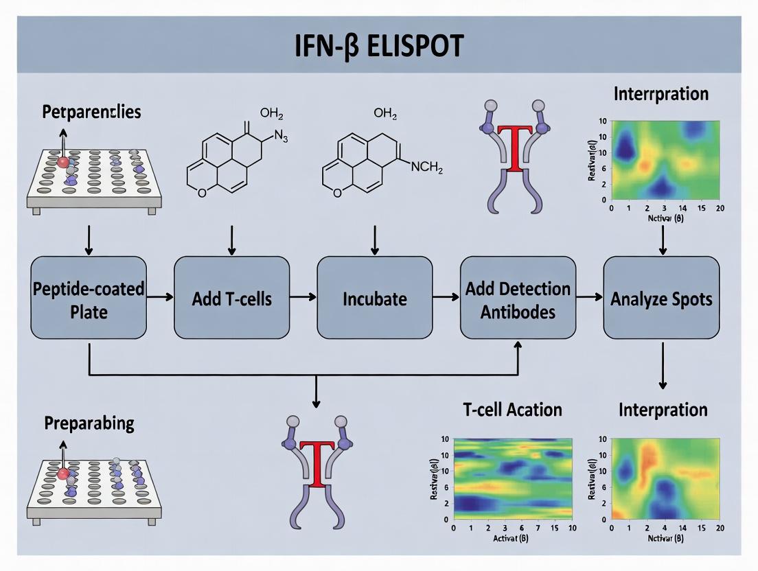
IFN-γ ELISPOT Experimental Workflow
This application note details the principles and protocols of the Enzyme-Linked Immunospot (ELISPOT) assay, framed within a broader thesis investigating peptide-specific T-cell responses via IFN-γ release. The assay's exceptional sensitivity (10-100 times more sensitive than conventional ELISA) allows for the direct ex vivo quantification of rare, antigen-specific T-cells from peripheral blood mononuclear cells (PBMCs) or tissue samples. Understanding the mechanistic journey from solid-phase capture to spot formation is critical for optimizing assays in vaccine development, cancer immunotherapy, and infectious disease research.
Core Technology: Principle of Operation
The IFN-γ ELISPOT assay is a solid-phase immunoassay. A capture antibody, specific for IFN-γ, is pre-coated onto a polyvinylidene difluoride (PVDF) or nitrocellulose-backed microplate. Upon stimulation with specific peptide antigens, responsive T-cells secrete IFN-γ cytokine. This cytokine is immediately captured by the surrounding antibodies on the membrane. After cell removal, a biotinylated detection antibody is added, followed by an enzyme-conjugated streptavidin (typically Alkaline Phosphatase-AP or Horseradish Peroxidase-HRP). Subsequent addition of a chromogenic or fluorogenic substrate results in a precipitate-forming reaction, generating a permanent, quantifiable spot at the location of each original cytokine-secreting cell.
Table 1: Key Performance Characteristics of Standard IFN-γ ELISPOT Assays
| Parameter | Typical Range / Specification | Notes |
|---|---|---|
| Sensitivity | 1 in 100,000 to 1 in 1,000,000 PBMCs | Can detect rare antigen-specific cells directly ex vivo. |
| Dynamic Range | ~10 to >1000 Spot Forming Cells (SFCs) per well | Linearity depends on cell density and spot confluence. |
| Assay Duration | 24-48 hour stimulation + 1 day detection | Total hands-on time is approximately 6-8 hours over 2-3 days. |
| Well Format | 96-well (standard), 24-well, 384-well | PVDF membranes are most common for 96-well format. |
| Spot Size | 50 - 200 μm in diameter | Size can vary with secretion rate and substrate incubation time. |
| Background Signal | Typically <5 spots per well in negative controls | Mitigated by using serum-free media and plate blocking. |
Detailed Protocols
Protocol 1: Coating and Plate Preparation
Objective: To immobilize anti-IFN-γ capture antibody on the PVDF membrane.
- Pre-wet the PVDF membrane of the ELISPOT plate with 15 μL/well of 35% ethanol for 1 minute.
- Wash the plate 3 times with 200 μL/well of sterile PBS.
- Dilute the anti-IFN-γ capture antibody in sterile PBS to the manufacturer's recommended concentration (e.g., 5-15 μg/mL).
- Add 100 μL of the coating antibody solution to each well. Seal the plate and incubate overnight at 4°C or for 2 hours at 37°C.
- Decant the antibody solution. Block the plate with 200 μL/well of complete cell culture medium (e.g., RPMI-1640 with 5-10% human AB serum or fetal bovine serum) for at least 1 hour at 37°C.
Protocol 2: Cell Stimulation and Incubation
Objective: To activate antigen-specific T-cells and allow localized cytokine capture.
- Prepare peptide pools or single peptides (typically at 1-10 μg/mL final concentration). Positive control: Phytohemagglutinin (PHA, 1-5 μg/mL) or anti-CD3 antibody. Negative control: Cells alone with DMSO (peptide solvent).
- Decant the blocking medium from the prepared ELISPOT plate.
- Add 100 μL of antigen solution or control solutions to appropriate wells in triplicate.
- Isolate PBMCs via density gradient centrifugation (Ficoll-Paque). Resuspend in complete medium.
- Add PBMCs to the plate at a density optimized for the expected frequency (e.g., 100,000 – 400,000 cells per well in 100 μL). Centrifuge briefly (100 x g, 1 min) to settle cells.
- Incubate the plate for 24-48 hours at 37°C, 5% CO₂ in a humidified incubator. Do not move or disturb the plate.
Protocol 3: Detection of Captured IFN-γ
Objective: To visualize captured cytokine as distinct spots.
- Discard cell suspension and lyse cells by washing the plate 5 times with 200 μL/well of PBS containing 0.05% Tween 20 (PBST).
- Dilute biotinylated anti-IFN-γ detection antibody in PBST with 1% BSA (e.g., 1-2 μg/mL). Add 100 μL/well. Incubate for 2 hours at room temperature or overnight at 4°C.
- Wash plate 5 times with PBST.
- Dilute enzyme-streptavidin conjugate (AP- or HRP-conjugated) per manufacturer's instructions in PBST/BSA. Add 100 μL/well. Incubate for 1-2 hours at room temperature, protected from light.
- Wash plate 5 times with PBST, then 2 times with PBS (to remove Tween before substrate).
- Prepare precipitating substrate (e.g., BCIP/NBT for AP, AEC for HRP). Add 100 μL/well. Develop at room temperature for 5-30 minutes, monitoring spot formation.
- Stop development by rinsing extensively with deionized water. Air-dry the plate completely in the dark.
Protocol 4: Spot Enumeration and Analysis
Objective: To quantify antigen-specific T-cell responses.
- Analyze the dried plate using an automated ELISPOT reader system.
- Key parameters are set: size and intensity gradient thresholds to distinguish true spots from background noise.
- Results are expressed as Spot Forming Cells (SFCs) per million input cells or per well. Standard calculation: Mean SFCs in antigen wells minus mean SFCs in negative control wells.
Visualizing the ELISPOT Workflow and Signaling
Diagram 1: IFN-γ ELISPOT Assay Workflow
Diagram 2: T-Cell Activation Signaling to IFN-γ Secretion
The Scientist's Toolkit: Key Research Reagent Solutions
Table 2: Essential Materials for IFN-γ ELISPOT Assays
| Reagent / Material | Function & Role in Assay | Critical Considerations |
|---|---|---|
| PVDF-Backed 96-Well Plate | Solid-phase matrix for antibody coating. Membrane porosity traps secreted cytokine locally. | Must be pre-wet with ethanol. Pre-coated plates are available for standardization. |
| Anti-IFN-γ Paired Antibodies | Capture and detection antibody pair. Must recognize different epitopes on IFN-γ. | Low cross-reactivity, high affinity. Validated for ELISPOT. Carrier protein-free (BSA) detection Ab is ideal. |
| Peptide Pools / Antigens | Stimulates T-cells via TCR engagement. | Length (15-mers for CD4+, 8-11-mers for CD8+), purity (>70%), solubility in DMSO/PBS. |
| Human AB Serum | Serum supplement in cell culture medium. Provides essential nutrients and factors. | Reduces background vs. FBS for human cells. Must be screened for low endotoxin and virus-free. |
| Biotinylated Detection Antibody | Binds captured IFN-γ, provides link for streptavidin-enzyme. | Concentration must be titrated to optimize signal-to-noise. |
| Streptavidin-AP/HRP Conjugate | Amplification system. Enzyme catalyzes substrate precipitation. | High specific activity. Must be titrated; excess causes high background. |
| BCIP/NBT or AEC Substrate | Chromogen precipitates at enzyme site, forming a visible spot. | Precipitating type is mandatory. Solution must be fresh and filtered. |
| Automated ELISPOT Reader | Imaged-based system for objective, high-throughput spot counting. | Calibrated with size/intensity algorithms to exclude artifacts and merged spots. |
This application note details the implementation of IFN-γ ELISPOT assays within the broader thesis research on peptide-specific T-cell responses. The assay's superior sensitivity, single-cell resolution, and functional protein readout make it indispensable for quantifying antigen-specific T-cells in vaccine development, immunomonitoring, and immunotherapy research. This document provides updated protocols and analyses based on current methodologies.
Table 1: Comparative Performance of T-Cell Assays
| Assay Format | Functional Readout | Sensitivity (Detection Limit) | Single-Cell Resolution | Throughput |
|---|---|---|---|---|
| IFN-γ ELISPOT | Cytokine Secretion (IFN-γ) | 1 in 300,000 PBMCs | Yes | Medium-High |
| Intracellular Cytokine Staining (ICS) | Cytokine Production (IFN-γ, TNF-α, etc.) | 1 in 10,000 PBMCs | Yes | Medium |
| Peptide-MHC Multimer Staining | TCR Binding | 1 in 1,000 PBMCs | Yes | High |
| 51Cr-Release Cytotoxicity | Target Cell Lysis | 1 in 100 PBMCs | No | Low |
Table 2: Typical IFN-γ ELISPOT Results from Peptide Stimulation
| Stimulus Condition | Mean Spot Forming Units (SFU) per 10^6 PBMCs | Standard Deviation | Significance (p-value vs. Unstimulated) |
|---|---|---|---|
| Unstimulated (Media) | 5 | 2 | -- |
| Positive Control (PMA/Ionomycin) | 850 | 120 | <0.0001 |
| CMV pp65 Peptide Pool | 250 | 45 | <0.0001 |
| Candidate Vaccine Peptide A | 180 | 30 | <0.001 |
| Candidate Vaccine Peptide B | 15 | 5 | 0.12 (NS) |
Detailed Protocols
Protocol 1: IFN-γ ELISPOT Assay for Peptide-Specific T-Cell Responses
Research Reagent Solutions & Essential Materials:
- ELISPOT Plate: 96-well PVDF-backed plate. Function: Provides matrix for antibody coating and spot development.
- Anti-IFN-γ Coating Antibody (Clone 1-D1K): Function: Captures secreted IFN-γ at the site of release.
- Anti-IFN-γ Detection Antibody, Biotinylated (Clone 7-B6-1): Function: Binds captured IFN-γ; conjugated for enzymatic detection.
- Streptavidin-ALP (Alkaline Phosphatase): Function: Binds biotin on detection antibody for colorimetric reaction.
- BCIP/NBT Substrate: Function: ALP catalyzes its conversion to an insoluble purple precipitate, forming a spot.
- Peptide Libraries/Epitope Pools: Function: Antigenic stimulus to activate specific T-cells.
- PBMCs (Peripheral Blood Mononuclear Cells): Function: Source of responder T-cells.
- RPMI-1640 Complete Media: Function: Cell culture medium supporting cell viability.
Methodology:
- Plate Coating: Coat plate wells with 100 µL of anti-IFN-γ coating antibody (5 µg/mL in sterile PBS). Incubate overnight at 4°C or 2 hours at 37°C.
- Plate Blocking: Aspirate coating antibody. Block plates with 200 µL/well of RPMI-1640 containing 10% FBS for 2 hours at 37°C.
- Cell Seeding and Stimulation: Prepare PBMCs. Add 100 µL of cell suspension (2-5 x 10^5 cells/well) to blocked plates. Add 100 µL of peptide solution (final concentration 1-2 µg/mL per peptide), positive control (PMA/Ionomycin), or media alone (negative control). Incubate for 24-48 hours at 37°C, 5% CO2.
- Cell Removal and Detection: Decant cells. Wash plates 6x with PBS, then 3x with PBS containing 0.05% Tween-20 (PBST). Add 100 µL of biotinylated anti-IFN-γ detection antibody (1 µg/mL in PBST with 1% BSA). Incubate 2 hours at room temperature (RT).
- Streptavidin Conjugate: Wash plates 3x with PBST. Add 100 µL of Streptavidin-ALP (diluted per manufacturer in PBST/BSA). Incubate 1 hour at RT.
- Substrate Development: Wash plates 4x with PBST and 2x with PBS. Add 100 µL of BCIP/NBT substrate. Develop for 5-20 minutes at RT in the dark. Stop reaction by rinsing with distilled water.
- Plate Reading and Analysis: Air-dry plates completely. Enumerate spots using an automated ELISPOT reader. Results are expressed as Spot Forming Units (SFU) per million input cells.
Protocol 2: Validation of Single-Cell Resolution via Serial Dilution
Methodology:
- Prepare a high-responder PBMC sample.
- Perform a limiting dilution series (e.g., from 200,000 to 3,125 cells/well) in quadruplicate with optimal peptide stimulus.
- Run the IFN-γ ELISPOT assay as described in Protocol 1.
- Plot the number of input cells against the mean SFU per well. A linear correlation (R^2 > 0.95) across dilutions confirms single-cell resolution and quantitative accuracy.
Diagrams
Title: ELISPOT Experimental Workflow
Title: T-cell Activation & Detection Principle
Introduction Within the thesis on IFN-γ ELISPOT assay for peptide-specific T-cell responses, the assay’s utility as a cornerstone in translational immunology is paramount. This Application Notes and Protocols document details its critical role in three primary domains: quantifying vaccine immunogenicity, monitoring adoptive T-cell therapies, and elucidating T-cell immunity in infectious diseases. The protocols herein standardize the measurement of antigen-specific effector T-cells, providing a quantitative foundation for immunological research and development.
Application Note 1: Vaccine Development – Immunogenicity Assessment In vaccine development, the IFN-γ ELISPOT assay is the gold standard for evaluating cell-mediated immunogenicity. It quantitatively measures the frequency of antigen-specific T-cells induced by vaccine candidates, directly informing vaccine efficacy and guiding adjuvant selection. The assay's sensitivity allows for detection even in early-phase clinical trials with limited sample volumes.
Table 1: Representative IFN-γ ELISPOT Data from Vaccine Trials
| Vaccine Target | Candidate Type | Mean SFC/10⁶ PBMCs (Post-Vaccination) | Key Peptide Pool | Reference |
|---|---|---|---|---|
| SARS-CoV-2 | mRNA (BNT162b2) | 280 - 550 SFC | Spike protein overlapping peptides | Goel et al., Cell, 2021 |
| Influenza | Recombinant HA | 120 - 300 SFC | Hemagglutinin peptides | Nayak et al., NPJ Vaccines, 2020 |
| HIV | Mosaic Immunogen | 150 - 400 SFC | Gag/Pol/Env peptide pools | Barouch et al., Lancet, 2018 |
| Malaria (RTS,S) | Protein-subunit | 50 - 200 SFC | CSP-derived peptides | Kester et al., J Infect Dis, 2016 |
Protocol 1.1: Assessment of Vaccine-Induced T-Cell Responses
- Sample Preparation: Isolate PBMCs from vaccinated donors via density gradient centrifugation. Cryopreserve or use fresh cells. Count and resuspend in complete RPMI medium.
- Plate Coating: Coat 96-well PVDF-plate with anti-human IFN-γ capture antibody (e.g., 1-D1K, Mabtech) at 15 µg/mL in sterile PBS overnight at 4°C.
- Blocking & Seeding: Block plate with complete RPMI for 2 hours at 37°C. Seed PBMCs at 2.5 x 10⁵ cells/well in triplicate. Include positive control (PHA/SEB) and negative control (cells + medium only).
- Antigen Stimulation: Add vaccine-relevant peptide pools (e.g., overlapping 15-mers) at a final concentration of 2 µg/mL per peptide. Incubate for 24-48 hours at 37°C, 5% CO₂.
- Detection: Discard cells. Add biotinylated detection antibody (e.g., 7-B6-1, Mabtech) at 1 µg/mL for 2 hours. Add Streptavidin-ALP for 1 hour.
- Spot Development: Add BCIP/NBT chromogenic substrate. Develop until spots are clearly visible. Stop reaction by washing with tap water.
- Analysis: Enumerate spots using an automated ELISPOT reader. Report results as Spot-Forming Cells (SFC) per 10⁶ input cells. Subtract mean background from negative control.
Application Note 2: Immunotherapy Monitoring – Adoptive Cell Therapy For cancer immunotherapies like TCR-T or CAR-T cells, the IFN-γ ELISPOT assay monitors the functional persistence and antigen specificity of infused products. It is used pre-clinically to validate engineered T-cell function and clinically to correlate post-infusion T-cell activity with patient outcomes.
Table 2: ELISPOT in Immunotherapy Monitoring
| Therapy Type | Target Antigen | Application Stage | Typical Readout (SFC/10⁶ cells) | Functional Correlation |
|---|---|---|---|---|
| TCR-T Cell | NY-ESO-1 | Pre-infusion Product Potency | >1000 SFC | In vivo tumor regression |
| CAR-T Cell | CD19 | Post-infusion Monitoring | Variable over time | B-cell aplasia & CRS |
| TIL Therapy | Tumor Lysate | Reactivity Screening | >500 SFC (to lysate) | Clinical response |
Protocol 2.1: Potency Assay for Engineered T-Cell Products
- Effector Cells: Use the final engineered T-cell product or co-culture cells post-expansion.
- Antigen-Presenting Cells (APCs): Use HLA-matched antigen-positive target cells (e.g., tumor cell lines) or peptide-pulsed T2 cells. For peptide-specific readout, pulse APCs with 10 µg/mL target peptide for 2 hours.
- Co-culture Setup: Seed IFN-γ pre-coated ELISPOT plate with 1 x 10⁴ target cells/well. Add effector T-cells at varying Effector:Target ratios (e.g., 10:1, 5:1, 1:1) in triplicate.
- Incubation & Development: Incubate for 18-24 hours. Follow standard detection and development steps as in Protocol 1.1 (steps 5-7).
- Interpretation: A positive potency result is defined as a significant IFN-γ response above the negative control (no peptide/unpulsed targets) at the time of product release.
Application Note 3: Infectious Disease Research – T-Cell Epitope Mapping In infectious disease research, the IFN-γ ELISPOT is indispensable for identifying immunodominant T-cell epitopes, understanding cross-reactivity, and assessing long-term immune memory in convalescent or exposed individuals.
Protocol 3.1: Epitope Mapping and Specificity Screening
- Peptide Library: Utilize overlapping peptide libraries (e.g., 15-mers overlapping by 11 amino acids) spanning the pathogen's proteome.
- High-Throughput Screening: Seed PBMCs from infected/recovered donors into pre-coated plates. Stimulate with individual peptides or peptide pools in a matrix format. Initial screening often uses peptide pools, followed by deconvolution with individual peptides.
- Enhanced Sensitivity: For low-frequency responses, increase incubation time to 40-48 hours and/or increase PBMC input to 5 x 10⁵ cells/well.
- Data Analysis: Identify positive peptides by comparing SFC counts to the mean + 2-3 standard deviations of the negative control wells. Confirm immunodominant epitopes with HLA restriction assays.
The Scientist's Toolkit: Key Research Reagent Solutions
| Item | Function & Importance |
|---|---|
| Anti-IFN-γ Coating Antibody (Clone 1-D1K) | High-affinity capture antibody immobilized on PVDF membrane; defines assay specificity. |
| Biotinylated Anti-IFN-γ Detection Antibody (Clone 7-B6-1) | Second antibody for detection; binds a distinct epitope on captured IFN-γ. |
| PVDF-Backed Microplates | Membranes facilitate high local contrast for spot formation; preferred over nitrocellulose. |
| Peptide Pools (15-mers, overlapping) | Stimulus for antigen-specific T-cells; critical for vaccine and epitope mapping studies. |
| RPMI-1640 with 5-10% Human AB Serum | Low-background culture medium maintains cell viability without stimulating immune cells. |
| Streptavidin-Alkaline Phosphatase (ALP) | Enzyme conjugate that binds biotinylated detection Ab; catalyzes insoluble precipitate formation. |
| BCIP/NBT Substrate | Chromogenic substrate for ALP; yields dark purple, stable spots at cytokine secretion sites. |
| Automated ELISPOT Reader | Provides objective, high-throughput spot enumeration and size analysis. |
Visualizations
IFN-γ ELISPOT Assay Principle
ELISPOT Experimental Workflow
Application Notes
The Enzyme-Linked Immunospot (ELISPOT) assay is a cornerstone technique for quantifying peptide-specific T-cell responses by measuring cytokine secretion (e.g., IFN-γ) at the single-cell level. Its high sensitivity makes it indispensable in vaccine development, cancer immunotherapy, and infectious disease research. The reliability and reproducibility of the assay are critically dependent on the quality and compatibility of core reagents and equipment.
- Plates: Polyvinylidene difluoride (PVDF)-backed 96-well plates are the standard. The PVDF membrane must be pre-wetted with ethanol to activate its protein-binding capacity. The plate's low background and high binding affinity are essential for clear spot formation.
- Antibodies: Paired monoclonal antibodies (capture and detection) with high specificity and affinity for IFN-γ are required. The capture antibody is coated onto the membrane. The detection antibody, typically biotinylated, must recognize a different epitope. Optimal concentrations must be determined via titration to maximize signal-to-noise.
- Peptide Libraries: These are collections of synthetic peptides spanning target antigens (e.g., viral proteins, tumor-associated antigens). They can be pooled or used individually to map T-cell epitopes. Purity (>70%) and solubility are crucial to avoid nonspecific activation or toxicity to cells.
- Readers: Automated ELISPOT readers use sophisticated image analysis algorithms to count spots and measure their size and intensity. Consistency in counting parameters (e.g., minimum and maximum spot size, intensity threshold) across experiments is vital for comparative analysis.
Protocol: IFN-γ ELISPOT for Peptide-Specific T-Cell Response
Day 1: Plate Coating
- Pre-wet wells with 15 μL of 35% ethanol (in sterile water) for 1 minute.
- Wash wells 4 times with 200 μL sterile PBS.
- Coat each well with 100 μL of anti-human IFN-γ capture antibody diluted in sterile PBS to the optimal concentration (typically 2-10 μg/mL). Seal plate and incubate overnight at 4°C.
Day 2: Cell Plating and Stimulation
- Aspirate coating solution. Block wells with 200 μL of complete culture medium (RPMI-1640 + 10% FBS) for 2 hours at 37°C.
- Prepare peripheral blood mononuclear cells (PBMCs) or other lymphocyte samples. Plate cells in duplicate/triplicate wells at densities of 1x10⁵ to 3x10⁵ cells/well in 100 μL medium.
- Add peptide library pools or individual peptides. Common final concentrations range from 1-10 μg/mL per peptide. Include positive control (e.g., PHA or PMA/Ionomycin) and negative control (cells + medium only).
- Incubate plate for 24-48 hours at 37°C, 5% CO₂ in a humidified incubator. Do not move the plate.
Day 3 or 4: Detection
- Discard cell suspension. Wash plate 4 times with 200 μL/well of PBS, then 4 times with PBS containing 0.05% Tween-20 (PBST).
- Add 100 μL/well of biotinylated anti-human IFN-γ detection antibody (typical concentration 0.5-2 μg/mL in PBST + 1% BSA). Incubate 2 hours at room temperature (RT).
- Wash plate 4 times with PBST.
- Add 100 μL/well of Streptavidin-Horseradish Peroxidase (HRP) conjugate diluted per manufacturer's instructions in PBST + 1% BSA. Incubate 1 hour at RT.
- Wash plate 4 times with PBST, then twice with PBS.
- Prepare AEC (3-amino-9-ethylcarbazole) substrate solution. Add 100 μL to each well. Develop for 5-30 minutes until distinct red spots appear. Stop reaction by rinsing extensively with tap water.
- Air-dry plate completely in the dark before reading.
Data Analysis
- Count spots using an automated ELISPOT reader. A positive response is typically defined as a well where the mean spot count exceeds the mean of the negative control wells by at least 2 standard deviations and has a minimum of 5-10 spot-forming units (SFU) per well.
- Results are expressed as SFU per million input cells.
Quantitative Data Summary
Table 1: Recommended Concentrations for Key Reagents
| Reagent | Typical Concentration Range | Purpose | Critical Notes |
|---|---|---|---|
| Capture Antibody | 2 - 10 μg/mL in PBS | Coats membrane to bind secreted IFN-γ | Sterile filtration recommended for overnight coating. |
| Detection Antibody | 0.5 - 2 μg/mL in PBST/BSA | Binds captured IFN-γ; biotinylated. | Must be titrated against capture antibody. |
| Streptavidin-HRP | 1:500 - 1:2000 dilution | Binds biotin; enables chromogenic detection. | Follow manufacturer's datasheet. |
| Peptide Stimulants | 1 - 10 μg/mL per peptide | Antigen-specific T-cell activation. | Solubilize in DMSO or PBS; ensure final DMSO <0.5%. |
| Cell Seeding Density | 1x10⁵ - 3x10⁵ cells/well | Balance between sensitivity and overcrowding. | Must be optimized for each cell type and antigen. |
Table 2: Key Equipment and Software Settings
| Equipment/Parameter | Specification/Setting | Function |
|---|---|---|
| ELISPOT Plate | 96-well, PVDF membrane | Platform for assay; binds capture antibody. |
| Automated Plate Washer | Program for 4x PBST washes | Ensures consistent, thorough washing. |
| Humidified CO₂ Incubator | 37°C, 5% CO₂ | Cell stimulation environment. |
| Automated ELISPOT Reader | N/A | Captures high-resolution images of wells. |
| Analysis Software | Spot Size: 50-500 μm² Intensity: User-defined | Accurately distinguishes true spots from background. |
Visualization
IFN-γ ELISPOT Assay Principle
ELISPOT Experimental Workflow
The Scientist's Toolkit: Research Reagent Solutions
- Anti-Human IFN-γ Antibody Pair (Coated/Biotinylated): Pre-optimized, matched pair to ensure high sensitivity and low background. Function: Specific capture and detection of human IFN-γ.
- PVDF-Backed 96-Well ELISPOT Plates: Sterile, ready-to-use plates with high protein binding capacity. Function: Solid-phase support for the immunoassay.
- Peptide Library (e.g., SARS-CoV-2 Overlapping Peptides): Lyophilized pools spanning the entire proteome of a target pathogen. Function: Stimulate a broad range of antigen-specific T-cells for immune response profiling.
- AEC Substrate Set (with Peroxide): Ready-to-use chromogenic substrate for HRP. Function: Produces an insoluble red precipitate at the site of cytokine secretion.
- RPMI-1640 Complete Medium: Cell culture medium supplemented with L-glutamine, HEPES, and heat-inactivated FBS. Function: Supports viability and function of lymphocytes during incubation.
- Streptavidin-HRP Conjugate: Highly purified conjugate for signal amplification. Function: Binds to biotinylated detection antibody, enabling colorimetric detection.
- ELISPOT Plate Sealers: Gas-permeable, adhesive seals. Function: Maintain sterility during long incubation while allowing gas exchange.
- Automated ELISPOT Analyzer & Software: Hardware and software for image acquisition and spot analysis. Function: Provides objective, quantitative spot counts and size analysis.
Step-by-Step IFN-γ ELISPOT Protocol: From PBMC Isolation to Data Acquisition
Ethical Considerations in Human T-Cell Research
The use of human biological samples for IFN-γ ELISPOT assays is governed by strict ethical and regulatory frameworks. Key principles include:
- Informed Consent: Donors must provide voluntary, informed, and documented consent for the use of their blood or PBMCs in research. The consent form should detail the purpose, procedures, risks, benefits, and data handling/storage plans.
- Institutional Review Board (IRB) / Ethics Committee (EC) Approval: All study protocols involving human samples must be reviewed and approved by an IRB/EC prior to initiation.
- Donor Anonymization/Pseudonymization: Personally identifiable information must be stripped from samples and data, using unique codes to protect donor privacy.
- Data Security: Research data must be stored securely in compliance with regulations (e.g., GDPR, HIPAA).
- Benefit-Risk Assessment: The potential scientific benefit of the research must justify the minor risks associated with blood draw.
Table 1: Essential Components of an IRB/EC Protocol Submission for ELISPOT Studies
| Component | Description |
|---|---|
| Study Rationale & Objectives | Clear hypothesis and scientific justification for using human T-cells. |
| Donor Recruitment Criteria | Inclusion/exclusion criteria (age, health status, prior exposure). |
| Informed Consent Document | Lay-language form explaining the study to potential donors. |
| Sample Collection Procedure | Details of blood draw volume (typically 20-100mL) and safety procedures. |
| Sample Processing & Storage | Methods for PBMC isolation, cryopreservation, and long-term storage. |
| Data Management Plan | Procedures for anonymization, analysis, storage, and potential sharing. |
| Biosafety Considerations | Handling of human-derived materials (BSL-2 standards). |
Antigen Selection Strategy
The choice of antigen is fundamental to detecting relevant T-cell responses.
- Defined Antigens: Recombinant proteins, viral lysates, or synthetic peptides. Ideal for vaccines or diseases with known immunodominant epitopes.
- Peptide Libraries: Overlapping peptide pools spanning entire proteins of interest. Essential for discovery-phase research where epitopes are unknown.
- Negative & Positive Controls:
- Negative Control: Unstimulated cells (medium alone) or an irrelevant peptide.
- Positive Control: Mitogens like PHA (polyclonal T-cell activator) or SEB (staphylococcal enterotoxin B) to confirm cell viability and assay performance.
Table 2: Quantitative Comparison of Antigen Types for ELISPOT
| Antigen Type | Typical Working Concentration | Advantages | Limitations |
|---|---|---|---|
| Synthetic Peptide Pools (15-mers) | 1-2 µg/mL per peptide | Major histocompatibility complex (MHC)-agnostic; customizable; minimal background. | May miss conformational epitopes; cost for large libraries. |
| Recombinant Protein | 5-20 µg/mL | Contains native conformational epitopes; can be processed and presented naturally. | Requires antigen-presenting cell (APC) processing; potential non-specific stimulation. |
| Viral/Bacterial Lysate | 1-10 µg/mL | Contains full antigenic repertoire of the pathogen. | High background risk; donor may have cross-reactive memory cells. |
| Positive Control (PHA) | 1-5 µg/mL | Strong polyclonal stimulus; validates assay platform. | Non-physiological; can mask antigen-specific responses if overused. |
Peptide Pool Design Protocol
Objective: To design and prepare a pool of synthetic peptides for screening T-cell responses against a target protein.
Materials:
- Target protein sequence (FASTA format).
- Peptide design software (e.g., PepTrace, in-house scripts).
- Peptide synthesis vendor specifications.
Detailed Methodology:
Step 1: Determine Peptide Length.
- For CD4+ T-helper cell responses, use 15-amino-acid (15-mer) peptides. These are optimally processed by APCs and presented on MHC class II molecules.
- For CD8+ cytotoxic T-cell responses, use 8-10-mer peptides. However, as MHC I binding prediction is allele-specific, 15-mers with 11-aa overlap are commonly used as they contain potential 9-mer cores and are processed and presented by APCs.
Step 2: Set Overlap Length.
- A standard 11-amino-acid overlap between successive 15-mer peptides ensures every possible linear epitope (including every potential 9-mer and 10-mer core) is contained within at least one peptide.
- Formula to calculate number of peptides: Number of peptides = [(Protein length - Peptide length) / (Peptide length - Overlap)] + 1.
Step 3: Generate Peptide Sequence List.
- Using the formula, generate the list of all peptide sequences. Example for a 100-aa protein: [(100-15)/(15-11)] + 1 = (85/4) + 1 = ~22 peptides.
- Peptide 1: aa 1-15
- Peptide 2: aa 5-19
- Peptide 3: aa 9-23 ... etc.
Step 4: Pooling Strategy.
- Single Large Pool: All peptides are combined into one pool. Efficient for screening, but a positive response requires deconvolution (testing sub-pools or individual peptides).
- Matrix Pooling: Peptides are arranged in a grid (e.g., 8x12 for 96 peptides), pooled by row and column. A positive response at the intersection of a positive row and column identifies the single reactive peptide without testing all individually.
- Sub-pools: Protein is divided into logical regions (e.g., domains), and peptides from each region are pooled separately to map responses.
Step 5: Preparation of Working Peptide Solution.
- Obtain lyophilized peptides (typically >70% purity).
- Resuspend each peptide in DMSO to a stock concentration of 10-20 mg/mL.
- Combine equal volumes (or masses) of each peptide stock into a master pool. Ensure the final DMSO concentration in the cell culture well does not exceed 0.5-1.0% (v/v), as it can be toxic to cells.
- Dilute the master pool in sterile, serum-free culture medium to create a 10X intermediate stock. Aliquot and store at ≤ -20°C.
- In the assay, dilute the 10X stock 1:10 in cell culture medium to achieve the final recommended concentration of 1-2 µg/mL per peptide.
Title: Peptide Pool Design & Preparation Workflow
Key Experimental Protocol: IFN-γ ELISPOT Setup with Peptide Pools
Title: Activation of Peptide-Specific T-Cells and IFN-γ Capture on ELISPOT Plate.
Principle: PBMCs are cultured with the peptide pool. Reactive T-cells are activated, secreting IFN-γ, which is captured by antibodies on the membrane. After removal of cells, a detection antibody and enzyme conjugate reveal spots, each representing a single reactive T-cell.
The Scientist's Toolkit: Key Research Reagent Solutions
| Reagent/Material | Function in the Protocol |
|---|---|
| Pre-coated IFN-γ ELISPOT Plates | 96-well plates with PVDF or nitrocellulose membrane coated with anti-IFN-γ capture antibody. Provides the solid phase for cytokine capture. |
| RPMI-1640 + 5-10% Human AB Serum | Complete cell culture medium. Provides nutrients and serum factors for cell viability without introducing exogenous cytokines. |
| Peptide Pool (15-mers, 11-aa overlap) | Antigenic stimulus. Activates memory T-cells specific to epitopes within the target protein. |
| Cryopreserved Human PBMCs | Source of T-effector cells. Must have high viability (>90%) post-thaw. |
| Anti-CD28 Co-stimulatory Antibody | Provides essential secondary signal for optimal T-cell activation, mimicking APC function. |
| Biotinylated Anti-IFN-γ Detection Antibody | Binds to captured IFN-γ. Conjugated to biotin for subsequent amplification. |
| Streptavidin-Alkaline Phosphatase (SA-AP) | Binds to biotin. Conjugated to AP enzyme for colorimetric detection. |
| BCIP/NBT Chromogenic Substrate | AP substrate that yields an insoluble purple precipitate at the site of cytokine secretion, forming a "spot." |
| ELISPOT Plate Reader | Automated microscope and image analysis system to count spots and quantify spot size/intensity. |
Detailed Protocol Steps:
- Plate Preparation: Bring pre-coated plate to room temperature. Block wells with 200 µL/well of complete medium for at least 1 hour at 37°C.
- Cell & Stimulant Preparation:
- Thaw and rest PBMCs for 2-4 hours.
- Prepare 2X peptide pool solution in complete medium (final in-well concentration: 1-2 µg/mL per peptide).
- Prepare positive control (PHA at 2-4 µg/mL final) and negative control (medium alone).
- Assay Setup: Aspirate blocking medium.
- Add 100 µL/well of 2X peptide solution or controls to designated wells.
- Add 100 µL/well of PBMC suspension (2-5 x 10^5 cells in complete medium) to all wells. Final volume = 200 µL/well.
- Add anti-CD28 antibody (final 0.5-1 µg/mL) to all test and positive control wells.
- Incubation: Incubate plate for 18-24 hours at 37°C, 5% CO₂ in a humidified incubator. Do not move or disturb the plate.
- Detection (Post-Incubation): a. Discard cell suspension. Wash plate 5x with PBS + 0.05% Tween-20. b. Add 100 µL/well of biotinylated anti-IFN-γ detection antibody (diluted per manufacturer's instructions). Incubate 2 hours at RT. c. Wash plate 5x. d. Add 100 µL/well of Streptavidin-Alkaline Phosphatase. Incubate 1 hour at RT. e. Wash plate 5x. f. Add 100 µL/well of BCIP/NBT substrate. Develop for 5-20 minutes until spots appear. Stop reaction by rinsing with distilled water.
- Analysis: Air-dry plate completely in the dark. Count spots using an automated ELISPOT reader. Data is expressed as Spot Forming Units (SFU) per 10^6 input cells.
Title: IFN-γ ELISPOT Assay Workflow Principle
The reliability of an IFN-γ ELISPOT assay for detecting peptide-specific T-cell responses is fundamentally dependent on the quality and viability of the starting lymphocyte population. Isolating peripheral blood mononuclear cells (PBMCs) or lymphocytes from other sources (e.g., tissues, apheresis products) is the critical first step. Consistent, high-yield cell preparation ensures that subsequent ELISPOT results accurately reflect the frequency and functionality of antigen-responsive T-cells, directly impacting data interpretation in vaccine development, cancer immunotherapy, and infectious disease research.
Table 1: Expected Yield and Viability from Common Lymphocyte Sources
| Cell Source | Typical Starting Volume/Amount | Expected PBMC/Lymphocyte Yield | Target Viability (Trypan Blue) | Key Consideration |
|---|---|---|---|---|
| Human Peripheral Blood | 10 mL (sodium heparin/CPT tube) | 10-20 x 10^6 PBMCs | ≥ 95% | Avoid EDTA anticoagulant; process within 24-32h. |
| Leukapheresis Product | 1-5 mL of product | 200-1000 x 10^6 PBMCs | ≥ 90% | High cell density; may require dilution prior to isolation. |
| Mouse Spleen | One whole spleen | 50-100 x 10^6 lymphocytes | ≥ 85% | Requires mechanical dissociation and RBC lysis. |
| Mouse Lymph Nodes | Pooled axial/inguinal nodes | 5-20 x 10^6 lymphocytes | ≥ 90% | Minimal erythrocyte contamination. |
| Human Tissue (e.g., Tumor) | 1 g of tissue | 1-10 x 10^6 lymphocytes (variable) | ≥ 70% | Requires enzymatic digestion (e.g., collagenase/DNase). |
Table 2: Comparison of Common PBMC Isolation Methods
| Method | Principle | Average Purity (CD45+) | Average Recovery | Throughput | Cost |
|---|---|---|---|---|---|
| Density Gradient Centrifugation | Buoyant density separation using Ficoll-Paque. | 95-99% | 60-85% | Medium | Low |
| Magnetic-Activated Cell Sorting (MACS) | Negative or positive selection via magnetic beads. | 95-99.5% (neg. selection) | 50-80% | Medium to High | High |
| Automated Cell Separators | Integrated density gradient or centrifugation. | 95-99% | 70-90% | High | Very High |
| Lysis-Based Methods (for RBC) | Ammonium-Chloride-Potassium (ACK) lysis buffer. | N/A (for RBC removal) | >90% of WBCs | High | Very Low |
Detailed Experimental Protocols
Protocol 1: Standard PBMC Isolation from Human Blood via Density Gradient
Objective: To isolate high-viability PBMCs from fresh human blood for use in IFN-γ ELISPOT assays.
Materials: See "The Scientist's Toolkit" below.
Procedure:
- Blood Dilution: Dilute heparinized blood 1:1 with room temperature Dulbecco's PBS (DPBS) or RPMI-1640 without serum.
- Gradient Setup: Carefully layer 5 mL of diluted blood over 3 mL of Ficoll-Paque PLUS in a 15 mL conical centrifuge tube. Avoid mixing the layers.
- Centrifugation: Centrifuge at 400 x g for 30-35 minutes at 20°C with the brake OFF. This is critical for preserving gradient integrity.
- Harvest PBMCs: After centrifugation, identify the opaque PBMC layer at the plasma-Ficoll interface. Using a sterile pipette, carefully aspirate the layer and transfer to a new 15 mL tube.
- Wash Cells: Fill the tube with wash buffer (DPBS + 2% FBS/BSA) to 15 mL. Centrifuge at 300 x g for 10 minutes at 20°C. Aspirate supernatant.
- Lysis (Optional): If erythrocyte contamination is high, resuspend pellet in 2-5 mL of ACK lysis buffer for 3-5 minutes at RT. Quench with 10 mL wash buffer.
- Final Wash: Resuspend pellet in 15 mL wash buffer. Centrifuge at 200 x g for 10 minutes at 20°C. Aspirate supernatant.
- Resuspension & Counting: Resuspend cell pellet in 1-5 mL of complete ELISPOT culture medium (e.g., RPMI-1640 + 10% FBS). Proceed to counting and viability assessment.
Protocol 2: Manual Cell Counting and Viability Assessment via Trypan Blue Exclusion
Objective: To accurately determine the concentration and viability of isolated PBMCs prior to plating in the ELISPOT assay.
Procedure:
- Prepare Cell Suspension: Ensure cells are in a single-cell suspension. Mix gently by pipetting.
- Dilution: Mix 10 µL of cell suspension with 10 µL of 0.4% Trypan Blue stain (1:1 dilution). Incubate for 1-2 minutes at RT. Do not exceed 5 minutes.
- Load Hemocytometer: Carefully pipette 10-15 µL of the mixture into one chamber of a hemocytometer, allowing capillary action to fill it.
- Counting: Using a light microscope at 10X magnification, count the live (unstained) and dead (blue-stained) cells in the four outer 1 mm² grids, each containing 16 smaller squares.
- Calculation:
- Total Cells Counted = Sum of live and dead cells from all 4 grids.
- Cell Concentration (cells/mL) = (Total Cells Counted / 4) x Dilution Factor (2) x 10^4.
- Viability (%) = (Total Live Cells / Total Cells Counted) x 100.
Workflow and Relationship Diagrams
Title: PBMC Isolation and QC Workflow for ELISPOT
Title: Cell Prep's Role in the ELISPOT Assay Chain
The Scientist's Toolkit: Essential Research Reagents & Materials
Table 3: Key Reagents and Materials for PBMC Isolation and Counting
| Item/Category | Specific Example(s) | Function & Critical Notes |
|---|---|---|
| Anticoagulant Blood Collection Tubes | Sodium Heparin tubes; CPT tubes. | Prevents coagulation and preserves cell viability. CPT tubes contain Ficoll and a gel barrier for simplified isolation. |
| Density Gradient Medium | Ficoll-Paque PLUS; Lymphoprep. | Polysaccharide solution with defined density (1.077 g/mL) for separating PBMCs from other blood components. |
| Wash/Cell Suspension Buffer | DPBS (Ca2+/Mg2+-free) + 2% Fetal Bovine Serum (FBS) or BSA. | Provides isotonic environment; serum/BSA reduces cell clumping and loss during washes. |
| Erythrocyte Lysis Buffer | Ammonium-Chloride-Potassium (ACK) lysis buffer. | Selectively lyses red blood cells without significantly harming lymphocytes. Use judiciously. |
| Complete Cell Culture Medium | RPMI-1640 + 10% FBS + 1% Penicillin/Streptomycin. | Medium for final cell resuspension and ELISPOT assay. Supports short-term cell viability. |
| Viability Stain | 0.4% Trypan Blue solution. | Dye excluded by intact plasma membranes of live cells; dead cells take up the stain. |
| Counting Chamber | Hemocytometer (Neubauer improved). | Microscope slide with etched grid for manual cell counting and viability determination. |
| Centrifuge | Swing-bucket rotor, temperature-controlled. | Must allow for brake-off operation during density gradient centrifugation. |
| Sterile Consumables | Serological pipettes, 15/50 mL conical tubes, cell strainers (40-70 µm). | For sterile liquid and cell handling, and filtering out aggregates post-isolation. |
Within the context of IFN-γ ELISPOT assay development for detecting peptide-specific T-cell responses, the foundational steps of plate coating and blocking are paramount. These initial procedures dictate the efficiency and specificity of the capture antibody immobilization, directly impacting the assay's sensitivity, signal-to-noise ratio, and overall reproducibility. Proper execution ensures maximal availability of antibody binding sites for cytokine capture while minimizing non-specific binding of cells and proteins. This Application Note details optimized protocols and current best practices for these critical steps.
Research Reagent Solutions Toolkit
| Item | Function in Coating/Blocking for ELISPOT |
|---|---|
| High Protein-Binding PVDF Membranes | The standard 96-well plate format for ELISPOT. PVDF provides superior protein adsorption capacity compared to polystyrene. |
| Anti-Human IFN-γ Coating Antibody | The primary capture monoclonal antibody (e.g., clone 1-D1K), specific for human IFN-γ, immobilized during coating. |
| Sterile Coating Buffer | Typically PBS (pH 7.4), used to dilute the capture antibody to the optimal concentration for uniform plate coating. |
| Bovine Serum Albumin (BSA), Fraction V | The most common blocking agent. Saturates remaining protein-binding sites on the PVDF membrane to prevent non-specific adsorption. |
| Fetal Bovine Serum (FBS) | Often used in conjunction with BSA in blocking buffers to more closely mimic the protein composition of cell culture media. |
| Non-Fat Dry Milk | An alternative, cost-effective blocking agent containing casein; requires screening for lot-to-lot consistency. |
| Tween-20 | A mild non-ionic detergent added to wash buffers (e.g., PBS with 0.05% Tween-20) to reduce hydrophobic interactions and background. |
Table 1: Impact of Coating Antibody Concentration on Spot Characteristics.
| Coating [Ab] (µg/mL) | Mean Spot Number (SFU) | Spot Intensity | Background | Optimal? |
|---|---|---|---|---|
| 2.5 | 85 ± 12 | Faint, Diffuse | Low | No |
| 5.0 | 152 ± 18 | Clear, Defined | Low | Yes |
| 10.0 | 155 ± 20 | Very Dense | Moderate | Saturation |
| 15.0 | 148 ± 22 | Very Dense | High | No, High Background |
Table 2: Comparison of Blocking Buffer Efficacy.
| Blocking Buffer (2h at RT) | Mean SFU | Background Signal | CV (%) | Notes |
|---|---|---|---|---|
| 1% BSA in PBS | 150 ± 15 | Low | 10 | Standard, reliable |
| 10% FBS in RPMI | 145 ± 20 | Very Low | 14 | Physiological, cell-friendly |
| 2% Non-Fat Milk in PBS | 158 ± 25 | Moderate-High | 16 | Variable, risk of contamination |
| No Blocking | 90 ± 35 | Very High | 39 | Unacceptable background |
Detailed Protocols
Protocol 1: Plate Coating with Capture Antibody
Objective: To uniformly immobilize anti-IFN-γ monoclonal antibody onto PVDF membrane plates. Materials: Sterile PBS (pH 7.4), Anti-human IFN-γ coating antibody, 96-well PVDF-backed plates (sterile), pipettes, sterile reservoir. Procedure:
- Pre-wet the PVDF membrane by adding 15 µL of 35% ethanol per well for 1 minute. Immediately aspirate and wash wells 3x with 200 µL sterile PBS.
- Prepare the coating antibody solution by diluting the stock in sterile PBS to a final concentration of 5 µg/mL. Mix gently.
- Dispense 100 µL of the antibody solution into each well of the pre-wetted plate.
- Seal the plate with parafilm or a plate sealer to prevent evaporation. Incubate overnight (~16 hours) at 4°C.
- The following day, decant and flick out the coating solution. Wash the plate 3x with 200 µL sterile PBS per well. Bang the plate dry on clean paper towels.
- The plate is now ready for the blocking step. Do not let the membrane dry completely.
Protocol 2: Plate Blocking for ELISPOT
Objective: To saturate non-specific protein-binding sites to minimize background. Materials: Sterile PBS, Bovine Serum Albumin (BSA), Fetal Bovine Serum (FBS), RPMI-1640 media. Procedure:
- Prepare blocking buffer: 1% BSA (w/v) in sterile PBS. Alternatively, use 10% FBS in RPMI-1640. Filter sterilize (0.22 µm).
- Add 200 µL of the chosen blocking buffer to each coated well.
- Incubate the plate for 2 hours at room temperature (or 37°C if using serum-based buffers for cell-specific adaptation).
- Decant the blocking buffer. Do not wash the plate.
- The plate is now ready for cell seeding. To prevent drying, proceed immediately to adding cells suspended in culture medium, or temporarily store the plate with 100 µL of culture medium per well.
Visualization of Workflows
Title: ELISPOT Plate Coating and Blocking Workflow
Title: Impact of Coating & Blocking on ELISPOT Results
Within the context of IFN-γ ELISPOT assay development for detecting peptide-specific T-cell responses, the optimization of ex vivo cell stimulation parameters is critical. The sensitivity and specificity of the assay depend on finely tuned conditions that balance sufficient antigen presentation with T-cell receptor engagement while minimizing background noise and non-specific activation. This application note details the systematic optimization of three interdependent variables: peptide antigen concentration, the number of peripheral blood mononuclear cells (PBMCs) plated, and the duration of incubation. The goal is to establish a robust protocol for research in vaccine development, oncology immunology, and infectious disease.
Key Optimization Variables & Rationale
A successful ELISPOT requires a careful equilibrium. Excessive peptide can lead to non-specific stimulation or toxicity, while insufficient peptide fails to activate low-frequency T-cells. Too many cells cause over-confluent spots that are impossible to enumerate; too few may miss rare responses. Incubation time must allow for cytokine secretion and capture without exhausting the cells or degrading the captured cytokine.
Table 1: Core Variables for ELISPOT Stimulation Optimization
| Variable | Typical Test Range | Rationale for Optimization |
|---|---|---|
| Peptide Concentration | 0.1 - 20 µg/mL | To find the saturating dose for TCR engagement without inducing toxicity or non-specific effects. |
| Cell Number per Well | 1x10^5 - 4x10^5 PBMCs/well | To ensure detection of low-frequency T-cells while preventing spot overlap. |
| Incubation Duration | 18 - 48 hours | To allow adequate cytokine (IFN-γ) production and capture before cell exhaustion or cytokine degradation. |
Detailed Optimization Protocols
Protocol 3.1: Titration of Peptide Antigen Concentration
Objective: To determine the optimal peptide concentration that elicits a maximal antigen-specific signal with minimal background. Materials: Peptide pool (e.g., CEF pool, viral peptide pools, or tumor-associated antigen peptides), PBMCs from a donor with known reactivity, IFN-γ ELISPOT kit, sterile 96-well PVDF-plate. Procedure:
- Prepare a serial dilution of the peptide stock in complete RPMI medium (e.g., 20, 10, 5, 2.5, 1, 0.5, 0.1 µg/mL). Include a negative control (medium only) and a positive control (e.g., PHA or SEB).
- Coat and block the ELISPOT plate according to the manufacturer's instructions.
- Seed PBMCs at a fixed density (e.g., 2x10^5 cells/well) in triplicate for each peptide concentration.
- Add 100 µL of each peptide dilution to the respective wells. Final volume per well: 200 µL.
- Incubate plate for 24 hours at 37°C, 5% CO2 in a humidified incubator.
- Develop the plate following kit protocol (plate washing, detection antibody, enzyme conjugate, substrate).
- Count spots using an automated ELISPOT reader.
- Analysis: Plot Spot Forming Units (SFU) per million cells against peptide concentration. The optimal concentration is the lowest dose that gives a plateauing maximal response.
Protocol 3.2: Determination of Optimal Cell Number
Objective: To identify the cell density that provides a linear readout for spot counts without confluence. Materials: PBMCs from a donor with known reactivity, optimal peptide concentration (from Protocol 3.1), IFN-γ ELISPOT kit. Procedure:
- Prepare a dilution series of PBMCs in complete RPMI: 4x10^5, 3x10^5, 2x10^5, 1x10^5, and 0.5x10^5 cells/well (in 100 µL).
- Add 100 µL of the optimal peptide concentration (2X final desired concentration) to each well. Include cell-only negative controls.
- Incubate and develop the plate as in Protocol 3.1.
- Analysis: Plot SFU/well against the number of cells plated. The ideal range is where the relationship is linear (SFU increases proportionally with cell number). Avoid the plateau region where spot merging occurs.
Protocol 3.3: Kinetics of IFN-γ Secretion (Incubation Duration)
Objective: To establish the incubation time that yields the highest signal-to-noise ratio. Materials: PBMCs, optimal peptide and cell number (from prior protocols), IFN-γ ELISPOT kit. Procedure:
- Plate PBMCs at the optimal density with the optimal peptide concentration in multiple identical plates (or use a time-course plate developer if available).
- Incubate separate plates for 18, 24, 36, and 48 hours under standard conditions.
- Develop all plates simultaneously using the same reagent batches.
- Analysis: Plot SFU/10^6 cells versus time. The optimal duration is typically at the peak of the curve before signal decline due to cytokine degradation or consumption.
Data Presentation from Optimization Experiments
Table 2: Representative Data from a Peptide Concentration Titration (24h incubation, 2x10^5 PBMCs/well)
| Peptide Concentration (µg/mL) | Mean SFU/Well (Triplicate) | SD | SFU/10^6 Cells | Signal-to-Noise (vs. Media) |
|---|---|---|---|---|
| 0 (Media Control) | 5 | 2 | 25 | 1.0 |
| 0.1 | 12 | 3 | 60 | 2.4 |
| 0.5 | 45 | 10 | 225 | 9.0 |
| 1.0 | 98 | 15 | 490 | 19.6 |
| 2.5 | 210 | 25 | 1050 | 42.0 |
| 5.0 | 225 | 30 | 1125 | 45.0 |
| 10.0 | 215 | 28 | 1075 | 43.0 |
| 20.0 | 205 | 35 | 1025 | 41.0 |
Conclusion: Optimal concentration = 2.5 - 5 µg/mL.
Table 3: Representative Data from Cell Number Titration (Optimal Peptide, 24h)
| PBMCs Plated (x10^5) | Mean SFU/Well | SD | Spot Morphology Assessment |
|---|---|---|---|
| 0.5 | 52 | 8 | Discrete, easily countable |
| 1.0 | 108 | 12 | Discrete, easily countable |
| 2.0 | 210 | 25 | Discrete, ideal density |
| 3.0 | 290 | 40 | Some spot merging begins |
| 4.0 | 310 | 55 | Significant confluence, hard to count |
Conclusion: Optimal cell number = 2x10^5 cells/well.
Table 4: Representative Data from Incubation Duration Kinetics (Optimal Peptide & Cell Number)
| Incubation Time (h) | Mean SFU/10^6 Cells | SD | Background (SFU/10^6) |
|---|---|---|---|
| 18 | 800 | 95 | 30 |
| 24 | 1050 | 120 | 25 |
| 36 | 1150 | 150 | 40 |
| 48 | 900 | 200 | 80 |
Conclusion: Optimal duration = 24-36 hours.
Visualization of Processes
Diagram Title: ELISPOT Optimization Decision Workflow
Diagram Title: T-cell Activation & IFN-γ Secretion in ELISPOT
The Scientist's Toolkit: Essential Research Reagent Solutions
Table 5: Key Reagents and Materials for ELISPOT Optimization
| Item | Function in Experiment | Key Consideration |
|---|---|---|
| Synthetic Peptide Pools | Antigen source for T-cell stimulation. Can be overlapping peptides covering a protein of interest. | Ensure >70% purity. DMSO concentration in well should be ≤0.5%. |
| Pre-coated IFN-γ ELISPOT Plates (PVDF) | Solid phase for cytokine capture. Pre-coated plates ensure consistency. | Check lot-specific certificate of analysis for binding capacity. |
| RPMI-1640 with L-Glutamine | Base medium for cell culture and incubation. | Supplement with 5-10% heat-inactivated human AB serum or FBS, and antibiotics. |
| Ficoll-Paque PLUS | Density gradient medium for isolation of viable PBMCs from whole blood. | Use fresh blood samples (<8h old) for optimal PBMC yield and viability. |
| Recombinant Human IL-2 (optional) | Can be added at low dose (e.g., 10 IU/mL) to enhance survival of activated T-cells during long incubations. | May increase background; requires validation. |
| Detection Antibody (Biotinylated) | Binds to captured IFN-γ for subsequent amplification. | Must be monoclonal and pair with the capture antibody. |
| Streptavidin-Alkaline Phosphatase (AP) | Enzyme conjugate that binds to biotinylated detection antibody. | Alternative: Streptavidin-HRP. AP typically yields sharper spots. |
| BCIP/NBT Chromogen Substrate | Precipitating substrate for AP, forms insoluble purple spots. | Protect from light during development. Stop reaction with water. |
| Automated ELISPOT Reader & Software | Objective, high-throughput spot enumeration and size analysis. | Critical for consistent analysis; calibration with control plates is essential. |
Within IFN-γ ELISPOT assay research for peptide-specific T-cell responses, the detection phase is critical for accurate, quantitative spot visualization. This protocol details optimized conjugate and substrate development steps, focusing on high signal-to-noise ratios and precise spot morphology for reliable enumeration of antigen-responsive T-cells.
The visualization of spots in an ELISPOT assay represents the culmination of a complex immunological reaction. Each spot corresponds to the secreted IFN-γ from a single activated T-cell, captured on a membrane. The fidelity of this visualization hinges entirely on the precision of the detection steps—specifically, the application of the detection antibody conjugate and the subsequent enzymatic substrate reaction. Inaccuracies here can lead to high background, poorly defined spots, or false negatives, compromising the entire assay's validity for vaccine or therapeutic development.
Core Principles of Detection
The Conjugate Step
The biotinylated detection antibody is bound by a streptavidin-enzyme conjugate (typically Streptavidin-Alkaline Phosphatase, SA-AP, or Streptavidin-Horseradish Peroxidase, SA-HRP). This amplification step is crucial for sensitivity.
The Substrate Step
The enzyme catalyzes the conversion of a colorimetric or fluorogenic precipitating substrate into an insoluble product at the site of cytokine capture, forming a permanent spot.
Table 1: Comparison of Common Conjugate-Substrate Systems
| System | Enzyme Conjugate | Substrate (Example) | Precipitating Color | Sensitivity | Development Time | Key Consideration |
|---|---|---|---|---|---|---|
| Colorimetric (AP) | Streptavidin-Alkaline Phosphatase (SA-AP) | 5-Bromo-4-chloro-3-indolyl phosphate / Nitro Blue Tetrazolium (BCIP/NBT) | Dark Purple/Black | High | 5-30 minutes | Can over-develop; requires water stop. |
| Colorimetric (HRP) | Streptavidin-Horseradish Peroxidase (SA-HRP) | 3-Amino-9-ethylcarbazole (AEC) | Red | Moderate | 3-15 minutes | Light sensitive; requires organic solvent stop. |
| Fluorogenic | Streptavidin-Alkaline Phosphatase (SA-AP) | AttoPhos / Vector Red | Fluorescent Red | Very High | 2-10 minutes | Requires fluorescence plate reader; less permanent. |
Detailed Protocol: Conjugate and Substrate for BCIP/NBT (SA-AP System)
Reagent Preparation
- Conjugate Diluent: PBS with 0.5% BSA (w/v), 0.025% Tween-20 (v/v), pH 7.4. Filter (0.2 µm).
- Streptavidin-AP Conjugate: Dilute in pre-chilled conjugate diluent to the manufacturer's optimal concentration (typically 1:1000 to 1:5000) immediately before use. Keep on ice.
- Substrate Buffer: For BCIP/NBT: 0.1M Tris-HCl, 0.1M NaCl, 5mM MgCl₂, pH 9.5. Warm to room temperature (RT).
- BCIP/NBT Stock: Use commercial ready-to-use solution or prepare from tablets. Protect from light.
Conjugate Incubation Workflow
- Following plate washing after the biotinylated antibody step, tap the plate vigorously on absorbent paper to remove all residual wash buffer.
- Add the prepared, chilled SA-AP conjugate (100 µL/well for a 96-well PVDF plate).
- Incubate at room temperature for 60 minutes on a bench rocker (gentle agitation). Do not incubate at 37°C, as this increases non-specific binding.
- Wash the plate 4 times with PBS-Tween (0.05%) and twice with PBS alone. For the final wash, wash 3 times with the substrate buffer (pH 9.5) to equilibrate the plate for the enzymatic reaction.
Substrate Development Workflow
- Prepare the BCIP/NBT working solution by adding 1 tablet to 10 mL of substrate buffer, or follow commercial instructions.
- Add the substrate solution (100 µL/well) promptly.
- Monitor spot development closely. Place the plate in a dark drawer or cabinet at RT.
- Critical Monitoring: Periodically (every 5-8 minutes) observe the plate against a white background. Optimal development is reached when distinct, dark purple spots are clearly visible against a clean, light-tan background, but before a general background tint appears.
- Stop the reaction by rinsing the plate extensively under cold, running deionized water (2-3 minutes). Tap the plate dry and allow to air-dry completely in the dark.
- Once bone-dry, store plates in the dark at room temperature until analysis.
The Scientist's Toolkit: Essential Reagents
Table 2: Research Reagent Solutions for ELISPOT Detection
| Reagent / Material | Function & Critical Specification |
|---|---|
| Biotinylated Anti-IFN-γ Antibody | Primary detection antibody. Must be high-affinity, paired with the capture antibody, and biotinylated at an optimal ratio. |
| Streptavidin-AP Conjugate | Amplification linker. Must be high-purity, free of aggregates, and have a high specific activity. Low non-specific binding is critical. |
| BCIP/NBT Substrate (Ready-to-Use) | Pre-mixed, stable precipitate-forming chromogen. Ensures consistency, saves time, and reduces variability between experiments. |
| PVDF-Backed 96-Well Plates | Assay plate. PVDF membrane must be pretreated with ethanol for hydrophilicity. Low autofluorescence background is essential. |
| Plate Washer (Automated or Manual) | For consistent, thorough washing. Must deliver consistent flow to each well to prevent cross-contamination or drying. |
| ELISPOT Plate Reader & Analysis Software | For automated spot enumeration. Calibration with control wells is mandatory for accurate size and intensity thresholding. |
Signaling & Workflow Visualization
ELISPOT Detection Steps from Capture to Spot
Molecular Basis of BCIP/NBT Spot Formation
Troubleshooting Table
Table 3: Common Detection Issues and Solutions
| Problem | Potential Cause | Corrective Action |
|---|---|---|
| High Uniform Background | Conjugate concentration too high; insufficient washing; substrate over-development. | Titrate conjugate; increase wash cycles/volume; shorten development time. |
| Fuzzy, Diffuse Spots | Substrate development too long; membrane too wet during substrate addition. | Stop reaction earlier; ensure plate is fully drained after final wash step. |
| No or Faint Spots | Conjugate inactive (old, improper storage); wrong substrate buffer pH; omitted detection Ab. | Use fresh conjugate aliquots; verify buffer pH is 9.5 for AP; check protocol steps. |
| Speckled Background | Bacterial or enzymatic contamination of buffers; precipitate in conjugate. | Filter all buffers (0.2µm); centrifuge conjugate before dilution. |
| Spots with "Halos" | Enzyme conjugate diffusing from spot center during development. | Ensure substrate is added immediately after final wash; do not let plate dry partially. |
Within the context of IFN-γ ELISPOT assay development for peptide-specific T-cell responses, accurate spot enumeration and the definition of a positive response are critical endpoints. This application note details protocols for utilizing automated ELISPOT readers and establishing statistically robust criteria for positive responses, essential for vaccine and immunotherapy research.
Automated ELISPOT Reader Systems: Principles and Comparison
Automated readers capture high-resolution images of ELISPOT plates and use sophisticated algorithms to distinguish true cytokine spots from background artifacts, debris, or well imperfections. Key parameters analyzed include spot size, circularity, intensity gradient, and contrast.
Table 1: Comparison of Common Automated ELISPOT Reader Features
| Feature | System A (Cellular Technology Ltd) | System B (AID GmbH) | System C (BioSpot Analyzer) |
|---|---|---|---|
| Image Capture | Color CCD, up to 3.2 MP | Monochrome/Color CCD, 9 MP | Color CCD, 5 MP |
| Analysis Algorithm | Adaptive thresholding, artifact recognition | Grayscale morphology, background flattening | User-defined size/intensity filters |
| Spot Sensitivity | 0.001 – 2.0 mm² | 0.01 – 3.0 mm² | 0.005 – 1.5 mm² |
| Throughput (96-well plate) | ~5 minutes | ~3 minutes | ~7 minutes |
| Key Metric Outputs | Spot count, size (area, diameter), intensity | Spot count, total area, intensity integral | Spot count, size distribution, contrast |
| Compliance | 21 CFR Part 11 optional | 21 CFR Part 11 optional | 21 CFR Part 11 ready |
Protocol: Standardized Workflow for IFN-γ ELISPOT Analysis with an Automated Reader
Materials:
- Processed IFN-γ ELISPOT plate (cells stimulated, developed, and dried).
- Automated ELISPOT imaging system (calibrated).
- Associated analysis software.
Procedure:
- Plate Loading: Place the completely dry ELISPOT plate into the plate stage of the reader. Ensure the plate barcode (if used) is oriented for scanning.
- Software Initialization: Launch the analysis software. Create a new experiment file, specifying the plate type (e.g., 96-well PVDF membrane).
- Plate Layout Definition: Input the experimental plate map into the software, designating wells as: Test (peptide-stimulated), Positive Control (PHA/SEB), Negative Control (cells + media only), and Blank (media only).
- Image Acquisition Settings:
- Select the appropriate focus mode (e.g., auto-focus per well, grid-based).
- Set illumination to achieve even background without saturation. A typical starting exposure for developed spots is 10-50 ms.
- Initiate automated scanning of all wells.
- Parameter Calibration & Analysis:
- Manually inspect several representative wells (high, low, and negative responses) to train the software.
- Set Discrimination Parameters: Adjust the following to optimally capture true spots while excluding artifacts:
- Size Range: Typically 0.001 mm² to 0.5 mm² for a single T-cell spot.
- Intensity Threshold: Set relative to the local background of the well.
- Gradient & Circularity: Adjust to exclude fibrous artifacts from membrane or irregular debris.
- Apply the parameter set to all wells in the plate.
- Review & Validation: Software presents counts per well. Manually review wells flagged for high artifact count or ambiguous spots. Accept or modify counts as necessary.
- Data Export: Export the final spot counts, well images, and analysis parameters to a spreadsheet or database for statistical evaluation.
Defining a Positive Response: Statistical Frameworks
A positive antigen-specific response must be distinguished from background noise in negative controls. Two common statistical methods are used:
A. Frequency-Based Threshold: Mean_negative + (x * SD_negative), where x is typically 2, 3, or determined by receiver operating characteristic (ROC) analysis.
B. Density-Based Threshold: Requires a minimum number of Spot Forming Cells (SFC) per million cells (e.g., >50 SFC/10⁶ PBMCs) AND exceeds the frequency-based threshold.
Protocol: Establishing Response Thresholds
- Calculate Background: From the exported data, compute the mean and standard deviation (SD) of spot counts from all replicate Negative Control wells (unstimulated cells).
- Set Preliminary Threshold: Calculate:
Threshold = Mean_negative + (3 * SD_negative). This captures >99.7% of the background distribution if normally distributed. - Apply Minimum SFC Criterion: Establish a minimum meaningful response level based on empirical data. A common cutoff in vaccine studies is ≥20 SFC/10⁶ PBMCs after background subtraction.
- Final Positive Call Rule: A test well is considered positive if:
(Test well SFC count) ≥ ThresholdAND(Test well SFC count - Mean_negative) ≥ 20 SFC/10⁶ PBMCsAND- The response is at least 2-fold greater than the mean negative control.
- Validation with Positive Control: Ensure positive control wells (e.g., SEB-stimulated) yield robust, analyzable spot counts as an assay validity criterion.
Table 2: Example Positive Response Determination
| Well Type | Spot Count (Replicates) | Mean SFC | SD | Mean + 3SD | SFC/10⁶ (bg sub) | Positive? |
|---|---|---|---|---|---|---|
| Negative Control | 4, 6, 5, 3 | 4.5 | 1.3 | 8.4 | -- | -- |
| Test Peptide A | 45, 52 | 48.5 | 4.9 | -- | 44.0 | Yes |
| Test Peptide B | 10, 8 | 9.0 | 1.4 | -- | 4.5 | No |
| Positive Control (SEB) | >500 | TNTC | -- | -- | >495 | Assay Valid |
The Scientist's Toolkit: Key Research Reagent Solutions
Table 3: Essential Materials for IFN-γ ELISPOT Analysis
| Item | Function & Rationale |
|---|---|
| Automated ELISPOT Reader | High-throughput, objective, and reproducible image capture and spot enumeration. Eliminates inter-operator variability. |
| Pre-coated IFN-γ ELISPOT Plates (PVDF) | Ensure consistent antibody coating and membrane quality, critical for spot morphology and low background. |
| Quality-Controlled Fetal Bovine Serum (FBS) | Serum lot must be tested for low background stimulation and support of T-cell viability. |
| cGMP-grade Peptide Pools | Overlapping peptide pools (e.g., for viral antigens) or defined epitopes. High purity reduces non-specific stimulation. |
| Mitogen Positive Control (e.g., PHA, SEB) | Validates overall assay functionality, cell viability, and detection sensitivity in each experiment. |
| Streptavidin-ALP/BCIP-NBT Substrate | Common high-sensitivity detection system producing stable, dark blue spots. ALP avoids endogenous peroxidase in PBMCs. |
| Plate Sealers | Gas-permeable seals prevent contamination and evaporation during 24-48h incubation without creating hypoxic conditions. |
| Analysis Software (Validated) | Software must allow parameter adjustment, manual review, audit trails, and compliant data export for regulatory submissions. |
Visualization of Workflows and Pathways
Diagram Title: Automated ELISPOT Analysis and Positive Call Workflow
Diagram Title: Logic for Defining a Positive ELISPOT Response
Diagram Title: T-cell Activation to Spot Formation Pathway
Solving Common IFN-γ ELISPOT Problems: A Troubleshooting and Optimization Manual
Within IFN-γ ELISPOT assay research for peptide-specific T-cell responses, low spot-forming unit (SFU) counts are a common but critical challenge. A systematic diagnostic approach is required to differentiate between root causes: poor cell viability, inefficient antigen presentation, or impaired cytokine secretion. This application note provides a structured framework and protocols to identify and resolve these issues, ensuring assay robustness and data reliability.
The primary variables affecting SFU counts can be categorized and measured. The following table summarizes key quantitative benchmarks for expected performance and common failure points.
Table 1: Key Performance Indicators and Troubleshooting Benchmarks
| Diagnostic Focus | Optimal/Expected Value | Sub-Optimal Range Indicating Issue | Common Cause & Solution |
|---|---|---|---|
| Cell Viability (Pre-assay) | >90% viability (e.g., Trypan Blue) | <80% viability | Apoptosis during thaw/culture; optimize thaw media, reduce serum lot variability. |
| Positive Control (PMA/lonomycin) SFU | High, confluent spots or >1000 SFU/10⁶ PBMCs | <500 SFU/10⁶ PBMCs | General T-cell dysfunction or assay execution error (e.g., plate coating, detection). |
| Negative Control (No Antigen) SFU | <10 SFU/10⁶ PBMCs (background) | >20 SFU/10⁶ PBMCs | Non-specific activation or contamination. |
| Antigen/Pep tide Response | Signal >2x background (and >50 SFU/10⁶) | Signal <2x background | Low T-cell frequency or antigen presentation failure. |
| Antigen-Presenting Cell (APC) Function | CD80/86 MFI >10³ (flow cytometry) | Low MHC-II/CD80 expression | Immature or inactivated APCs; check differentiation/activation protocols. |
| Secretory Pathway Health (Brefeldin A Test) | >70% intracellular IFN-γ+ in CD8+ with PMA/lonomycin | <40% intracellular IFN-γ+ | Impaired cytokine production/secretion; check cell health, inhibit Golgi transport correctly. |
Detailed Diagnostic Protocols
Protocol 1: Comprehensive Cell Viability and Function Assessment
Objective: Rule out general T-cell dysfunction as the cause of low spot counts. Materials: Fresh or properly thawed PBMCs, RPMI-1640+10% FBS, PMA (e.g., 5 ng/mL), Ionomycin (e.g., 500 ng/mL), 96-well ELISPOT plate pre-coated with anti-IFN-γ.
- Prepare Cell Suspensions: Count PBMCs and confirm viability >90% via Trypan Blue exclusion.
- Stimulate Cells: Seed 2.5x10⁵ PBMCs/well in triplicate under conditions:
- Test Antigen: Peptide pool (e.g., 1-2 µg/mL per peptide).
- Positive Control: PMA + Ionomycin.
- Negative Control: Media alone.
- Incubate: 24-48 hours at 37°C, 5% CO₂.
- Develop Plate: Follow manufacturer's protocol for biotinylated detection antibody, streptavidin-ALP/HRP, and substrate.
- Analyze: Use an automated ELISPOT reader. Interpretation: If PMA/lonomycin counts are low (<500 SFU/10⁶ cells), the entire T-cell pool or assay execution is compromised. Proceed to Protocol 2.
Protocol 2: Antigen Presentation Efficiency Assay
Objective: Determine if low SFUs are due to deficient antigen processing/presentation. Materials: Antigen-presenting cells (e.g., monocyte-derived dendritic cells), peptide pools, MHC blocking antibody (e.g., anti-HLA-ABC), T-cell line with known antigen specificity.
- Generate APCs: Differentiate monocytes with IL-4 (50 ng/mL) and GM-CSF (100 ng/mL) for 5-7 days.
- Pulse APCs: Incubate APCs with test peptide (2 µg/mL) for 2 hours. Include an unpulsed control and a MHC-blocked control (pre-treat APC with 10 µg/mL anti-HLA-ABC for 30 min).
- Co-culture: Wash APCs, co-culture with known antigen-specific T-cells (e.g., 1:10 APC:T-cell ratio) in an ELISPOT plate for 24h.
- Develop and Count. Interpretation: If the unpulsed or MHC-blocked controls show similar low counts to the peptide-pulsed condition, but the T-cells respond to directly peptide-pulsed (bypassing processing) T2 cells, the issue is antigen presentation.
Protocol 3: Intracellular Cytokine Staining (ICS) Confirmatory Test
Objective: Differentiate between impaired cytokine secretion vs. lack of T-cell activation. Materials: Brefeldin A (5 µg/mL), anti-CD3/CD28 antibodies, flow cytometry antibodies (anti-CD4, CD8, IFN-γ).
- Stimulate: Incubate PBMCs (1x10⁶/mL) with:
- Test peptide
- Positive Control: Anti-CD3/CD28 beads.
- Negative Control: Media. Add Brefeldin A after 2 hours.
- Incubate: 6-12 hours total at 37°C.
- Stain: Perform surface staining (CD4, CD8), then fix/permeabilize and stain for intracellular IFN-γ.
- Analyze by Flow Cytometry. Interpretation: High %IFN-γ+ cells by ICS but low ELISPOT SFU indicates a secretory pathway or capture efficiency issue. Low %IFN-γ+ confirms a true lack of activation/viability issue.
Visualizing the Diagnostic Workflow
Title: Diagnostic Decision Tree for Low ELISPOT Counts
The Scientist's Toolkit: Key Research Reagent Solutions
Table 2: Essential Reagents for ELISPOT Troubleshooting
| Item | Function & Rationale for Use |
|---|---|
| High-Grade Fetal Bovine Serum (FBS) | Provides essential growth factors and cytokines. Lot variability can drastically impact cell viability and background. Use characterized lots for immune assays. |
| PMA (Phorbol 12-Myristate 13-Acetate) & Ionomycin | Pharmacologic T-cell activators bypassing TCR. Serves as a critical positive control to test maximum cytokine secretion capacity. |
| Brefeldin A (or Monensin) | Golgi transport inhibitors. Used in ICS to accumulate cytokine intracellularly, allowing distinction between secretion failure and lack of production. |
| Recombinant Human IL-2 | Enhances survival and expansion of antigen-specific T-cells during pre-assay culture, improving low-frequency detection. |
| MHC Class I/II Blocking Antibodies | Confirm antigen-specificity of response by inhibiting peptide presentation, a key control for Protocol 2. |
| Anti-Human CD28 Co-stimulatory Antibody | Provides critical Signal 2 during antigen stimulation. Its omission can lead to anergy and low spot counts. |
| Pre-Coated, Quality-Controlled ELISPOT Plates | Ensure consistency in antibody coating and well morphology, eliminating a major variable in capture efficiency. |
| Defined Peptide Pools (e.g., CEF/CEFX) | Control peptides from CMV, EBV, Flu viruses. Test CD8+ T-cell function in donor PBMCs, verifying overall assay competency. |
Within the context of IFN-γ ELISPOT assay research for peptide-specific T-cell responses, high background noise manifests as diffuse, non-distinct spots or high overall signal, obscuring true antigen-specific responses. This compromises data quantification and statistical power. This application note systematically details the primary causes—contamination, non-specific binding, and inadequate blocking—and provides validated protocols to mitigate them.
Primary Causes of High Background Noise
High background in ELISPOT assays arises from three interconnected pillars. The logical relationship between them and their sub-causes is summarized in the following diagram.
Diagram 1: Core Causes of High Background Noise
Quantitative Impact Assessment
The following table summarizes typical background spot counts under various failure conditions versus an optimized protocol.
Table 1: Impact of Failure Modes on ELISPOT Background
| Condition | Mean Background Spots/Well (PBMC Control) | Coefficient of Variation (CV%) | Key Observable Artefact |
|---|---|---|---|
| Optimized Assay | 0 - 5 | < 15% | Clear, sharp negative control well. |
| High Endotoxin (>0.1 EU/mL) | 20 - 100+ | > 50% | Diffuse, irregular spots; false positives. |
| Inadequate Blocking | 15 - 50 | 30-40% | Hazy background, indistinct spot edges. |
| Antibody Aggregation | 10 - 30 | > 40% | Large, speckled clusters; uneven distribution. |
| Cell Overloading/Apoptosis | 10 - 40 | 25-35% | Confluent spots, streaking. |
Detailed Mitigation Protocols
Protocol 1: Systematic Decontamination & Reagent Validation
Objective: Eliminate contamination from reagents, cells, and equipment. Workflow:
- Endotoxin Testing: Use LAL chromogenic assay on all critical reagents (capture antibody, peptides, serum, media). Reject lots with >0.05 EU/mL.
- Antibody Preparation: Centrifuge lyophilized antibodies at 12,000 x g for 5 min before reconstitution to pellet aggregates. Filter sterilize (0.22 µm) all antibody stocks.
- Cell Wash: Wash PBMCs three times in endotoxin-free, protein-free PBS before plating.
- Positive Control Mitigation: Use a defined mitogen (e.g., PHA-L) instead of potentially variable and high-endotoxin PHA-P. Include an internal plate-level positive control.
Protocol 2: Optimized Blocking & Coating Procedure
Objective: Maximize blocking of non-specific protein binding sites. Materials: See "The Scientist's Toolkit" below. Procedure:
- Coating: Dilute anti-IFN-γ capture antibody in sterile PBS (pH 7.4) to 5-15 µg/mL. Add 100 µL/well to PVDF plate. Incubate overnight at 4°C (humid chamber).
- Blocking: Decant coating solution. Add 200 µL/well of pre-warmed (37°C) complete blocking buffer (RPMI-1640 + 10% human AB serum + 1% L-glutamine). Do not use FBS for human cytokine assays.
- Incubation: Block for minimum 2 hours at 37°C in a CO₂ incubator. For high-density membranes, extend to 3 hours.
- Wash: Wash plate 6x with 200 µL/well sterile PBS. Tap plate firmly on absorbent paper after final wash. Do not let wells dry before adding cells.
Protocol 3: Cell Plating & Viability Assessment
Objective: Minimize non-specific binding from dead/apoptotic cells. Procedure:
- Viability Stain: Assess PBMC viability using Trypan Blue or AO/PI staining. Accept only preparations with >95% viability.
- Cell Density Optimization: Titrate cell number. For memory recall assays, start with 2.5 x 10⁵ PBMCs/well. Overloading increases background.
- Plating Technique: Resuspend cells thoroughly in pre-warmed, complete assay medium (with 1% human AB serum). Add peptides/mitogens before adding cells to the plate. Gently pipette mix in the well without touching the membrane.
- Incubation: Incubate for 24-48h (for IFN-γ) at 37°C, 5% CO₂ in a level, humidified incubator to prevent uneven cell distribution.
The experimental workflow integrating these protocols is depicted below.
Diagram 2: High-Stringency ELISPOT Workflow
The Scientist's Toolkit: Key Research Reagent Solutions
Table 2: Essential Materials for Low-Noise IFN-γ ELISPOT
| Item | Function & Rationale | Example/Specification |
|---|---|---|
| PVDF-Backed Plates | Hydrophobic PVDF membrane enables high protein binding capacity and sharp spot formation. Must be pre-wet with ethanol. | Millipore MSIPS4W10 or equivalent. |
| Endotoxin-Free anti-IFN-γ mAb Pair | Matched antibody pair validated for ELISPOT. Low endotoxin (<0.05 EU/mL) is critical to prevent monocyte activation. | Mabtech, R&D Systems, or U-CyTech pairs. |
| Human AB Serum | Species-specific serum for blocking and assay medium. Reduces NSB versus FBS. Must be heat-inactivated and screened for low background. | Pooled, male, heat-inactivated. |
| LAL Endotoxin Assay Kit | Quantifies endotoxin in all reagents (peptides, antibodies, serum). Essential for QC. | Chromogenic, sensitivity <0.01 EU/mL. |
| Peptide Libraries/Single Peptides | Stimulus for T-cells. Must be >85% purity, dissolved in endotoxin-free DMSO/pBS, and filter-sterilized. | JPT, Mimotopes, Genscript. |
| Cell Viability Stain | Accurately assess PBMC health prior to plating. High viability (>95%) reduces NSB from apoptotic cells. | Trypan Blue or Acridine Orange/Propidium Iodide. |
| Sterile, Protein-Free PBS | For all washes and dilutions. Must be Ca²⁺/Mg²⁺ free and validated for low endotoxin. | Corning 21-040-CV or equivalent. |
| ELISPOT Reader & Software | Automated image capture and analysis with adjustable size and intensity thresholds to discriminate true spots from noise. | AID, CTL, or Biosys readers. |
Application Notes
Within the broader thesis on utilizing the IFN-γ ELISPOT assay for characterizing peptide-specific T-cell responses, a central pillar for success is the empirical optimization of key reagent concentrations. Suboptimal concentrations of cells, peptides, or detection antibodies can lead to false negatives, high background, or non-linear results, compromising data reliability for critical applications in vaccine development and immunotherapy monitoring. These Application Notes detail a systematic titration approach to establish the optimal signal-to-noise ratio, ensuring the assay is both sensitive and robust for detecting low-frequency antigen-specific T-cells.
Core Principle: The goal is to identify the "sweet spot" where the specific signal (peptide-stimulated spots) is maximized while the background signal (negative control spots) is minimized. Each component interacts dynamically:
- Cell Number: Too few cells may miss low-frequency responders; too many can cause confluence or increased non-specific background.
- Peptide Concentration: Suboptimal concentration may not fully activate T-cells; saturation can lead to toxicity or non-specific stimulation.
- Antibody Concentration: Insufficient capture/detection antibody lowers sensitivity; excess antibody increases background and cost.
A matrix-based titration experiment is the most efficient path to optimization.
Protocols
Protocol 1: Matrix Titration of Cells and Peptides
Objective: To determine the optimal combination of effector cell number and peptide antigen concentration.
Materials: (See Scientist's Toolkit) Procedure:
- Prepare a serial dilution of the peptide pool or individual peptide in complete T-cell medium (e.g., 20 µg/mL, 10 µg/mL, 5 µg/mL, 1 µg/mL, 0.2 µg/mL, and a no-peptide control).
- Prepare a serial dilution of peripheral blood mononuclear cells (PBMCs) or isolated CD8+ T-cells (e.g., 300,000, 100,000, 50,000, 25,000 cells per well).
- Coat an ELISPOT plate with anti-IFN-γ capture antibody (pre-optimized concentration, typically 5-10 µg/mL) overnight at 4°C.
- Block the plate with serum-containing medium for 1-2 hours at 37°C.
- In a systematic matrix, add 100 µL of each cell concentration to wells containing 100 µL of each peptide concentration. Perform in triplicate.
- Incubate for 24-48 hours at 37°C, 5% CO₂.
- Proceed with standard ELISPOT development: biotinylated detection antibody, enzyme conjugate, and precipitation chromogen.
- Analyze spots using an automated ELISPOT reader. Calculate the mean Spot Forming Units (SFU) per well for each condition.
Data Analysis: Plot SFU against peptide concentration for each cell density. The optimal point is where SFU is high and linear, with minimal background.
Protocol 2: Titration of Capture and Detection Antibodies
Objective: To determine the optimal concentration of the matched antibody pair for maximal specific signal with minimal background.
Procedure:
- Coat the ELISPOT plate with varying concentrations of capture antibody (e.g., 15, 10, 5, 2.5, 1 µg/mL) in PBS overnight at 4°C.
- Block plate.
- Seed a constant, optimal number of PBMCs (determined from Protocol 1) with an optimal and a suboptimal concentration of a known immunogenic peptide (positive control) and a negative control.
- After cell incubation and plate washing, titrate the biotinylated detection antibody (e.g., 2, 1, 0.5, 0.25, 0.1 µg/mL) in the recommended buffer.
- Complete development with a fixed concentration of streptavidin-enzyme conjugate.
- Analyze spots. The optimal pair is the lowest concentration of both antibodies that yields the highest signal-to-noise ratio (Positive Control SFU / Negative Control SFU).
Data Presentation
Table 1: Sample Data from Cell and Peptide Matrix Titration
| PBMC/Well | No Peptide (SFU) | 0.2 µg/mL Peptide (SFU) | 1 µg/mL Peptide (SFU) | 5 µg/mL Peptide (SFU) | 10 µg/mL Peptide (SFU) |
|---|---|---|---|---|---|
| 25,000 | 2 ± 1 | 5 ± 2 | 15 ± 3 | 22 ± 4 | 25 ± 5 |
| 50,000 | 5 ± 2 | 12 ± 3 | 45 ± 6 | 85 ± 8 | 88 ± 9 |
| 100,000 | 8 ± 2 | 25 ± 4 | 105 ± 10 | 210 ± 15 | 215 ± 18 |
| 200,000 | 20 ± 5 | 50 ± 7 | 180 ± 20 | 350 ± 25 | 355 ± 30 |
| 300,000 | 35 ± 8 | 90 ± 10 | 200 ± 22 | 340 ± 30 | 320 ± 28 |
SFU = Mean Spot Forming Units ± SD. Optimal combination highlighted.
Table 2: Sample Data from Antibody Pair Titration
| Capture Ab (µg/mL) | Detection Ab (µg/mL) | Positive Control (SFU) | Negative Control (SFU) | Signal-to-Noise Ratio |
|---|---|---|---|---|
| 10 | 1 | 320 ± 30 | 15 ± 3 | 21.3 |
| 5 | 1 | 310 ± 25 | 8 ± 2 | 38.8 |
| 5 | 0.5 | 295 ± 28 | 7 ± 2 | 42.1 |
| 2.5 | 1 | 280 ± 26 | 10 ± 2 | 28.0 |
| 2.5 | 0.5 | 250 ± 22 | 9 ± 2 | 27.8 |
Optimal combination providing high signal and best ratio highlighted.
The Scientist's Toolkit
| Item | Function in IFN-γ ELISPOT Optimization |
|---|---|
| PVDF-Backed Microplates | Provide a high-protein binding surface for efficient capture antibody immobilization. |
| Anti-IFN-γ Coating Antibody | Mouse or rat monoclonal; captures secreted IFN-γ cytokine locally. |
| Biotinylated Anti-IFN-γ Detection Ab | Second antibody, specific to a different IFN-γ epitope, reveals captured cytokine. |
| Peptide Pools (e.g., CEF Pool) | A known mix of viral peptides, serves as a reliable positive control for CD8+ T-cells. |
| RPMI-1640 + 5-10% Human AB Serum | Optimal culture medium for human T-cell activation and viability. |
| Streptavidin-Alkaline Phosphatase (AP) | High-affinity conjugate that binds biotin, enabling enzymatic detection. |
| BCIP/NBT Chromogen Solution | AP substrate that forms an insoluble, dark blue precipitate at the site of cytokine secretion. |
| Automated ELISPOT Reader | Provides objective, high-throughput counting and size analysis of spots. |
Visualizations
IFN-γ ELISPOT Assay Workflow
Assay Problem & Solution Mapping
IFN-γ ELISPOT Detection Pathway
Within the context of IFN-γ ELISPOT assay optimization for peptide-specific T-cell responses, reproducibility is the cornerstone of valid preclinical and clinical research. This document provides standardized application notes and protocols to minimize inter-laboratory variability and ensure reliable, comparable data in T-cell immunology and immunotherapy development.
Table 1: Impact of Key Pre-Analytical Variables on Spot Formation
| Variable | Tested Range | Optimal Value/Standard | Effect on Spot Count (Mean ± SD% Change from Optimal) | Reference |
|---|---|---|---|---|
| PBMC Cryopreservation | Fresh vs. Frozen | Use within 8h of draw vs. Controlled-rate freeze | -25 ± 10% (Frozen, suboptimal thaw) | Janetzki et al., 2015 |
| Peptide Stimulation Concentration | 0.1 - 10 µg/mL | 2 µg/mL for most peptides | <1 µg/mL: -60 ± 15%; >5 µg/mL: Plateau or +5 ± 8% | Currier et al., 2002 |
| Cell Seeding Density | 50,000 - 500,000 cells/well | 200,000 - 300,000 cells/well | 50k: -40 ± 12%; 500k: Confluence, +unreadable | Immune Assay Guidelines |
| Assay Duration (Incubation) | 12 - 48 hours | 20 - 24 hours | <18h: -35 ± 10%; >36h: Increased background +30 ± 12% | Cox et al., 2006 |
| Development Time | 1 - 10 minutes | 2 - 5 minutes (kinetic monitor) | <2min: -50 ± 20%; >7min: High background, merging spots | Manufacturer Data |
Table 2: Rigorous Control Wells: Types and Acceptability Criteria
| Control Well Type | Purpose | Expected Result | Acceptance Criterion for Assay Validity |
|---|---|---|---|
| Negative Control (Media only) | Background noise assessment | Low, discrete spots (non-specific secretion) | Mean spots ≤ 10 per well AND ≤ 5% of positive control |
| Positive Control (Mitogen, e.g., PHA) | Assay performance & cell viability | High, often confluent spot formation | Mean spots ≥ 200 per well (for 2.5e5 PBMCs) |
| Peptide Solvent Control (e.g., DMSO) | Control for solvent toxicity | Spot count comparable to negative control | Not significantly different from Neg. Control (p>0.05) |
| Reference Peptide Pool (e.g., CEF) | Inter-assay comparability | Consistent spot range established in lab | Within historical lab mean ± 3SD |
Detailed Experimental Protocols
Protocol 1: Standardized PBMC Thawing and Resting for ELISPOT Objective: To maximize cell viability and minimize background activation prior to peptide stimulation.
- Quickly thaw cryopreserved PBMC vial in a 37°C water bath (<2 minutes).
- Transfer cells dropwise to 10mL pre-warmed complete RPMI-1640 medium (10% FBS, 1% Pen/Strep).
- Centrifuge at 300 x g for 10 minutes. Discard supernatant.
- Resuspend pellet gently in 10mL complete medium. Perform a viable cell count using Trypan Blue.
- Adjust cell concentration to 2-3 x 10^6 cells/mL and place in a tissue culture flask.
- Incubate overnight (16-20 hours) at 37°C, 5% CO₂ in a humidified incubator.
- The next day, collect cells, recount, and resuspend at the desired final density for plating.
Protocol 2: IFN-γ ELISPOT Assay for Peptide-Specific T-Cells Objective: To detect and quantify peptide-reactive T-cells via IFN-γ secretion. Day 1: Plate Coating
- Dilute anti-human IFN-γ capture antibody in sterile PBS to the manufacturer's recommended concentration (typically 5-15 µg/mL).
- Add 100 µL/well to a PVDF-backed microplate.
- Seal plate and incubate overnight at 4°C. Day 2: Cell Plating and Stimulation
- Discard coating solution. Wash plate once with 200 µL/well sterile PBS.
- Block plate with 200 µL/well complete RPMI-1640 medium for at least 2 hours at 37°C.
- Prepare peptides and controls in complete medium at 2x final concentration.
- Discard blocking medium. Add 100 µL/well of peptide solution or control.
- Add 100 µL/well of rested PBMC suspension (prepared in Protocol 1) to achieve final density of 2-3 x 10^5 cells/well.
- Incubate plates for 20-24 hours at 37°C, 5% CO₂. Day 3: Detection
- Discard cell suspension. Lyse cells by washing 5x with 200 µL/well deionized water.
- Wash 5x with 200 µL/well PBS-T (0.05% Tween-20).
- Add detection antibody (biotinylated anti-human IFN-γ) diluted in PBS/1% BSA. Incubate 2 hours at RT.
- Wash 5x with PBS-T.
- Add Streptavidin-ALP (or HRP) conjugate diluted per manufacturer's instructions. Incubate 1 hour at RT.
- Wash 5x with PBS-T. Develop using BCIP/NBT substrate until spots emerge (2-10 minutes).
- Stop reaction by rinsing with tap water. Air-dry plate in the dark.
- Enumerate spots using an automated ELISPOT reader.
Mandatory Visualizations
Standardized IFN-γ ELISPOT Workflow
T-Cell Activation to IFN-γ Secretion Pathway
The Scientist's Toolkit: Research Reagent Solutions
Table 3: Essential Materials for Reproducible IFN-γ ELISPOT
| Item | Function & Criticality | Standardization Note |
|---|---|---|
| PVDF-Backed Microplates | Provide matrix for antibody coating and spot formation. | Use plates pre-activated with 35% ethanol; lot consistency is key. |
| Paired IFN-γ Antibodies | Matched monoclonal capture/detection pair for specificity. | Validate new lots against established controls; use same clone. |
| Peptide Pools/Libraries | Antigenic stimuli to activate specific T-cells. | Use GMP-grade or highly purified peptides; standardize solvent and storage. |
| Fetal Bovine Serum (FBS) | Supports cell viability during assay. | Heat-inactivate; use the same lot for a study series to minimize variability. |
| Cell Culture Medium | Base nutrient solution (e.g., RPMI-1640). | Use phenol-red-free medium for better spot contrast; supplement consistently. |
| BCIP/NBT Substrate | Chromogenic precipitating substrate for ALP. | Develop for a standardized, timed period; protect from light. |
| Automated ELISPOT Reader | Objective, high-throughput spot enumeration. | Calibrate regularly; use identical analysis settings across an experiment. |
| Cryopreservation Medium | For viable long-term PBMC storage. | Use defined serum-free medium with 10% DMSO; control freezing rate. |
Within the context of IFN-γ ELISPOT assay development for peptide-specific T-cell responses, the translation from controlled model systems to real-world clinical and research samples presents significant hurdles. Two predominant challenges are the routine use of cryopreserved peripheral blood mononuclear cells (PBMCs) and the detection of low-frequency antigen-specific T-cells, common in chronic infections, cancer, and autoimmune disorders. This application note details protocols and optimizations to adapt the standard IFN-γ ELISPOT assay for these demanding sample types, ensuring data reliability and sensitivity.
Table 1: Impact of Cryopreservation on PBMC Viability and Function
| Parameter | Fresh PBMCs | Cryopreserved PBMCs (Standard Thaw) | Cryopreserved PBMCs (Optimized Thaw) |
|---|---|---|---|
| Average Viability (Trypan Blue) | >95% | 70-85% | >90% |
| Monocyte Recovery | 100% (Baseline) | 40-60% | 70-85% |
| T-cell Function (SFU/million)* | 100% (Baseline) | 50-80% | 85-95% |
| Key Optimization | N/A | Rapid thaw, Benzonase use | Rest period (6-24h), IL-7/IL-15 priming |
*SFU: Spot Forming Units; relative to fresh PBMC response.
Table 2: Strategies for Low-Frequency T-Cell Detection
| Strategy | Principle | Typical Fold-Increase in Sensitivity | Key Consideration |
|---|---|---|---|
| Increased Cell Number/Well | Higher input increases target T-cell probability. | 2-5x | Limited by well size, confluence, and background. |
| Extended Antigen Stimulation (48-60h) | Allows for greater cytokine accumulation per cell. | 1.5-3x | Requires anti-cytokine Ab coating stability; risk of spot merging. |
| Cytokine Capture Enhancement (e.g., anti-CD28/anti-CD49d) | Co-stimulation lowers activation threshold. | 2-4x | Must be titrated to avoid non-specific activation. |
| Pre-stimulation/Culture (e.g., 10-14 days with IL-2) | In vitro expansion of rare antigen-specific clones. | 10-100x | Alters original T-cell repertoire phenotype. |
| IFN-γ/IL-7/IL-15 Pre-incubation (6-24h) | Enhances T-cell responsiveness and survival. | 1.5-2.5x | Simple to implement with cryopreserved samples. |
Detailed Protocols
Protocol 1: Optimized Thawing and Resting of Cryopreserved PBMCs for ELISPOT
Objective: Maximize viability, recovery, and functionality of cryopreserved PBMCs. Reagents: Pre-warmed complete RPMI (cRPMI), DNase I (e.g., Benzonase) or DNAse-containing media, Fetal Bovine Serum (FBS).
- Rapid Thaw: Remove vial from liquid nitrogen and immediately place in a 37°C water bath until only a small ice crystal remains (~1-2 min).
- DNase Step: Transfer cell suspension to a 15mL tube. Slowly add 10mL of pre-warmed cRPMI containing DNase I (50 U/mL) dropwise while gently swirling.
- Wash: Centrifuge at 300 x g for 10 minutes. Aspirate supernatant.
- Resuspend & Rest: Resuspend cell pellet gently in 10mL warm cRPMI. Count cells and assess viability via trypan blue.
- Critical Rest Period: Adjust cell concentration to 2-4 x 10^6 viable cells/mL in cRPMI. Place in a tissue culture flask or non-treated plate. Incubate overnight (12-24h) at 37°C, 5% CO₂.
- Pre-Stimulation (Optional for Low-Frequency Cells): Add human IL-7 (5-10 ng/mL) and IL-15 (5-10 ng/mL) to the resting culture.
- Post-Rest: The next day, gently collect cells, wash once with cRPMI, recount, and proceed with ELISPOT plate seeding.
Protocol 2: Enhanced Sensitivity ELISPOT for Low-Frequency T-Cells
Objective: Detect antigen-specific T-cells with frequencies <0.001%. Reagents: IFN-γ ELISPOT kit, co-stimulatory antibodies (anti-human CD28, anti-human CD49d), recombinant human IL-2. Modified Assay Workflow:
- Plate Coating: Standard coating with anti-IFN-γ capture antibody.
- Cell Input: Seed 2.0-3.0 x 10^5 PBMCs/well for high-probability samples. For ultra-sensitive detection, seed up to 5.0 x 10^5 cells/well in 96-well plates, or use 24-well plate format ELISPOT.
- Stimulation & Co-stimulation: Add peptide pools/specific antigens. Include co-stimulatory antibodies (e.g., 1 µg/mL each of anti-CD28 and anti-CD49d) to all antigen and positive control (PMA/Ionomycin) wells. Do not add to negative control wells.
- Extended Incubation: Incubate plate at 37°C, 5% CO₂ for 48 hours.
- Detection: Proceed with standard biotinylated detection antibody, streptavidin-AP/HRP, and substrate steps.
- Analysis: Use an automated ELISPOT reader with adjusted size and intensity thresholds to account for larger, more diffuse spots from prolonged secretion.
Visualizations
Title: Optimized Workflow for Cryopreserved PBMC Recovery
Title: Enhanced Activation Pathway for Rare T-Cells
The Scientist's Toolkit
Table 3: Essential Research Reagent Solutions
| Item | Function in Adapted ELISPOT |
|---|---|
| DNase I (e.g., Benzonase) | Degrades DNA released from dead cells post-thaw, reducing cell clumping and improving viability and recovery. |
| Recombinant Human IL-7 & IL-15 | Cytokines used during PBMC rest to enhance T-cell survival, maintain homeostasis, and prime for antigen responsiveness without driving differentiation. |
| Co-stimulatory Antibodies (anti-CD28/anti-CD49d) | Provides critical Signal 2 to T-cells, lowering the activation threshold and increasing assay sensitivity for weak or rare responses. |
| High-Performance FBS (Lot-Selected) | Supports optimal cell viability and function during the resting phase; critical for reducing background noise. |
| cRPMI with HEPES | Provides superior pH buffering during extended 48-60 hour incubations outside a CO₂ incubator during plate setup/processing. |
| Pre-coated/Pre-validated ELISPOT Plates | Ensures consistency, reduces protocol steps, and is critical for maintaining coating integrity during prolonged stimulations. |
| Automated ELISPOT Reader & Analysis Software | Essential for objective, quantitative analysis of high cell density plates and complex spot morphologies from enhanced protocols. |
Validating and Comparing ELISPOT Data: Benchmarks and Alternative Assays
1. Introduction In the context of thesis research on peptide-specific T-cell responses, robust validation of the IFN-γ ELISPOT assay is paramount. This assay is a cornerstone for quantifying antigen-specific immune cells in vaccine development, oncology immunotherapy, and infectious disease research. This document details the application notes and protocols for establishing the three core validation criteria—Specificity, Sensitivity, and Precision—ensuring data reliability for scientific and regulatory decision-making.
2. Application Notes & Core Criteria
2.1 Specificity Specificity defines the assay's ability to measure solely the analyte of interest (IFN-γ from T-cells) without interference from non-target cells or cytokines.
- Key Challenge: Non-specific IFN-γ secretion from NK cells or background "spots" from serum components.
- Validation Approach: Use of negative controls and cross-reactivity panels.
- Data Interpretation: A validated assay must show minimal response in negative control wells.
2.2 Sensitivity Sensitivity refers to the lowest number of peptide-responsive T-cells the assay can reliably detect. It is a function of the signal-to-noise ratio.
- Key Challenge: Distinguishing a low-frequency antigen-specific T-cell response from background.
- Validation Approach: Limiting dilution assays using defined T-cell populations.
- Data Interpretation: The detection limit is typically expressed as the minimum number of spot-forming cells (SFC) per million peripheral blood mononuclear cells (PBMCs) that can be distinguished from zero with 95% confidence.
2.3 Precision Precision describes the reproducibility of the assay results under defined conditions, encompassing repeatability (intra-assay) and intermediate precision (inter-assay, inter-operator, inter-day).
- Key Challenge: Variability introduced by PBMC isolation, cell counting, and plate reader instrumentation.
- Validation Approach: Repeated testing of identical samples across multiple runs.
- Data Interpretation: Expressed as the coefficient of variation (%CV) for SFC counts across replicates.
3. Experimental Protocols
3.1 Protocol for Specificity Assessment Title: Evaluation of IFN-γ ELISPOT Specificity Using Control Wells. Materials: Pre-coated IFN-γ ELISPOT plates, PBMCs from healthy donor, test peptide, irrelevant peptide, positive control (PMA/Ionomycin or PHA), cell culture medium. Procedure:
- Isolate PBMCs via density gradient centrifugation (e.g., Ficoll-Paque).
- Plate PBMCs (2 x 10^5 cells/well) in quadruplicate for each condition:
- Test: PBMCs + immunogenic peptide (e.g., CEF peptide pool).
- Negative Control 1: PBMCs + irrelevant peptide (e.g., HIV gag if donor is HIV-naïve).
- Negative Control 2: PBMCs + medium only.
- Positive Control: PBMCs + PMA (5 ng/mL) / Ionomycin (250 ng/mL).
- Incubate plate for 24-48 hours at 37°C, 5% CO₂.
- Develop plate per manufacturer's instructions (biotinylated detection Ab, Streptavidin-ALP, BCIP/NBT substrate).
- Enumerate spots using an automated ELISPOT reader. Analysis: Calculate mean SFC for each condition. Specificity is confirmed if Negative Control wells yield SFC counts below the pre-defined threshold (e.g., ≤10 SFC/10^6 PBMCs or ≤2 times the medium-only background).
3.2 Protocol for Sensitivity (Detection Limit) Determination Title: Limiting Dilution Assay for Sensitivity. Materials: T-cell line/clone specific for a known peptide, antigen-presenting cells (APCs), serial dilutions of T-cells. Procedure:
- Generate a peptide-specific CD8+ T-cell clone (e.g., against CMV pp65 peptide).
- Irradiate autologous PBMCs to serve as APCs. Load APCs with specific peptide.
- Perform a limiting dilution of the T-cell clone into wells containing peptide-pulsed APCs. Start at 1000 T-cells/well and perform 1:2 serial dilutions down to ~1 cell/well. Use at least 24 replicate wells per dilution.
- Include control wells with APCs only (no T-cells).
- Perform IFN-γ ELISPOT as standard.
- Count positive wells (any detectable spots) at each dilution. Analysis: Apply Poisson distribution analysis. The frequency of negative wells is used to calculate the estimated cell number. The minimum detectable frequency is the lowest cell number where the response is statistically significant (p<0.05) versus the no-cell control.
3.3 Protocol for Precision Evaluation Title: Assessment of Intra- and Inter-Assay Precision. Materials: Cryopreserved PBMC aliquots from a donor with known reactivity to a target peptide. Procedure:
- Intra-Assay (Repeatability): Thaw one PBMC vial. Plate the same cell suspension across 10 replicate wells on one plate with the target peptide. Perform the ELISPOT assay.
- Inter-Assay (Intermediate Precision): Using the same donor's cryopreserved PBMCs from the same bulk isolation, repeat the assay with the target peptide on three different days, by two different operators, using different reagent lots. Use at least 6 replicates per run.
- Enumerate all spots. Analysis: Calculate the mean, standard deviation (SD), and %CV [(SD/Mean)*100] for SFC/10^6 PBMCs for both intra- and inter-assay experiments.
4. Data Summary Tables
Table 1: Specificity Validation Data
| Condition | Mean SFC / 10^6 PBMCs (n=4) | SD | Pass/Fail Criteria (Example) | Result |
|---|---|---|---|---|
| Test Peptide | 450 | 35 | >50 and >2x Negative Control | Pass |
| Irrelevant Peptide | 12 | 5 | ≤20 SFC | Pass |
| Medium Only | 8 | 3 | ≤20 SFC | Pass |
| Positive Control | >1000 | 120 | >500 SFC | Pass |
Table 2: Sensitivity (Limiting Dilution) Data
| Input T-cell Clone #/Well | Positive Wells / Total Wells | Response Frequency (95% CI) | Statistical Significance (vs. 0 cells) |
|---|---|---|---|
| 1000 | 24/24 | 1/42 (1/38 - 1/47) | p < 0.0001 |
| 125 | 18/24 | 1/287 (1/217 - 1/408) | p < 0.0001 |
| 15.6 | 6/24 | 1/2731 (1/1450 - 1/8570) | p = 0.002 |
| 1.95 | 1/24 | Not Calculable | p = 0.32 |
| 0 (APCs only) | 0/24 | - | - |
Table 3: Precision Validation Data (%CV)
| Precision Type | Target (Mean SFC/10^6 PBMCs) | SD | %CV | Acceptable Limit (Example) |
|---|---|---|---|---|
| Intra-Assay (n=10) | 255 | 18.5 | 7.3% | ≤20% |
| Inter-Assay (n=6 per run) | ||||
| - Day 1, Operator A | 248 | 22.1 | 8.9% | ≤25% |
| - Day 2, Operator A | 231 | 25.3 | 11.0% | ≤25% |
| - Day 3, Operator B | 262 | 28.9 | 11.0% | ≤25% |
| Overall Inter-Assay | 247 | 22.8 | 9.2% | ≤25% |
5. Diagrams
ELISPOT Validation Workflow
T-Cell Activation to Spot Formation
6. The Scientist's Toolkit: Key Research Reagent Solutions
| Item | Function in IFN-γ ELISPOT Validation |
|---|---|
| Pre-coated IFN-γ ELISPOT Plates | Provides the immobilized capture antibody for specific IFN-γ binding, standardizing the initial step. |
| CEF Peptide Pool | A well-characterized mix of peptides from CMV, EBV, and Flu viruses; serves as a universal positive control for T-cell responses in most donors. |
| PMA (Phorbol Myristate Acetate) / Ionomycin | Pharmacological T-cell activators that bypass the TCR; used as a maximum positive control for cell viability and IFN-γ production capacity. |
| Ficoll-Paque Density Gradient Medium | Essential for the isolation of viable PBMCs from whole blood or leukapheresis samples. |
| Human AB Serum | Preferred supplement for assay medium to reduce non-specific background compared to fetal bovine serum. |
| Biotinylated Anti-IFN-γ Detection Antibody | Second antibody that binds captured IFN-γ; conjugate allows amplification via streptavidin-enzyme. |
| Streptavidin-Alkaline Phosphatase (ALP) | High-affinity conjugate that binds biotin, linking detection to the enzymatic reaction for spot development. |
| BCIP/NBT Substrate | Colorimetric substrate for ALP; precipitates as a dark blue-purple spot at the site of cytokine secretion. |
| Automated ELISPOT Reader | Critical for objective, high-throughput image capture and spot enumeration, reducing analyst bias. |
| Cryopreserved PBMC Reference Samples | Vials from a characterized donor are essential for longitudinal precision testing and assay performance tracking. |
Application Notes
Thesis Context: Optimizing the IFN-γ ELISPOT Assay in T-Cell Research
Within the framework of a broader thesis on peptide-specific T-cell response research using the IFN-γ ELISPOT assay, the implementation of rigorous controls is not merely a procedural formality but a fundamental scientific requirement. These controls are critical for validating assay performance, interpreting biological significance, and ensuring the reliability of data used in vaccine development, immunotherapy, and infectious disease research. The positive (mitogen) control confirms the functional competency of the isolated T-cells and the technical execution of the assay. The negative (media-only) control establishes the baseline spontaneous secretion, defining the assay's noise floor. Finally, the peptide-specific control(s) are the experimental core, identifying antigen-reactive T-cells. Accurate quantification of spot-forming units (SFU) hinges on the correct interpretation of data against these controls.
The following tables summarize expected outcomes and acceptance criteria for essential controls in a standard human PBMC IFN-γ ELISPOT assay.
Table 1: Expected Response Ranges for Key Controls
| Control Type | Stimulus Example | Expected SFU/10⁶ PBMCs | Purpose & Acceptance Criteria |
|---|---|---|---|
| Positive (Mitogen) | PHA (1-5 µg/mL) or SEB (0.1-1 µg/mL) | 500 - 2000 | Validates cell viability/functionality. Should yield a high, confluent or too-numerous-to-count response. Failure indicates technical or cell viability issues. |
| Negative (Media) | Culture media alone | Typically 0-20, lab-defined threshold (e.g., <50) | Defines background. SFU should be minimal. Used to establish the threshold for positivity in experimental wells. |
| Peptide Pool | Viral peptide pool (e.g., CEF, 1-2 µg/mL/peptide) | 100 - 1000 (donor-dependent) | Acts as a reference positive for antigen recall. Validates assay sensitivity for detecting antigen-specific memory T-cells. |
| Peptide-Specific | Target antigen peptides (e.g., 2-10 µg/mL) | Variable; positive response is statistically above negative control. | Identifies specific T-cell reactivity. Responses must be significantly higher than the negative control (e.g., 2x background or statistically defined). |
Table 2: Common Pitfalls and Troubleshooting Based on Control Results
| Control Result | Potential Cause | Corrective Action |
|---|---|---|
| Low/No Positive Control Response | Cell viability low, improper cytokine detection Ab, expired reagents, insufficient incubation time. | Check cell viability (>90% pre-assay), verify reagent concentrations/expiry, confirm incubation conditions (37°C, 5% CO₂). |
| High Negative Control Background | Non-sterile reagents, contaminated cells, plate washing issues, high edge effect. | Use fresh, sterile-filtered media/reagents, practice aseptic technique, ensure proper washing, pre-wet plates to avoid edge effect. |
| High Peptide Background, Low Signal | Peptide toxicity, non-specific stimulation, suboptimal peptide concentration. | Titrate peptides, check peptide solubility/DMSO concentration (<0.1%), use HLA-matched peptides. |
Experimental Protocols
Protocol 1: Setup of Control Wells for Human PBMC IFN-γ ELISPOT
Objective: To correctly plate control wells alongside experimental peptides for a standardized assay.
Materials (Research Reagent Solutions):
- Pre-coated IFN-γ ELISPOT plates (commercial or lab-coated).
- Isolated PBMCs (cryopreserved or fresh, viability >90%).
- Complete RPMI-1640 media (with L-glutamine, 10% FBS, 1% Pen/Strep).
- Positive Control Stock: Phytohemagglutinin (PHA) at 1 mg/mL or Staphylococcal Enterotoxin B (SEB) at 100 µg/mL in PBS.
- Peptide Pool Control: e.g., CEF Pool (CMV, EBV, Flu) at 2 mg/mL total in DMSO/PBS.
- Experimental Peptides: Individual peptides or pools at 1-2 mg/mL in DMSO.
- Sterile phosphate-buffered saline (PBS) for washing.
- Cell counting equipment and reagent.
Procedure:
- Plate Blocking: Add 100 µL of complete RPMI-1640 media to all wells of the pre-coated ELISPOT plate. Incubate at 37°C for 10-30 minutes to block non-specific binding.
- Cell Preparation: Thaw or isolate PBMCs. Wash twice and resuspend in complete RPMI. Count and adjust cell concentration to 1-2.5 x 10⁶ cells/mL based on optimization.
- Control Stimulus Preparation:
- Negative Control (Media): Aliquot 1 mL of complete RPMI.
- Positive Control (Mitogen): Dilute PHA stock in complete RPMI to a 10X working concentration (e.g., 10 µg/mL for a final well concentration of 1 µg/mL).
- Peptide Control(s): Dilute peptide pool and experimental peptides in complete RPMI to a 10X working concentration.
- Plate Layout & Stimulation: Aspirate blocking media from the plate. Set up wells in triplicate or duplicate as per statistical needs.
- Add 100 µL of the appropriate 10X stimulus (media for negative, PHA, peptides) to the designated wells.
- Add 100 µL of cell suspension (containing 1-2.5 x 10⁵ cells) to every well, including controls. The final volume is now 200 µL/well.
- Final concentrations: PHA (1 µg/mL), peptides (typically 1-2 µg/mL per peptide).
- Incubation: Place the plate in a humidified 37°C, 5% CO₂ incubator for 16-48 hours (24h for mitogen, 24-48h for peptides). Do not move or disturb the plate.
- Detection: Follow manufacturer's protocol for plate washing, addition of detection antibody, enzyme conjugate, and chromogenic substrate (e.g., BCIP/NBT).
- Analysis: Enumerate spots using an automated ELISPOT reader. Export SFU counts for each well.
Protocol 2: Data Analysis and Validation Using Controls
Objective: To process raw SFU data and validate the assay based on control performance.
Procedure:
- Calculate Mean SFU: For each condition (Negative, Positive, Peptide X, etc.), calculate the mean SFU from replicate wells.
- Assay Validation Check:
- Positive Control: The mean SFU must be high (confluent or >500 SFU/well). If not, the assay is invalid.
- Negative Control: The mean SFU should be low (<50 SFU/well, lab-dependent). Calculate the mean + 2 or 3 standard deviations (SD) to establish a "background threshold."
- Determine Peptide-Specific Response:
- Background Subtraction: Subtract the mean negative control SFU from the mean peptide well SFU.
- Significance: A response is typically considered positive if: a) The net SFU (peptide - negative) is ≥ 2x the mean negative control SFU. b) The net SFU is > the statistically defined threshold (e.g., mean negative + 3 SD). c) For low backgrounds, a minimum net SFU (e.g., >10-20 SFU/10⁶ cells) is often applied.
- Report Results: Express final data as Net SFU per 10⁶ cells or per well, with clear indication of which control criteria were used.
Visualization
Title: ELISPOT Control Workflow & Validation Logic
Title: Essential Reagents for ELISPOT Control Implementation
Within the broader thesis on peptide-specific T-cell response research using the IFN-γ ELISPOT assay, accurate data interpretation is paramount. The assay's raw output, spot-forming units (SFU), must be standardized and evaluated against statistically defined thresholds to distinguish true antigen-specific responses from background noise. This application note details the protocols for data normalization, threshold determination, and reporting to ensure reproducible and reliable quantification of T-cell immunity, critical for vaccine and therapeutic development.
Standardizing SFU: From Raw Counts to Interpretable Data
Raw SFU counts must be normalized to a standard unit of measurement to allow cross-experiment and cross-laboratory comparison.
Table 1: Standard Units for IFN-γ ELISPOT Data Reporting
| Normalization Factor | Standard Unit | Calculation Formula | Primary Use Case |
|---|---|---|---|
| Cells per well | SFU per 10⁶ cells | (SFU / Number of cells plated) × 10⁶ | Most common; normalizes for input cell number. |
| Volume of blood | SFU per mL blood | (SFU / Cells plated) × (Cells per mL blood) | For direct PBMC assays; relates response to blood volume. |
| Surface area | SFU per cm² | SFU / Well growth area | Rare; for comparing different plate formats. |
Protocol 2.1: SFU Normalization to per 10⁶ Cells
- Input: Obtain raw SFU count for the test well (e.g., peptide-stimulated) and the cell count plated into that well.
- Calculation: Apply the formula: Normalized SFU = (Raw SFU / Number of Cells Plated) × 1,000,000.
- Example: A well plated with 200,000 PBMCs producing 55 spots yields: (55 / 200,000) × 1,000,000 = 275 SFU/10⁶ cells.
- Reporting: Always report the normalized value alongside the cell input number (e.g., 275 SFU/10⁶ PBMCs).
Establishing Response Thresholds: Statistical Definitions of Positivity
A positive antigen-specific response is defined as a statistically significant increase over the background (unstimulated control). Multiple statistical methods are employed.
Table 2: Common Statistical Thresholds for IFN-γ ELISPOT Positivity
| Threshold Method | Calculation | Typical Cut-off | Advantages | Disadvantages |
|---|---|---|---|---|
| Mean + nSD | Background Mean + (n × Standard Deviation) | n=2, 3, or 4 | Simple, widely used. | Sensitive to background variance; assumes normal distribution. |
| Distribution-based | Percentile of background response distribution (e.g., 99th) | ≥ 99th percentile | Non-parametric; robust to non-normal data. | Requires high number of background replicates (>24). |
| Fisher's Exact Test | Statistical test comparing SFU in test vs. background wells. | p-value < 0.05 | Rigorous statistical significance. | Requires replicate wells for both test and background. |
| Minimum Spot Count | Absolute minimum SFU per well above background. | e.g., ≥ 10 SFU/well, and >2x background | Simple, prevents very low positives. | Arbitrary; may lack statistical basis alone. |
Protocol 3.1: Determining Positivity Using Mean + 3SD (Recommended Minimum)
- Experimental Design: Include a minimum of 3-6 replicate negative control wells (cells + media only).
- Data Collection: Count SFU for all negative control (NC) and peptide-stimulated (TEST) wells.
- Calculate Background: Compute the Mean (µ) and Standard Deviation (σ) of SFU counts from all NC wells.
- Set Threshold: Threshold (T) = µ(NC) + (3 × σ(NC)).
- Apply Criteria: A TEST well is considered positive if:
- a) Its SFU count ≥ T.
- b) Its SFU count ≥ (Mean(NC) × 2). (Dual criterion increases specificity).
- Normalize: Apply the threshold to normalized data (SFU/10⁶ cells) by performing steps 3-5 on normalized NC values.
Visualization of Data Analysis Workflow
IFN-γ ELISPOT Data Analysis Decision Workflow
The Scientist's Toolkit: Essential Reagents and Materials
Table 3: Key Research Reagent Solutions for IFN-γ ELISPOT
| Item | Function in Assay | Critical Consideration |
|---|---|---|
| Anti-IFN-γ Coated Plates | Capture antibody bound to membrane to immobilize secreted cytokine. | Pre-coated plates ensure consistency; batch-to-batch validation advised. |
| Peptide Pools/Libraries | Antigenic stimulation of T-cells. | Solubility, purity (>70%), and DMSO concentration in final culture (<0.5%). |
| Cell Culture Medium | Environment for cell viability and stimulation. | Must include serum (e.g., 5-10% human AB serum) and be antibiotic-free for optimal responses. |
| Detection Antibody (Biotinylated) | Binds captured IFN-γ for visualization. | Must be a matched pair, specific to a different epitope than the capture antibody. |
| Enzyme Conjugate (Streptavidin-ALP/HRP) | Links detection antibody to enzymatic colorimetric reaction. | ALP (BCIP/NBT) or HRP (AEC) substrates available; ALP often preferred for sensitivity. |
| Substrate Solution | Precipitates upon enzymatic reaction to form visible spots. | Must be fresh and filtered; development time must be standardized. |
| Positive Control (PMA/Ionomycin or PHA) | Non-specific stimulator to confirm cell functionality. | Used to verify assay performance; titrate to avoid excessive spot confluence. |
| Plate Reader/Analyzer | Automated imaging and SFU counting. | Consistent size and intensity gating across experiments is crucial. |
Within the broader thesis on IFN-γ ELISPOT assay for peptide-specific T-cell responses, selecting the optimal T-cell immunomonitoring technique is critical. Two cornerstone methodologies are the Enzyme-Linked ImmunoSpot (ELISPOT) assay and Intracellular Cytokine Staining (ICS) coupled with flow cytometry. This document provides detailed application notes, comparative data, and protocols to guide researchers in choosing and implementing these techniques.
Comparative Analysis: ELISPOT vs. ICS
Table 1: Core Comparison of IFN-γ ELISPOT and ICS for T-Cell Analysis
| Feature | IFN-γ ELISPOT | Intracellular Cytokine Staining (ICS) with Flow Cytometry |
|---|---|---|
| Primary Readout | Discrete spots representing cytokine secretion from individual cells. | Fluorescence intensity measuring cytokine accumulation inside individual cells. |
| Key Measured Parameter | Frequency of cytokine-secreting cells (spots per well). | Frequency of cytokine-positive cells (% of parent population) and cytokine intensity (MFI). |
| Sensitivity | Very high (1 in 300,000 to 1 in 1,000,000 PBMCs). Can detect low-frequency responses. | High (typically 0.01% - 0.1% of CD4+ or CD8+ T cells). May be slightly lower than ELISPOT for very rare cells. |
| Multiplexing Capacity | Limited. Typically single cytokine per well. Duplicate wells or colorimetric multiplexing (2-3 cytokines) is possible but complex. | High. Simultaneous measurement of 6+ cytokines, plus surface immunophenotyping (CD4, CD8, memory subsets, activation markers). |
| Throughput | High for sample number, lower for parameters per sample. Ideal for screening many samples for a single cytokine response. | High for parameters per sample, lower for sample number. Ideal for deep profiling of fewer samples. |
| Cell Viability Requirement | Cells must be viable and capable of secretion but are not recovered. Endpoint assay. | Cells must be viable for stimulation and fixation but are permeabilized. Cells are not recovered for culture. |
| Key Advantage | Superior sensitivity, simplicity, cost-effectiveness for frequency analysis. | Deep multiparametric phenotyping of responding cells, functional profiling of subsets. |
| Key Limitation | Minimal phenotypic data on the responding cell. | More complex protocol, requires expensive instrumentation, and advanced technical expertise. |
Table 2: Typical Experimental Outputs from a Peptide-Specific T-Cell Study
| Assay | Typical Positive Response Threshold | Data Output Example (Hypothetical Vaccinee vs. Control) |
|---|---|---|
| IFN-γ ELISPOT | ≥2-fold increase over background AND ≥50 SFC/10⁶ PBMCs. | Vaccinee: 250 SFC/10⁶ PBMCs to vaccine peptide pool. Control: 20 SFC/10⁶ PBMCs. Result: Positive, antigen-specific response detected. |
| ICS (Flow Cytometry) | ≥2-fold increase over background AND ≥0.05% of CD4+ or CD8+ T cells. | Vaccinee: 0.8% of CD8+ T cells are IFN-γ+CD107a+ (polyfunctional). Control: 0.03% of CD8+ T cells. Result: Positive, polyfunctional cytotoxic response characterized. |
Detailed Protocols
Protocol 1: IFN-γ ELISPOT for Peptide-Specific T-Cell Responses
Application Note: This protocol is optimized for detecting rare, peptide-reactive T cells from peripheral blood mononuclear cells (PBMCs), as commonly required in vaccine immunogenicity studies.
Key Research Reagent Solutions:
- ELISPOT Plates (PVDF-backed): Provide the membrane substrate for antibody coating and spot formation.
- Coating Antibody (Anti-IFN-γ, clone 1-D1K): Captures secreted cytokine at the site of release.
- Detection Antibody (Biotinylated Anti-IFN-γ, clone 7-B6-1): Binds to captured cytokine.
- Streptavidin-Enzyme Conjugate (ALP or HRP): Amplifies signal for spot detection.
- Chromogenic Substrate (e.g., BCIP/NBT or AEC): Precipitates upon enzymatic reaction to form visible spots.
- Peptide Pools (e.g., overlapping peptides spanning target antigen): Used to stimulate antigen-specific T cells.
- Positive Control (e.g., PHA or SEB): Non-specific mitogen to validate cell functionality.
- Cell Culture Medium (RPMI-1640 + 5-10% AB Serum): Supports cell viability and function during incubation.
Methodology:
- Plate Coating: Coat sterile PVDF plate wells with 100 µL of capture antibody (e.g., 15 µg/mL in sterile PBS). Incubate overnight at 4°C.
- Plate Blocking: Discard coating solution. Block wells with 200 µL of complete culture medium for at least 2 hours at 37°C.
- Cell Plating & Stimulation: Prepare PBMCs. Add peptides (typically 1-2 µg/mL per peptide), controls (medium only for background, mitogen for positive control), and cells (100,000 - 300,000 cells/well in triplicate) to blocked plates.
- Incubation: Incubate plates for 24-48 hours at 37°C, 5% CO₂. Do not move plates.
- Cell Removal & Detection: Decant cells. Wash wells thoroughly. Add detection antibody (1 µg/mL in PBS/0.5% BSA) for 2 hours at room temperature (RT).
- Signal Amplification & Development: Wash. Add Streptavidin-ALP (1:1000 dilution) for 1 hour at RT. Wash. Add BCIP/NBT substrate. Develop until spots are visible (5-30 minutes).
- Plate Analysis: Stop reaction by rinsing with water. Air-dry plates. Enumerate spots using an automated ELISPOT reader.
Protocol 2: Intracellular Cytokine Staining (ICS) for Flow Cytometry
Application Note: This protocol details the steps to phenotype and assess functionality of peptide-specific T cells, including polyfunctional cytokine profiles.
Key Research Reagent Solutions:
- Protein Transport Inhibitor (Brefeldin A/Monensin): Inhibits Golgi transport, causing cytokine accumulation intracellularly.
- Fluorochrome-conjugated Antibodies: Surface markers (CD3, CD4, CD8), intracellular cytokines (IFN-γ, IL-2, TNF-α), and viability dye.
- Cell Stimulation Cocktail (e.g., PMA/Ionomycin): Positive control for T-cell activation.
- Fixation/Permeabilization Buffer Kit: Fixes cells and permeabilizes membranes to allow intracellular antibody staining.
- Flow Cytometry Staining Buffer (PBS + 0.5-2% BSA/ FBS): For antibody dilution and washing to minimize non-specific binding.
- Flow Cytometer with 3+ Lasers: Essential for detecting multiple fluorochromes simultaneously.
Methodology:
- Cell Stimulation: Place PBMCs (1-2 x 10⁶ cells/tube) in culture with peptide antigens (1-2 µg/mL) or controls in a 96-well U-bottom plate. Add co-stimulatory antibodies (e.g., anti-CD28/CD49d). Add protein transport inhibitor (e.g., Brefeldin A, 1 µL/mL). Incubate 6-18 hours at 37°C, 5% CO₂.
- Surface Staining: Transfer cells to FACS tubes. Wash with cold buffer. Stain with surface marker antibodies (e.g., CD3, CD4, CD8, viability dye) for 30 minutes at 4°C in the dark. Wash.
- Fixation & Permeabilization: Resuspend cell pellet in fixation/permeabilization buffer (e.g., 250 µL of 4% paraformaldehyde). Incubate 20 minutes at RT. Wash. Resuspend in 1X permeabilization buffer.
- Intracellular Staining: Add intracellular antibodies (e.g., anti-IFN-γ, IL-2, TNF-α) in permeabilization buffer. Incubate 30-45 minutes at 4°C in the dark. Wash thoroughly with permeabilization buffer, then final wash with staining buffer.
- Acquisition & Analysis: Resuspend cells in fixation buffer. Acquire data on a flow cytometer within 24-48 hours. Use sequential gating (singlets → lymphocytes → live CD3+ → CD4+/CD8+ → cytokine+) to analyze antigen-specific populations.
Integrated Application within Thesis Research
For a comprehensive thesis on IFN-γ ELISPOT, the complementary use of ICS is highly recommended. ELISPOT serves as the high-throughput, sensitive screen to identify responding donor samples or optimal antigenic peptides. Subsequently, ICS can be deployed on ELISPOT-positive samples to perform deep immune profiling, characterizing the phenotype (e.g., effector memory, terminally differentiated), polyfunctional capacity, and precise lineage (Th1, Tc1, etc.) of the elicited peptide-specific T-cell response. This combined approach provides both quantitative frequency data and qualitative functional depth, offering a complete picture of cellular immunity.
The IFN-γ Enzyme-Linked Immunospot (ELISPOT) assay has been a cornerstone in peptide-specific T-cell response research for decades. Its strength lies in quantifying antigen-reactive T cells by measuring a single key cytokine, typically IFN-γ, which is a hallmark of Th1 and cytotoxic T-cell responses. However, T-cell efficacy, especially in vaccine development and immunotherapy, often depends on polyfunctionality—the ability to produce multiple cytokines (e.g., IL-2, TNF-α, Granzyme B) simultaneously. The standard IFN-γ ELISPOT, while sensitive and quantitative, cannot capture this multidimensional profile. This limitation has driven the evolution of the FluoroSpot assay, which, in its multiplex format, enables the concurrent detection of multiple cytokines from single cells, thereby assessing polyfunctionality within the same experimental framework.
Comparative Analysis: ELISPOT vs. Multiplex FluoroSpot
Table 1: Core Technical and Performance Comparison
| Feature | Traditional IFN-γ ELISPOT | Multiplex FluoroSpot |
|---|---|---|
| Detection Principle | Colorimetric (enzyme-substrate precipitate) | Fluorescent (fluorophore-conjugated detectors) |
| Multiplexing Capacity | Single analyte per well | Typically 2-4 analytes per well (up to 8 in advanced systems) |
| Key Output | Frequency of cells secreting a single cytokine (e.g., IFN-γ) | Frequency of cells secreting single or multiple cytokines (polyfunctional subsets) |
| Sensitivity | High (comparable to FluoroSpot for single analyte) | High, with potential for increased signal-to-noise due to fluorescence |
| Data Complexity | Simple, countable spots | Complex, requires spectral separation and analysis software |
| Primary Advantage | Proven, standardized, cost-effective for high-throughput single-cytokine screens | Unmatched ability to profile T-cell polyfunctionality and subset characterization |
| Cost per Well | Lower | Higher (reagents, imaging equipment) |
Table 2: Representative Polyfunctionality Data from a FluoroSpot Assay (Hypothetical CMV pp65 Stimulation)
| T-cell Subset Defined by Secretion Profile | Spot Count (per 250,000 PBMCs) | Percentage of Total Antigen-Specific Response |
|---|---|---|
| IFN-γ+ only | 450 | 56.3% |
| IL-2+ only | 120 | 15.0% |
| TNF-α+ only | 80 | 10.0% |
| IFN-γ+ & IL-2+ | 90 | 11.3% |
| IFN-γ+ & TNF-α+ | 40 | 5.0% |
| IL-2+ & TNF-α+ | 10 | 1.3% |
| IFN-γ+ & IL-2+ & TNF-α+ (Polyfunctional) | 10 | 1.3% |
| Total Antigen-Specific Cells | 800 | 100% |
Detailed Protocols
Protocol A: Standard IFN-γ ELISPOT for Peptide-Specific T-Cells
Objective: To enumerate IFN-γ-secreting cells in response to peptide pool stimulation.
Materials: See "The Scientist's Toolkit" below.
Procedure:
- Plate Coating: Coat PVDF-membrane 96-well plate with 100 µL/well of anti-human IFN-γ capture antibody (e.g., 1-D1K, 15 µg/mL in sterile PBS). Incubate overnight at 4°C.
- Plate Blocking: Aspirate antibody, wash plate twice with sterile PBS. Add 200 µL/well of complete RPMI-1640 culture medium. Block for a minimum of 2 hours at 37°C.
- Cell and Peptide Stimulation: Prepare PBMCs or isolated T-cells. Aspirate blocking medium. Add cells (100,000-250,000 per well) in 100 µL complete medium. Add peptide pools or single peptides (typical final concentration: 1-2 µg/mL per peptide) in 100 µL. Include positive (PHA/SEB) and negative (no peptide, DMSO control) controls. Perform in triplicate.
- Incubation: Incubate plate for 24-48 hours at 37°C, 5% CO₂ in a humidified incubator.
- Detection: Aspirate cells. Wash plate 5x with PBS/0.05% Tween-20 (PBST). Add 100 µL/well of biotinylated anti-human IFN-γ detection antibody (e.g., 7-B6-1, 1 µg/mL in PBST/1% BSA). Incubate 2 hours at RT.
- Streptavidin-Enzyme Conjugate: Wash 5x with PBST. Add 100 µL/well of Streptavidin-Alkaline Phosphatase (AP) (1:1000 dilution in PBST/1% BSA). Incubate 1 hour at RT.
- Spot Development: Wash 5x with PBST, then 2x with PBS. Add 100 µL/well of BCIP/NBT chromogenic substrate. Develop for 5-30 minutes at RT in the dark. Stop reaction by rinsing with tap water.
- Analysis: Air-dry plate completely. Count spots using an automated ELISPOT reader. Data expressed as Spot Forming Cells (SFC) per million input cells.
Protocol B: Multiplex FluoroSpot (IFN-γ/IL-2/TNF-α)
Objective: To simultaneously enumerate T-cells secreting IFN-γ, IL-2, and TNF-α, identifying mono- and polyfunctional subsets.
Materials: See "The Scientist's Toolkit" below.
Procedure:
- Plate Coating: Coat PVDF plate with a pre-mixed cocktail of anti-IFN-γ, anti-IL-2, and anti-TNF-α capture antibodies (each at manufacturer-recommended concentration in PBS). Incubate overnight at 4°C.
- Blocking and Stimulation: Follow steps 2-4 from Protocol A.
- Multiplex Detection: After cell incubation and washing, add a pre-mixed cocktail of fluorophore-conjugated detection antibodies (e.g., anti-IFN-γ-FITC, anti-IL-2-Cy3, anti-TNF-α-Cy5) in assay diluent. Incubate for 2 hours at RT in the dark.
- Amplification (Optional but Recommended): Wash plate. Add fluorophore-specific amplification reagents (e.g., anti-FITC-490, anti-Cy3-550, anti-Cy5-640) to enhance signal. Incubate for 1 hour at RT in the dark.
- Plate Finalization and Analysis: Wash plate extensively. Let plate dry completely in the dark. Read plate using a FluoroSpot reader equipped with specific filter sets for each fluorophore. Software performs spectral unmixing to assign spots to specific cytokines or combinations.
Visualizations
FluoroSpot Workflow & Outcomes
Assay Selection Decision Guide
The Scientist's Toolkit: Research Reagent Solutions
Table 3: Essential Materials for ELISPOT/FluoroSpot Assays
| Item | Function / Description | Example/Note |
|---|---|---|
| PVDF-Backed 96-Well Plates | Microplate with membrane to capture secreted cytokines. Essential for spot formation. | Merck Millipore MSIPS4W10, Mabtech 4HFPLATES |
| Cytokine-Specific Antibody Pairs | Matched capture and detection monoclonal antibodies for target cytokines. | Mabtech, R&D Systems, BD Biosciences |
| Peptide Pools (e.g., PepTivator) | Overlapping peptides spanning viral/tumor antigens for broad T-cell stimulation. | Miltenyi Biotec, JPT Peptide Technologies |
| Biotin-Streptavidin System (ELISPOT) | Signal amplification system for colorimetric detection. | Standard for ELISPOT; Streptavidin-AP or -HRP |
| Fluorophore-Conjugated Detectors (FluoroSpot) | Directly or indirectly labeled detection antibodies for multiplexing. | FITC, Cy3, Cy5, and proprietary fluorophores |
| FluoroSpot Amplification Kit | Enhances fluorescence signal, improving sensitivity and spot definition. | Mabtech FluoroSpot Amplification Kit |
| Automated ELISPOT/FluoroSpot Reader | Automated microscope and software for spot counting and spectral analysis. | AID iSpot, CTL ImmunoSpot S6 Ultra |
| Cell Culture Medium (Serum-Free) | Supports cell viability without interfering with detection. | TexMACS, AIM V, or X-Vivo 15 medium |
| Positive Control Stimuli | Non-specific T-cell activators for assay control. | PHA, SEB, or CD3/CD28 beads |
Application Notes: Standardizing IFN-γ ELISPOT within the MIATA Framework for T-Cell Research
The Minimal Information About T-cell Assays (MIATA) framework provides a structured approach to ensure the transparency, reproducibility, and comparability of data in immunomonitoring, essential for vaccine and immunotherapy development. For peptide-specific T-cell response research using IFN-γ ELISPOT, adherence mitigates inter-laboratory variability. The following application notes integrate MIATA with broader immunoassay standardization principles (e.g., from CLSI and ISO).
Core MIATA Modules for ELISPOT:
- Donor Information: Demographics, HLA type, treatment history, sample collection timepoints.
- Sample Characteristics: Cell type (e.g., PBMCs), isolation method, viability, storage conditions.
- Assay Procedure: Detailed protocol, including reagent vendors, plate type, cell numbers, peptide pools/individual peptides, positive/negative controls, incubation duration.
- Data Acquisition: Instrument (automated reader), analysis software, settings, raw image retention policy.
- Data Analysis & Reporting: Criteria for spot identification, background subtraction, positivity threshold (e.g., mean + 3 SD of negative control), data format (e.g., spot-forming units per million cells, SFU/10⁶).
Key Harmonization Benefits:
- Enables meta-analysis across clinical trials.
- Facilitates technology transfer between labs and to CROs.
- Provides clear audit trails for regulatory submissions.
Table 1: Impact of Standardized Protocols on Inter-Assay Variability
| Parameter | Non-Standardized Protocol (CV%) | MIATA-Adherent Protocol (CV%) | Improvement |
|---|---|---|---|
| Intra-lab Reproducibility | 25-40% | 10-15% | >60% reduction |
| Inter-lab Reproducibility | 50-70% | 20-25% | >60% reduction |
| Positive Control (PHA) Response | 500-2000 SFU/10⁶ | 1200-1500 SFU/10⁶ | Range narrowed by ~65% |
| Limit of Detection | Variable | Consistent 5-10 SFU/10⁶ | Reliable low-frequency detection |
Table 2: Essential Controls for MIATA-Compliant IFN-γ ELISPOT
| Control Type | Purpose | Acceptability Criteria |
|---|---|---|
| Negative Control | Unstimulated cells (media only). Defines background. | Typically <20 SFU/10⁶; used for threshold calculation. |
| Positive Control | Mitogen (e.g., PHA) or SEB. Assesses cell viability/function. | Strong response required (e.g., >500 SFU/10⁶). |
| Peptide-Specific | Test wells with antigen of interest. | Response > threshold (e.g., mean negative + 3SD) AND >2x mean negative. |
| No-Cell Control | Media & reagents only. Checks for reagent contamination. | Zero spots. |
| Reference Sample | Cryopreserved PBMCs from a well-characterized donor. | Tracks assay performance over time (e.g., 300-600 SFU/10⁶ for a specific antigen). |
Detailed Experimental Protocols
Protocol 1: MIATA-Compliant IFN-γ ELISPOT for Peptide-Specific T-Cell Responses
Objective: To quantify peptide-reactive, IFN-γ-secreting T-cells from human PBMCs with standardized reporting.
Materials: See "The Scientist's Toolkit" below.
Pre-Assay Notes:
- Ethics & Donor Info (MIATA Module 1): Document institutional review board approval, donor ID, HLA typing (if known), and clinical status.
- Sample Handling (MIATA Module 2): Record PBMC isolation method (e.g., Ficoll density gradient), viability (>90% recommended), cryopreservation details (freezing medium, rate), and thawing process.
Procedure:
- Plate Coating:
- Dilute anti-human IFN-γ monoclonal capture antibody in sterile PBS to 5-10 µg/mL.
- Add 100 µL/well to a PVDF-backed microplate.
- Seal plate and incubate overnight at 4°C or for 2 hours at 37°C.
- Wash plate 3x with sterile PBS. Block with 200 µL/well of complete RPMI culture medium for at least 2 hours at 37°C.
Cell Preparation & Plating (MIATA Module 3):
- Thaw cryopreserved PBMCs rapidly, wash twice in complete medium.
- Count and adjust cell density to 2.5-4.0 x 10⁶ cells/mL, depending on expected frequency.
- Prepare peptide solutions. Use individual peptides or pools (typically 1-2 µg/mL per peptide). Include controls: Media only (negative), PHA (1-5 µg/mL, positive).
- Aspirate blocking medium from plate. Add 100 µL of peptide solution or control to appropriate wells in triplicate.
- Add 100 µL of cell suspension to each well (final 2.5-4.0 x 10⁵ cells/well). Include a "no-cell" control well with medium/peptide only.
- Incubate plate for 20-24 hours at 37°C, 5% CO₂ in a humidified incubator. DO NOT move or disturb the plate.
Detection (MIATA Module 4):
- Discard cells by flicking plate. Wash 5x with PBS, then 3x with PBS containing 0.05% Tween-20 (PBST).
- Add biotinylated anti-human IFN-γ detection antibody at recommended concentration (e.g., 1 µg/mL in PBST + 1% BSA). Incubate 2 hours at room temperature (RT).
- Wash 3x with PBST.
- Prepare Streptavidin-ALP conjugate per manufacturer's instructions. Add to wells and incubate for 1-2 hours at RT.
- Wash 3x with PBST, then 2x with PBS.
- Prepare BCIP/NBT substrate. Add to wells and develop until distinct spots emerge (5-30 minutes). Stop reaction by rinsing extensively under deionized water.
- Air-dry plate completely in the dark.
Data Acquisition & Analysis (MIATA Modules 4 & 5):
- Acquire images using a calibrated automated ELISPOT reader.
- Archive raw image files for each well.
- Set analysis parameters consistently: minimum spot size, intensity difference from background. Apply the same settings across all plates in a study.
- For each test condition, calculate mean SFU from replicate wells. Subtract the mean SFU of the negative control (background).
- Apply positivity threshold: A response is positive if: (i) Mean SFU (test) ≥ Mean SFU (negative) + 3 SD (negative), AND (ii) Mean SFU (test) ≥ 2 x Mean SFU (negative).
- Report final data as Net SFU per 10⁶ input cells.
Visualizations
MIATA-Compliant ELISPOT Workflow
T-cell Activation to IFN-γ Detection Pathway
The Scientist's Toolkit: Key Research Reagent Solutions
Table 3: Essential Materials for Standardized IFN-γ ELISPOT
| Item | Function & Importance | Example/Notes |
|---|---|---|
| PVDF-Backed Microplate | Membrane substrate for antibody coating and spot formation. | Pre-wet with 35% ethanol for 1 min before coating. Critical for spot quality. |
| Paired IFN-γ Antibodies | Monoclonal Ab pair for capture and detection. Specificity is paramount. | Use validated, low-endotoxin pairs from major suppliers (e.g., Mabtech, BD, R&D). Document clone numbers. |
| Peptide Antigens | Stimulate TCR-specific response. Defines assay specificity. | Use >80% purity, preferably GMP-grade for trials. Document sequence, vendor, stock conc. |
| RPMI 1640 Medium | Cell culture medium supporting T-cell viability and function. | Must include L-glutamine, HEPES, and antibiotics. Use consistent serum batch (e.g., 10% human AB serum). |
| Bovine Serum Albumin (BSA) | Blocking agent to reduce non-specific binding. | Use low IgG, protease-free, ELISPOT-tested grade. |
| BCIP/NBT Substrate | Chromogenic precipitating substrate for Alkaline Phosphatase. | Yields insoluble purple spots. Must be fresh; document lot and development time. |
| Automated ELISPOT Reader | Objective, high-resolution image capture and spot counting. | Calibrate regularly. Use same instrument & software version for a study. |
| Cryopreserved PBMC Reference | Inter-assay control for longitudinal study monitoring. | Characterized donor PBMCs, aliquoted from single collection, tested for response to a common antigen (e.g., CEF pool). |
| Cell Viability Stain | Accurate assessment of live cell input. | Trypan Blue or automated cell counters. Viability >90% pre-stimulation recommended. |
Conclusion
The IFN-γ ELISPOT assay remains an indispensable, sensitive, and relatively simple tool for dissecting peptide-specific T-cell responses. Its strength lies in directly linking cellular function to a quantifiable output, making it critical for immunomonitoring in vaccine trials, cancer immunotherapy, and autoimmune research. Mastering the technique requires not only meticulous protocol execution but also a deep understanding of optimization, rigorous validation, and appropriate data analysis. Future directions point toward increased multiplexing (via FluoroSpot), further automation for high-throughput applications, and deeper integration with omics technologies to provide a more comprehensive view of immune responses. As personalized medicine advances, robust and standardized cellular assays like ELISPOT will be fundamental in translating immunology discoveries into effective clinical interventions.