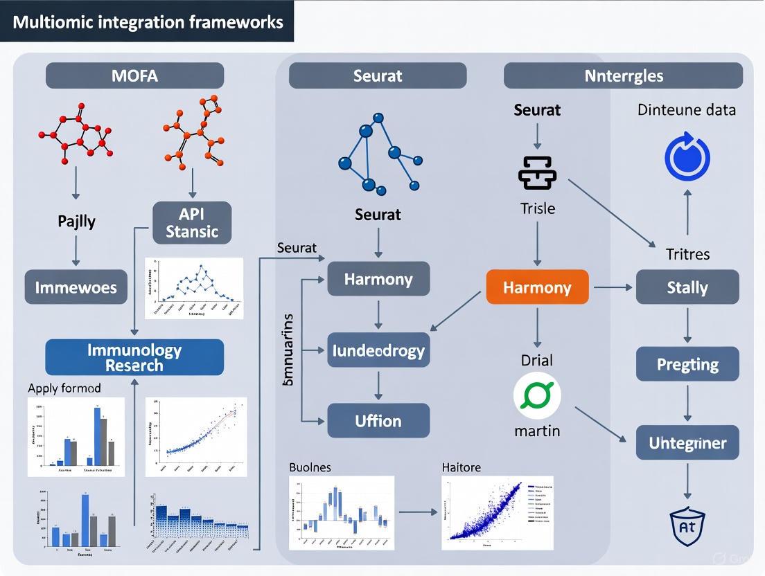
Integrating Multi-Omic Data in Immunology: A Comprehensive Guide to MOFA+, Seurat, and Harmony Frameworks
The complexity of the immune system, characterized by extraordinary cellular diversity and dynamic responses, demands analytical approaches that can holistically integrate multiple layers of molecular information.
Integrating Multi-Omic Data in Immunology: A Comprehensive Guide to MOFA+, Seurat, and Harmony Frameworks
Abstract
The complexity of the immune system, characterized by extraordinary cellular diversity and dynamic responses, demands analytical approaches that can holistically integrate multiple layers of molecular information. This article provides researchers, scientists, and drug development professionals with a comprehensive guide to three leading multi-omic integration frameworks—MOFA+, Seurat, and Harmony. We explore the foundational principles of these statistical and computational tools, detail their methodological application to immunological questions—from characterizing innate immune cells at CNS borders to profiling T-cell activation—and offer practical troubleshooting strategies for overcoming common bottlenecks related to data heterogeneity, computational scaling, and reproducibility. Finally, we present a comparative analysis of their performance against emerging methods and validate their utility through groundbreaking case studies in immunology and therapeutic development.
Understanding the Need for Multi-Omic Integration in Modern Immunology
Frequently Asked Questions (FAQs)
Q1: What are the main categories of data integration methods, and how do I choose? Data integration methods can be broadly divided into four categories, each with different strengths and resource requirements [1].
Table: Categories of Data Integration Methods
| Method Type | Key Principle | Examples | Best For |
|---|---|---|---|
| Linear Embedding Models | Uses singular value decomposition and local neighborhoods for correction [1]. | Harmony, Seurat, Scanorama [1] | Simple batch correction tasks [1]. |
| Deep Learning Approaches | Uses autoencoder networks, often conditioned on batch covariates [1]. | scVI, scANVI, scGen [1] | Complex data integration tasks [1]. |
| Graph-based Methods | Uses nearest-neighbor graphs, forcing connections between batches [1]. | BBKNN [1] | Fast runtime on smaller datasets [2]. |
| Global Models | Models batch effect as a consistent additive/multiplicative effect [1]. | ComBat [1] | - |
Q2: I'm getting a Harmony error in my Seurat v5 pipeline. What should I check?
A common error when using IntegrateLayers with HarmonyIntegration in Seurat v5 is: "Error in names(groups) <- "group" : attempt to set an attribute on NULL" [3]. This is often related to how cell identities are set before subsetting. To resolve this:
- Check Active Identities: Before subsetting your Seurat object, ensure that the active identities (
Idents) are set correctly. The error can occur if you have changed the active identity from the defaultseurat_clustersto another grouping (likeRNA_snn_res.0.3) and then attempt to subset and integrate [3]. - Verification Step: Re-run your subsetting and check the object's metadata to confirm that the intended clusters are present before proceeding to normalization and integration.
Q3: How does MOFA differ from standard PCA? MOFA is a statistically rigorous generalization of Principal Component Analysis (PCA) for multi-omics data [4]. While PCA reduces dimensionality for a single data matrix, MOFA infers a set of latent factors from multiple data matrices (omics layers) simultaneously. These factors capture the principal sources of variation, revealing which are shared across omics and which are specific to a single modality [4].
Q4: My Harmony algorithm isn't converging and shows a warning. What does this mean?
The warning "did not converge in 25 iterations" indicates that Harmony's iterative process has not stabilized [5]. The algorithm uses a soft k-means clustering approach and may require more iterations to find a stable solution where cell cluster assignments are no longer strongly dependent on dataset origin [5] [2]. You can typically increase the max.iter.harmony parameter to allow the algorithm more time to converge.
Troubleshooting Guides
Issue: Poor Mixing of Datasets After Integration
If cells still cluster by batch or dataset after integration instead of by biological cell type, consider the following steps:
- Verify Method Choice: Confirm you are using an appropriate method for your task's complexity. For complex integrations (large protocol differences, non-overlapping cell types), a deep learning method like scVI may be more effective than a linear embedding model [1].
- Check the Batch Covariate: The choice of "batch" covariate determines what variation is removed. Using a fine-grained covariate (e.g., individual samples) may remove meaningful biological signal, while a coarse covariate (e.g., sequencing platform) might leave technical artifacts [1].
- Inspect Input Data: Ensure your data preprocessing (normalization, highly variable feature selection) is consistent and appropriate across all batches.
Issue: Over-correction and Loss of Biological Variation
A key challenge is over-correction, where a method removes genuine biological variation along with the batch effect [1].
- Quantitative Evaluation: Use metrics like the k-nearest-neighbor Batch-Effect Test (kBET) to quantify batch mixing and metrics that measure the conservation of biological variation, which require ground-truth cell identity labels [1].
- Leverage Labels if Available: Some methods, like scANVI and scGen, can use cell identity labels to guide integration and prevent the removal of meaningful biological signal [1].
Issue: Computational Limitations with Large Datasets
When working with hundreds of thousands of cells, some methods become computationally infeasible.
- Benchmark for Scaling: Harmony has been demonstrated to integrate up to 500,000 cells on a personal computer, requiring significantly less memory and time than other methods like Seurat's MultiCCA or MNN Correct [2].
- Consider Federated Learning: For extremely large or privacy-sensitive distributed data, "Federated Harmony" provides a framework to perform integration without sharing raw data, reducing computational strain on any single machine [6].
Experimental Protocols
Protocol 1: Basic Multi-omics Integration Workflow with MOFA2 (R)
This protocol outlines the steps for an unsupervised integration of multiple omics data types (e.g., mutations, RNA expression, DNA methylation) using the MOFA2 R package [7].
- Data Preparation: Format your multi-omics data into a
MultiAssayExperimentobject or a list of matrices where features are rows and columns are samples. MOFA can handle missing data for samples not profiled with all omics [4]. - Model Training: Create a MOFA object and train the model. The core function will infer the latent factors.
- Downstream Analysis: Use the trained model for:
- Visualization: Plot the factor values for samples to identify continuous gradients or subgroups.
- Factor Interpretation: Examine the loadings to identify which features (e.g., genes, methylation sites) drive each factor.
- Annotation: Perform gene set enrichment analysis on the highly weighted features in relevant factors [7].
Protocol 2: Single-Cell RNA-Seq Integration with Seurat and Harmony
This protocol details the integration of multiple scRNA-seq datasets to correct for batch effects using Seurat and Harmony [5] [8].
- Preprocessing: Create a Seurat object for each dataset and perform standard preprocessing (normalization, identification of highly variable features) individually [8].
- PCA: Merge the objects and run PCA on the merged dataset to obtain an initial low-dimensional embedding [5] [8].
- Harmony Integration: Run Harmony on the PCA embedding to remove batch-specific effects.
- Clustering and UMAP: Use the Harmony-corrected embedding for downstream clustering and UMAP visualization. Cells should now group by cell type rather than batch [2].
Diagram: MOFA2 Workflow for Multi-omics Integration.
Diagram: Single-Cell Integration with Seurat and Harmony.
Research Reagent Solutions
Table: Essential Materials for Multi-omics Integration Experiments
| Reagent / Resource | Function in Analysis | Example or Note |
|---|---|---|
| 10x Genomics Chromium | Platform for generating single-cell or single-nucleus RNA-seq and multiome (ATAC + GEX) data [2]. | Used in PBMC benchmarks for Harmony [2]. |
| Seurat Object | Primary data container in Seurat; holds count matrix, metadata, and analysis results [8]. | - |
| Chronic Lymphocytic Leukaemia (CLL) Data | A common multi-omics benchmark dataset featuring mutations, RNA, DNA methylation, and drug response [4]. | Used in the original MOFA application [7] [4]. |
| PBMC (Peripheral Blood Mononuclear Cells) | A standard biological system for testing and benchmarking integration methods due to well-defined cell types [2]. | Freely available from 10X Genomics [8]. |
Frequently Asked Questions (FAQs)
Q1: What are the key differences between MOFA+, Seurat, and Harmony for multi-omics integration?
A1: These tools employ distinct mathematical approaches for data integration. MOFA+ is a factor analysis model that identifies latent factors representing shared and specific sources of variation across multiple omics layers [9]. It uses Bayesian framework with Automatic Relevance Determination priors to jointly model variation across sample groups and data modalities. Seurat employs an "anchor-based" integration workflow to find mutual nearest neighbors between datasets, enabling the creation of a shared embedding [10]. Harmony utilizes an iterative algorithm that projects cells into a shared embedding where cells group by cell type rather than dataset-specific conditions, using soft clustering and linear correction functions [2].
Q2: How do I choose between unsupervised (MOFA) and supervised (DIABLO) integration methods?
A2: The choice depends on your research question and whether you have predefined outcome variables. MOFA is ideal for exploratory analysis to discover inherent sources of variation without prior knowledge of sample groups [11]. DIABLO is preferable when you want to identify multi-omics patterns associated with specific clinical outcomes or predefined sample classes [11]. In practice, using both methods complementarily can provide comprehensive insights, as demonstrated in chronic kidney disease research where both approaches identified shared pathways despite different methodologies [11].
Q3: What are the minimum computational requirements for analyzing large-scale single-cell multi-omics datasets?
A3: Requirements vary by tool. Harmony is particularly efficient, capable of integrating ~10⁶ cells on a personal computer with minimal memory requirements (0.9GB for 30,000 cells to 7.2GB for 500,000 cells) [2]. MOFA+ implements stochastic variational inference enabling GPU acceleration, achieving up to 20-fold speed increases compared to conventional inference methods [9]. For very large datasets (>100,000 cells), Seurat and MOFA+ both offer scalable solutions, but may require high-performance computing resources for optimal performance [9] [10].
Q4: How can I validate that my multi-omics integration has preserved biological variation while removing technical artifacts?
A4: Use quantitative metrics like Local Inverse Simpson's Index (LISI) to assess integration quality [2]. Integration LISI (iLISI) measures dataset mixing, while cell-type LISI (cLISI) measures biological separation. Additionally, inspect known biological markers across integrated clusters to ensure they remain coherent. For factor models like MOFA+, examine the variance decomposition to verify that factors capture biologically meaningful patterns across modalities [9].
Q5: What strategies exist for integrating single-cell data with bulk omics measurements or spatial transcriptomics?
A5: Multiple approaches exist depending on data types. Harmony has demonstrated capabilities for cross-modality spatial integration [2]. MOFA+ can integrate single-cell modalities with bulk measurements when they share sample origins [9]. The emerging MEFISTO extension incorporates temporal or spatial dependencies in multi-omics integration [12]. For spatial transcriptomics integration with single-cell data, methods like Seurat's integration with spatial mapping enable cell-type localization in tissues [10].
Troubleshooting Guides
Issue 1: Poor Integration Quality Across Datasets
Symptoms: Cells cluster primarily by dataset origin rather than cell type; biological signals are obscured.
Solution:
- Preprocessing consistency: Ensure consistent normalization across all datasets
- Feature selection: Use highly variable features that are robust across batches
- Parameter optimization: Adjust the strength of integration parameters:
Verification: Calculate LISI metrics after integration. Successful integration should show high iLISI (dataset mixing) while maintaining low cLISI (cell-type separation) [2].
Issue 2: Failure to Identify Biologically Meaningful Factors in MOFA+
Symptoms: Factors explain technical variance but not biological processes; factors don't align with known biology.
Solution:
- Input normalization: Ensure proper normalization per modality (e.g., TF-IDF for chromatin data, log-normalization for RNA)
- Factor number selection: Use the evidence lower bound (ELBO) or automatic relevance determination to select appropriate factor numbers [9]
- Variance decomposition: Examine the variance explained by factors across modalities and groups to identify shared and specific signals
- Biological interpretation: Use known marker genes and pathway enrichment to annotate factors biologically
Advanced Tip: For temporal data, use MEFISTO which incorporates temporal smoothness constraints to improve factor interpretability [12].
Issue 3: Memory or Computational Limitations with Large Datasets
Symptoms: Analysis fails due to memory errors; excessive computation time.
Solution:
- Dataset size:
- Feature reduction:
- Pre-filter lowly abundant features
- Use highly variable features rather than all genes
- Computational resources:
- For MOFA+: Enable GPU acceleration when available [9]
- Increase memory allocation or use distributed computing
Table 1: Computational Requirements Comparison
| Tool | Maximum Dataset Size | Memory Efficiency | GPU Support | Integration Approach |
|---|---|---|---|---|
| Harmony | ~1 million cells | High (7.2GB for 500k cells) | No | Iterative linear correction |
| MOFA+ | Hundreds of thousands of cells | Moderate | Yes (Stochastic VI) | Bayesian factor analysis |
| Seurat | Hundreds of thousands of cells | Moderate | Limited | Anchor-based integration |
| DIABLO | Moderate (~100s of samples) | Moderate | No | Multivariate supervised |
Issue 4: Inconsistent Cell Type Annotation Across Modalities
Symptoms: Different modalities suggest different cell-type assignments; conflicting marker expression.
Solution:
- Cross-modality validation:
- Use conserved marker detection (e.g., Seurat's FindConservedMarkers) [10]
- Transfer labels from well-annotated modalities to others
- Employ symmetric integration approaches that weight modalities equally
- Reference mapping:
- Map to established references like Azimuth [13]
- Use manual annotation based on canonical markers from literature
- Multi-level annotation:
- Implement hierarchical annotation (L1, L2) as used in OASIS atlas [13]
- Distinguish broad cell types from fine-grained states
Verification: Check expression of known marker genes across modalities in integrated space. Use cross-validation with held-out datasets.
Issue 5: Handling Missing Data or Unbalanced Modalities
Symptoms: Some samples missing certain modalities; uneven quality across data types.
Solution:
- MOFA+ handling: MOFA+ naturally handles missing modalities by learning from available data [9]
- Imputation approaches: Use cross-modality imputation (e.g., Seurat's bridge integration) when appropriate
- Weighting strategies: Adjust modality weights based on data quality and biological importance
- Subsetting: Analyze complete cases only for certain questions, then validate findings in full dataset
Experimental Protocols
Protocol 1: Basic Multi-Omics Integration with MOFA+
Application: Integrating scRNA-seq with scATAC-seq or DNA methylation data from the same cells [9].
Workflow:
- Input preparation: Create a MOFA+ object with multiple assays (views) and sample groups
- Data preprocessing: Normalize each modality appropriately (e.g., log+CPM for RNA, TF-IDF for ATAC)
- Model training: Set up and train the MOFA+ model with appropriate options:
- Specify number of factors (start with 5-15)
- Enable stochastic inference for large datasets
- Set sparsity options to encourage interpretability
- Model evaluation: Examine variance explained, ELBO convergence, and factor correlation
- Downstream analysis:
- Use factors for visualization (UMAP/t-SNE)
- Identify driving features per factor
- Perform gene set enrichment on factor weights
MOFA+ Analysis Workflow
Protocol 2: Multi-Group Integration with Harmony
Application: Integrating multiple scRNA-seq datasets across conditions, batches, or technologies [2].
Workflow:
- Initial processing: Normalize and run PCA on each dataset separately
- Integration: Apply Harmony to remove dataset-specific effects:
- Specify batch covariates (e.g., technology, donor)
- Adjust theta (diversity penalty) and lambda parameters
- Run iterative correction until convergence
- Visualization: Generate UMAP/t-SNE embeddings from Harmony coordinates
- Validation:
- Calculate LISI metrics to quantify integration quality
- Check preservation of known biological states
- Verify removal of technical artifacts
Protocol 3: Cross-Modality Integration with Seurat
Application: Mapping scRNA-seq data to spatial transcriptomics or integrating CITE-seq data [10] [2].
Workflow:
- Layer separation: Split Seurat object by condition or modality using the
splitfunction [10] - Anchor identification: Find integration anchors between layers using
FindIntegrationAnchors - Data integration: Integrate layers using
IntegrateLayersorIntegrateData - Joint analysis:
- Perform PCA on integrated data
- Run UMAP for visualization
- Identify conserved markers across conditions
- Differential analysis: Compare conditions within integrated cell types
Table 2: Key Parameters for Multi-Omics Integration Tools
| Tool | Critical Parameters | Default Values | Adjustment Guidelines |
|---|---|---|---|
| MOFA+ | Number of factors, Sparsity, Training iterations | Factors: 5-25 | Increase factors for complex systems; adjust sparsity for interpretability |
| Harmony | Theta (θ), Lambda (λ), Max iterations | θ=2, λ=1 | Increase θ for stronger batch correction; λ prevents overcorrection |
| Seurat | k.anchor, k.filter, k.score | k.anchor=5, k.filter=200 | Increase for datasets with rare cell types; decrease for better integration |
| DIABLO | Number of components, KeepX parameters | Components: 2-5 | Optimize using cross-validation; adjust KeepX for feature selection |
Protocol 4: Multi-Omics eQTL Mapping at Single-Cell Resolution
Application: Identifying cell-type specific genetic regulation using single-cell data with genotype information [13].
Workflow:
- Cell typing: Annotate cell types using reference mapping or manual annotation
- Pseudobulk creation: Aggregate expression by donor and cell type
- cis-eQTL mapping: Test variant-gene associations within 1Mb of TSS
- Cell-state mapping: Extend analysis to continuous cell states
- Cross-population comparison: Compare eQTL effects across ancestries
Single-cell eQTL Mapping Workflow
The Scientist's Toolkit: Research Reagent Solutions
Table 3: Essential Multi-Omics Technologies and Their Applications
| Technology | Target Molecules | Key Applications in Immunology | Considerations |
|---|---|---|---|
| 10x Genomics Chromium | RNA, ATAC, protein (CITE-seq) | Immune cell profiling, receptor sequencing | Compatible with single-cell multiome kits |
| Olink Proteomics | 2,925 plasma proteins | Plasma protein quantification with high sensitivity | Requires only 1μL plasma per measurement |
| Single-cell bisulfite sequencing | DNA methylation | Epigenetic heterogeneity in immune cells | Can profile non-CpG methylation in neurons [9] |
| scNMT-seq | Nucleosome, transcriptome, methylation | Parallel multi-layer profiling from same cells | Technically challenging; lower throughput |
| CITE-seq | RNA + surface proteins | Immunophenotyping with transcriptomics | ~200 proteins simultaneously with transcriptome |
| SPLiT-seq | RNA (low-cost) | High-throughput screening applications | Fixed cells; lower sequencing depth |
| CRISPR-based screening | Functional genomics | Identify regulators of immune functions | Pooled screens with single-cell readout |
Advanced Integration Strategies
Strategy 1: Leveraging Complementary Integration Approaches
Rationale: Different integration methods have complementary strengths. Using both unsupervised (MOFA) and supervised (DIABLO) approaches on the same dataset can provide more comprehensive insights [11].
Implementation:
- Run MOFA for unsupervised discovery of sources of variation
- Use DIABLO for supervised analysis with clinical outcomes
- Compare results to identify robust signals across methods
- Prioritize pathways and features identified by both approaches
Example: In CKD research, both MOFA and DIABLO identified complement and coagulation cascades, cytokine-cytokine receptor interaction, and JAK/STAT signaling pathways, despite their different mathematical foundations [11].
Strategy 2: Dynamic Genetic Regulation Analysis
Rationale: Genetic effects on gene expression can vary across cell types and states, requiring single-cell resolution to fully capture [13].
Implementation:
- Map eQTLs across finely resolved cell types (L1 and L2 annotations)
- Assess sharing of eQTL effects across related cell types
- Compare eQTL effects across populations (e.g., Japanese vs. European)
- Integrate with GWAS findings through colocalization analysis
Key Finding: Statistical power for eQTL discovery depends more on cell counts per sample than total sample size, highlighting the importance of deep sequencing [13].
Strategy 3: Somatic Mutation Integration
Rationale: Somatic mutations in immune cells (mCAs, LOY, mt-heteroplasmy) can influence immune function and be detected at single-cell resolution [13].
Implementation:
- Detect somatic mutations from WGS or SNP array data
- Project mutations onto single-cell transcriptomes
- Identify immune features associated with somatic mutations
- Assess clonal expansion patterns in disease contexts
Application: In COVID-19, somatic mutations in expanded B cell clones were assessed for reactivity against SARS-CoV-2 antigens [13].
Frequently Asked Questions
Q1: I encounter the error "argument 3 matches multiple formal arguments" when running RunHarmony in Seurat. What should I do?
This error often arises from a parameter naming conflict, frequently with the reduction parameter [14]. The Seurat wrapper for Harmony has many function arguments, and a partial argument name might match more than one formal argument.
- Solution: The most straightforward fix is to explicitly name your arguments when calling the function. Ensure you are using the exact, full parameter names as defined in the
RunHarmonydocumentation. For example, if you were running: Double-check thatreductionis the correct parameter name for your Seurat version. In some cases, simply removing a conflicting argument likereductionhas resolved the issue, though this is not recommended as it may use a default setting you did not intend [14].
Q2: After updating Seurat, I get many warnings about deprecated parameters like tau and block.size in Harmony. Will this affect my results?
These warnings indicate you are using old parameter names that are no longer supported in the updated version of Harmony or its Seurat integration [15]. Your analysis will likely still run, as the warnings state the deprecated parameters will be ignored.
- Solution: Update your code to use the new parameter names. The warnings often specify the correct new parameter to use. For example:
Q3: I cannot reproduce my old Harmony clustering results on the same data. The number and structure of clusters are different. What could be wrong?
Reproducibility issues can stem from several factors, especially across different computing environments [16].
- Solution:
- Set Random Seeds: Use
set.seed()before runningRunHarmonyand any subsequent clustering steps (e.g.,FindClusters,RunUMAP) to ensure stochastic processes are reproducible. - Check Software Versions: Different versions of Seurat, Harmony, or even underlying R packages can alter results. Note the versions used in the original analysis. For instance, the default UMAP method in Seurat changed from a Python implementation to an R-native one, which can change visualization and downstream clustering [16].
- Processor Architecture: Evidence suggests that differences in processor chips (e.g., Intel vs. Apple M-series) can lead to floating-point arithmetic variations, affecting numerical results and thus clustering outcomes [16]. Try to run the analysis on the same architecture as the original.
- Parameter Consistency: Ensure all parameters (e.g.,
resolutionfor clustering, number of PCs,dimsforRunUMAPandFindNeighbors) are identical to your original run.
- Set Random Seeds: Use
Troubleshooting Guides
Problem: Harmony fails to converge or displays a "Quick-TRANSfer stage steps exceeded maximum" warning.
This warning relates to the internal k-means clustering algorithm and may indicate that the algorithm is struggling to find a stable solution [5] [15].
- Diagnosis: This is often a warning, not an error, and the algorithm may still produce usable results. However, it warrants attention.
- Solutions:
- Increase
max_iter: Allow the algorithm more iterations to converge. - Adjust Harmony Parameters: Parameters like
theta(diversity clustering penalty) can influence convergence. The deprecatedtauwas related to this penalty, and its modern equivalent can be set viaharmony_options()[15]. - Verify Input Data: Ensure your PCA input is appropriate and not containing extreme outliers.
- Increase
Problem: General challenges with multi-omics data integration.
The integration of disparate data types (e.g., transcriptomics, proteomics) is inherently complex [17] [18].
- Diagnosis: Common challenges include:
- Data Heterogeneity: Different omics layers have unique data structures, scales, noise profiles, and batch effects [18].
- Missing Values: Some features may be present in one data modality but absent in another [17].
- Lack of Standardization: No universal pre-processing protocols exist across all omics types [18].
- Solutions:
- Choose the Right Method: Select an integration framework suited to your data and question.
- Thorough Pre-processing: Normalize and scale each data type individually before integration.
- Utilize Supervised Integration: If you have a known outcome (e.g., disease state), methods like DIABLO can use this information to guide integration and feature selection [18].
Multi-Omics Integration Frameworks at a Glance
The following table compares three key integration tools, helping you select the right one for your research goals.
| Framework | Core Methodology | Primary Use Case | Integration Type | Key Feature |
|---|---|---|---|---|
| MOFA+ [18] | Unsupervised Bayesian factor analysis | Identifying hidden sources of variation across multiple omics datasets. | Vertical / Matched | Learns factors that can be shared or specific to data types. |
| Harmony [5] | Iterative clustering with integration | Removing dataset-specific effects for better joint clustering (e.g., batch correction). | Horizontal / Unmatched | Designed for single-cell RNA-seq data, often used within Seurat. |
| DIABLO [18] | Supervised multiblock PLS-DA | Classifying phenotypes using multi-omics data and selecting biomarkers. | Vertical / Matched | Uses phenotype labels to drive integration and feature selection. |
Experimental Workflow for Multi-Omics Integration
A generalized workflow for integrating matched multi-omics samples using a factor-based approach like MOFA+ is outlined below.
Diagram Title: MOFA+ Multi-Omics Integration Workflow
Methodology Details:
- Data Pre-processing: Each omics data matrix (e.g., from RNA-Seq, DNA methylation, proteomics) undergoes individual normalization and scaling to account for technical noise and different data distributions [18].
- Model Training: The MOFA+ model is trained in an unsupervised manner to decompose the multi-omics data into a set of latent factors. These factors represent the principal sources of variation across all datasets and samples [18].
- Variance Decomposition: The model quantifies the percentage of variance explained by each factor in each omics dataset. This helps identify factors that are shared across all modalities or specific to a single data type [18].
- Factor Interpretation: Researchers correlate the latent factors with sample metadata (e.g., clinical outcomes, cell type labels) or use gene set enrichment analysis to attach biological meaning to the discovered sources of variation [18].
The Scientist's Toolkit: Essential Research Reagents & Materials
| Item | Function in Multi-Omics Research |
|---|---|
| Single-Cell Multi-Omics Platform | Enables correlated measurements of genomics, transcriptomics, and epigenomics from the same cell, providing a matched data structure for vertical integration [19]. |
| High-Throughput Sequencing | Generates the foundational data for genomic (WGS), transcriptomic (RNA-seq), and epigenomic (ChIP-seq, ATAC-seq) analyses [19]. |
| Spatial Transcriptomics Platform | Provides RNA expression data within a morphological context, adding a spatial layer to multi-omics integration [19]. |
| Liquid Biopsy Assay | Allows non-invasive collection of multi-analyte biomarkers (e.g., cfDNA, RNA, proteins) from biofluids, crucial for clinical translation [19]. |
Technical Support Center
FAQs & Troubleshooting Guides
Q: My MOFA model fails to converge when integrating scRNA-seq and CITE-seq data. What could be the cause? A: Non-convergence often stems from improper data scaling or extreme outliers.
- Solution 1: Ensure each data modality is scaled to unit variance. For CITE-seq ADT data, use centered log ratio (CLR) transformation instead of log-normalization.
- Solution 2: Filter out low-quality cells and genes/features with excessive zeros before integration. Check for technical outliers using PCA on each view independently.
Q: After integrating datasets with Harmony, my T cell clusters are still batch-specific. How can I improve integration? A: This indicates strong batch effects that Harmony's default parameters cannot overcome.
- Solution 1: Increase the
thetaparameter, which controls the strength of batch correction. A highertheta(e.g., 3-5) applies more aggressive correction. - Solution 2: Pre-filter your features more stringently. Use highly variable genes (HVGs) for transcriptomics and high-variance proteins for proteomics to give the algorithm a stronger signal.
Q: I observe a poor correlation between transcriptomic and proteomic measurements for the same surface marker in my Seurat object. Why? A: This is a common biological and technical discrepancy.
- Solution 1 (Technical): Check for antibody non-specificity in your CITE-seq data. Compare to an isotype control or FMO (Fluorescence Minus One) control.
- Solution 2 (Biological): Account for post-transcriptional regulation and protein half-life. The mRNA level may not reflect the current protein abundance. Consider a time-course experiment.
Q: My metabolomics data has many missing values, causing issues in multi-omic integration. How should I handle this? A: Metabolomics data is inherently sparse due to limits of detection.
- Solution 1: Apply a minimum detection filter. Remove metabolites that are missing in >80% of samples.
- Solution 2: For remaining missing values, use imputation methods appropriate for metabolomics, such as k-nearest neighbors (KNN) or half-minimum imputation (replacing NA with half the minimum value for that metabolite).
Data Presentation
Table 1: Typical Single-Cell Multi-Omic Data Yields and QC Metrics
| Omics Layer | Assay | Typical Cells Post-QC | Median Features per Cell | Key QC Metric (Passing Threshold) |
|---|---|---|---|---|
| Genomics | scDNA-seq | 5,000 - 10,000 | 50,000 - 100,000 SNPs | Mean Read Depth > 20x |
| Epigenomics | scATAC-seq | 5,000 - 15,000 | 5,000 - 20,000 Peaks | TSS Enrichment Score > 8 |
| Transcriptomics | scRNA-seq (10x) | 8,000 - 15,000 | 2,000 - 5,000 Genes | Mitochondrial Read % < 20% |
| Proteomics | CITE-seq (100-plex) | 8,000 - 15,000 | 50 - 150 ADTs | Total ADT Counts > 1,000 |
| Metabolomics | scMetabolomics (MS) | 500 - 2,000 | 50 - 200 Metabolites | CV < 30% in QC Samples |
Experimental Protocols
Protocol 1: CITE-seq for Surface Protein and Transcriptome Co-Profiling
- Cell Preparation: Harvest and count cells. Aim for >90% viability.
- Antibody Staining: Incubate 1 million cells with a TotalSeq-B antibody cocktail (0.5-2 µg/mL per antibody) in 100 µL PBS + 0.04% BSA for 30 minutes on ice.
- Washing: Wash cells twice with 2 mL PBS + 0.04% BSA to remove unbound antibodies.
- Cell Barcoding: Proceed immediately to single-cell partitioning on the 10x Genomics Chromium Controller using the Single Cell 5' Reagent Kit v2.
- Library Construction: Generate cDNA, ADT (Antibody-Derived Tag), and HTO (Hashtag Oligo) libraries according to the manufacturer's protocol. Pool libraries equimolarly for sequencing.
Protocol 2: Multi-Omic Integration with MOFA+ on Seurat Objects
- Data Preprocessing: Create individual Seurat objects for RNA and ADT data. Normalize RNA using
SCTransformand ADT data usingCLRnormalization. - Feature Selection: Identify and retain the top 3,000 highly variable genes and all high-quality ADT features.
- MOFA Object Creation: Use the
create_mofa_from_seuratfunction to combine the RNA and ADT assays into a single MOFA object. - Model Training: Train the model with default options. Set
convergence_modeto "slow" for more robust results. - Downstream Analysis: Regress MOFA factors against cell metadata (e.g., disease status) to identify factors driving variation. Plot factor weights to see which genes/proteins define each factor.
Mandatory Visualizations
Multi-Omic Integration Pathway
T Cell Activation Multi-Omic View
The Scientist's Toolkit: Research Reagent Solutions
| Reagent / Material | Function in Multi-Omic Immunology |
|---|---|
| TotalSeq-B Antibodies | Antibody-derived tags (ADTs) for quantifying surface protein abundance alongside transcriptome in CITE-seq. |
| 10x Genomics Chromium | Microfluidic platform for partitioning single cells and barcoding nucleic acids (RNA, DNA, ATAC). |
| Tn5 Transposase | Enzyme for tagmenting accessible chromatin in scATAC-seq assays. |
| Hashtag Oligos (HTOs) | Sample-barcoding antibodies for multiplexing samples, reducing batch effects and costs. |
| Cell Hashing Antibodies | Antibodies conjugated to HTOs for pooling samples prior to scRNA-seq/CITE-seq. |
| MOFA+ R Package | Statistical framework for integrating multiple omics data sets to uncover latent factors of variation. |
| Harmony R Package | Algorithm for integrating single-cell data across multiple batches or conditions. |
| Seurat R Package | Comprehensive toolkit for single-cell genomics data analysis, including multi-omic integration. |
A Practical Workflow for Multi-Omic Data Integration and Analysis
Frequently Asked Questions (FAQs)
FAQ on Data Processing
Q: How should I normalize my data for MOFA+? Proper normalization is critical for the model to work correctly. For count-based data such as RNA-seq or ATAC-seq, it is strongly recommended to perform size factor normalization followed by variance stabilization. If this step is not done correctly, the model will learn a very strong Factor 1 that captures differences in total expression per sample, pushing more subtle sources of biological variation to subsequent factors [20].
Q: Should I filter features before using MOFA+? Yes. It is strongly recommended that you filter for highly variable features (HVGs) per assay. However, an important caveat applies when doing multi-group inference: you must regress out the group effect before selecting HVGs to avoid bias [20].
Q: How do I handle batch effects or other technical factors?
MOFA+ is designed to discover dominant sources of variation, whether biological or technical. If you have known, strong technical factors (e.g., batch effects), MOFA+ will likely dedicate initial factors to capturing this large variability. To prevent this, it is recommended to regress out these technical factors a priori using methods like linear models in limma. This allows MOFA+ to focus on discovering more subtle biological sources of variation [20].
Q: My data modalities have different numbers of features. Does this matter? Yes. Larger data modalities (those with more features) tend to be overrepresented in the inferred factors. It is good practice to filter uninformative features to bring the different views within the same order of magnitude. If the size disparity is unavoidable, be aware that the model might miss small sources of variation present in the smaller dataset [20].
Q: Can MOFA+ handle missing values? Yes. MOFA+ simply ignores missing values in the likelihood calculation, and there is no hidden imputation step. Matrix factorization models like MOFA+ are known for their robustness to missing values [20].
FAQ on Model Training and Setup
Q: How does the multi-group framework work? The multi-group framework in MOFA+ is designed to compare the sources of variability that drive each pre-defined group (e.g., different experimental conditions, batches, or donors). Its goal is not to capture simple differential changes in mean levels between groups. To achieve this, the group effect is regressed out from the data before fitting the model, allowing you to identify which factors are shared across groups and which are specific to a single group [20].
Q: How do I define groups for my analysis? Group definition is a hypothesis-driven step motivated by your experimental design. There is no single "right" way, but the definition should align with your biological question. Crucially, remember that the multi-group framework aims to dissect variability, not to find factors that separate groups. If your aim is to find a factor that cleanly "separates" groups, you should not use the multi-group framework [20].
Q: How many factors should I learn? The optimal number of factors is context-dependent. It is influenced by the aim of your analysis, the dimensions of your assays, and the complexity of the data. As a general guideline, if the goal is to identify major biological sources of variation, considering the top 10-15 factors is typical. For tasks like imputation of missing values, where even minor sources of variation can be important, training a model with a larger number of factors is advisable [20] [21].
Q: What types of data can MOFA+ handle? MOFA+ can cope with three main types of data via different likelihoods:
- Continuous data: Modelled using a Gaussian likelihood.
- Binary data: Modelled using a Bernoulli likelihood.
- Count data: Modelled using a Poisson likelihood. A critical best practice is that if your data can be safely transformed to meet the assumptions of a Gaussian likelihood (e.g., normalizing and variance-stabilizing RNA-seq counts), this is always recommended. Non-Gaussian likelihoods require statistical approximations and are generally less accurate [20].
FAQ on Interpretation and Downstream Analysis
Q: How do I interpret the factors? MOFA+ factors capture global sources of variability, ordinating cells along a one-dimensional axis centered at zero. The relative positioning of cells is what matters. Cells with different signs display opposite "effects" along the inferred axis, and a higher absolute value indicates a stronger effect. This interpretation is analogous to interpreting principal components in PCA [20].
Q: How do I interpret the weights? Weights indicate how strongly each feature relates to a factor, enabling biological interpretation. Features with weights near zero have little association with the factor. Features with large absolute weights are strongly associated. The sign of the weight indicates the direction of the effect: a positive weight means the feature has higher levels in cells with positive factor values, and vice versa [20].
Troubleshooting Common Experimental Issues
Model Fitting and Convergence Problems
Issue: The model converges very slowly or not at all.
- Potential Cause: The learning rate for the stochastic variational inference (SVI) may be set too high or too low.
- Solution: Adjust the
learning_rateanddrop_factorparameters. A common strategy is to start with a higher learning rate (e.g., 0.01) and a modestdrop_factor(e.g., 0.5) to allow the learning rate to decrease after a set number of iterations without improvement in the ELBO [9].
Issue: The model is not capturing known biological signal.
- Potential Cause 1: Overwhelming technical variation. As noted in the FAQs, strong technical effects like batch or library size can dominate the first factors.
- Solution: Revisit your data preprocessing. Ensure you have properly normalized and regressed out known technical covariates before training the model [20].
- Potential Cause 2: The number of factors (K) is too small.
- Solution: Increase the number of factors and use the model selection criteria (e.g., looking at the variance explained by additional factors) to choose an appropriate K [21].
Data Integration and Interpretation Challenges
Issue: One data modality is dominating the factors.
- Potential Cause: A large disparity in the number of features or the amount of intrinsic variability between modalities.
- Solution: Perform more aggressive feature filtering on the larger/more variable modality to balance its influence relative to the other modalities. The goal is to have all modalities contribute meaningfully to the latent space [20].
Issue: In multi-group mode, I cannot find any group-specific factors.
- Potential Cause: The groups may not have distinct internal structures, or the signal of group-specific variation may be weak compared to shared variation.
Experimental Protocols & Workflows
Standard Protocol for Multi-Group Single-Cell RNA-seq Integration
This protocol is adapted from the MOFA+ application on a time-course scRNA-seq dataset of mouse embryos [9].
1. Input Data Preparation:
- Data: 16,152 cells from mouse embryos at embryonic days E6.5, E7.0, and E7.25 (two biological replicates per stage).
- Group Definition: The six batches (two replicates per stage) were defined as distinct groups in the MOFA+ model.
2. Preprocessing and Normalization:
- Filtering: Cells and genes were filtered based on standard quality control metrics.
- Normalization: Size factor normalization and variance stabilization transformation were applied to the count data.
- Feature Selection: Highly variable genes were selected after regressing out the group effect.
3. Model Training:
- The MOFA+ model was trained using stochastic variational inference, which provided a ~20-fold speed increase on large datasets compared to standard variational inference.
- The model was run for a sufficient number of iterations until convergence of the Evidence Lower Bound (ELBO).
4. Downstream Analysis:
- Variance Decomposition: The model identified 7 factors explaining >1% of variance, collectively accounting for 35-55% of transcriptional variance per embryo.
- Factor Interpretation:
- Factor 1 and 2 were associated with extra-embryonic (ExE) cell types (marked by Ttr, Apoa1, Rhox5, Bex3).
- Factor 4 captured the transition of epiblast to nascent mesoderm (marked by Mesp1, Phlda2).
- Factor 3 was identified as a technical factor related to metabolic stress.
- Temporal Analysis: Inspection of factor activity over time showed that the variance explained by Factor 4 (mesoderm) increased from E6.5 to E7.25, consistent with developmental progression.
Protocol for Multi-Modal Single-Cell Data Integration
This protocol outlines the general workflow for integrating data from different molecular layers, such as RNA expression and DNA methylation [9] [4].
1. Data Input and Setup:
- Assemble your data matrices where features are aggregated into non-overlapping views (e.g., RNA expression, DNA methylation) and cells are aggregated into non-overlapping groups (e.g., different conditions or samples).
2. Model Configuration:
- Specify the appropriate likelihood for each data view (Gaussian for normalized continuous data, Bernoulli for binary data, etc.).
- Choose the number of factors. It is often practical to start with a slightly larger number and then prune factors that do not explain meaningful variation.
3. Model Inference:
- Use the GPU-accelerated training option for large datasets to significantly speed up computation [9].
4. Analysis of Results:
- Use the learned latent factors for a wide range of analyses: variance decomposition, inspection of feature weights, inference of trajectories, and clustering.
Table 1: Performance Comparison of MOFA+ and Other Integration Methods on a Single-Cell Multiome Dataset (69,249 BMMC cells) [22]
| Method | Running Time (Minutes) | Peak Memory Usage (GB) | Batch Correction Score (kBET) | Biological Conservation (ARI) |
|---|---|---|---|---|
| Smmit | <15 | 23.05 | Highest | Highest |
| Multigrate | ~167 | 217.29 | Intermediate | High |
| scVAEIT | >1690 | >230 | Intermediate | Intermediate |
| MOFA+ | Not Reported | Not Reported | Lower | Lower |
| MultiVI | Not Reported | Not Reported | Lower | Lower |
Table 2: Key Statistical Features of the MOFA+ Framework [9]
| Feature | Description |
|---|---|
| Core Model | Bayesian Group Factor Analysis |
| Inference | Stochastic Variational Inference (SVI) with GPU acceleration |
| Key Prior | Automatic Relevance Determination (ARD) for structured sparsity across views and groups |
| Handling of Missing Data | Ignored in the likelihood (no imputation) |
| Data Types | Supports Gaussian (continuous), Bernoulli (binary), and Poisson (count) likelihoods |
Research Reagent Solutions
Table 3: Essential Materials and Computational Tools for MOFA+ Analysis
| Item / Software | Function / Purpose |
|---|---|
| MOFA2 (R Package) | The primary R package for running MOFA+, including data input, model training, and comprehensive downstream analysis. |
| mofapy2 (Python Package) | The Python core that performs the model inference. Can be used independently for model training. |
| Seurat | A toolkit for single-cell genomics; often used in conjunction with MOFA for upstream filtering or downstream analysis like UMAP visualization and clustering [22]. |
| Harmony | An integration algorithm that can be used as an alternative or complement to MOFA+ for batch correction [22]. |
| Scanpy | A Python-based toolkit for analyzing single-cell gene expression data; can be used for preprocessing steps. |
| NVIDIA GPU with CuPy | Recommended hardware and software configuration to enable accelerated model training for large datasets [9] [20]. |
| OpenBLAS / Intel MKL | Optimized math libraries to speed up CPU-based linear algebra operations during model training [20]. |
Workflow and Conceptual Diagrams
MOFA+ Core Workflow and Downstream Analysis
Multi-Group Integration Logic in MOFA+
Leveraging Seurat v5 for Integrative Multimodal Analysis and Bridge Integration
Frequently Asked Questions
Q: What are the key integration methods available in Seurat v5 and how do I choose between them?
Seurat v5 provides streamlined access to multiple integration methods through the IntegrateLayers function. The available methods include Anchor-based CCA integration (CCAIntegration), Anchor-based RPCA integration (RPCAIntegration), Harmony (HarmonyIntegration), FastMNN (FastMNNIntegration), and scVI (scVIIntegration). RPCA integration is faster and more conservative than CCA, while scVI requires additional Python dependencies but can be particularly powerful for complex integration tasks. The choice depends on your data characteristics and computational resources [23].
Q: How does Seurat v5 handle multimodal data like CITE-seq with simultaneous RNA and protein measurements?
Seurat v5 stores multimodal data in separate assays within the same object. For CITE-seq data, you would typically have an RNA assay for transcriptomic measurements and an ADT assay for antibody-derived tags. The CreateAssay5Object function allows you to add additional modalities, and you can switch between assays using DefaultAssay() or specify modalities using assay keys in feature names (e.g., "adtCD19" for protein data vs "rnaCD19" for RNA data) [24].
Q: What should I do if my FeaturePlot color palette is only showing two bins in Seurat v5?
This appears to be a known issue in Seurat v5 where supplying a custom color vector may cause the data to be binned into only two categories corresponding to the minimal and maximal values. As a workaround, you can use the built-in color palettes like CustomPalette(), BlackAndWhite(), BlueAndRed(), or PurpleAndYellow() which are designed to work properly with the visualization functions [25] [26].
Q: How do I properly normalize different data modalities in Seurat?
Different modalities require different normalization approaches. For RNA data, standard log-normalization or SCTransform is appropriate. For ADT data from CITE-seq, use NormalizeData with normalization.method = "CLR" and margin = 2 to perform centered log-ratio normalization across cells. For count-based data like ATAC-seq, size factor normalization with variance stabilization is recommended [24] [20].
Q: What are the key considerations for successful multi-omic integration?
Proper normalization is critical to prevent any single modality from dominating the integration. Filter highly variable features per assay and ensure different modalities have comparable numbers of informative features. For multi-group inference, regress out group effects before selecting variable features. Larger data modalities tend to be overrepresented, so balancing informative features across modalities is important [20].
Troubleshooting Guides
Integration and Clustering Issues
Problem: Poor integration results with batch effects still visible after running IntegrateLayers
- Cause: Insufficient correction of technical variability or improper normalization
- Solutions:
- Ensure proper normalization of each modality separately before integration
- Check that you have regressed out known technical factors like batch effects prior to integration
- Try different integration methods (RPCA is more conservative than CCA)
- Increase the number of variable features used for integration
- Verify that the data layers are properly split by batch before integration [20] [23]
Problem: Integration removes biological signal along with technical noise
- Cause: Over-correction during the integration process
- Solutions:
- Use the more conservative RPCA integration method instead of CCA
- Reduce the strength of the integration parameters (
integration_args) - Include known biological covariates in the downstream analysis rather than relying solely on integration to preserve them
- Compare integrated results with unintegrated analysis to verify biological signals are maintained [23]
Multimodal Data Analysis Issues
Problem: Difficulty visualizing both RNA and protein markers in the same analysis
- Cause: Default assay setting may not match the modality you want to visualize
- Solutions:
- Use assay keys in feature names (e.g., "adtCD3" for protein, "rnaCD3E" for RNA) to explicitly specify modalities
- Switch default assays using
DefaultAssay(object) <- "ADT"orDefaultAssay(object) <- "RNA" - Use the
FeaturePlotfunction with specified assay keys to visualize both modalities side-by-side [24]
Problem: One data modality dominates the integrated analysis
- Cause: Imbalance in dimensionality or information content between modalities
- Solutions:
- Filter uninformative features from the larger modality to balance dimensions
- Apply more stringent variable feature selection to prevent overrepresentation
- Consider down-weighting the influence of the dominant modality in the integration parameters
- Ensure proper normalization specific to each data type [20]
Visualization Problems
Problem: Custom color palettes not working properly in FeaturePlot
- Cause: Changed behavior in Seurat v5 regarding color palette handling
- Solutions:
- Use built-in palettes like
BlueAndRed(k = 50)orCustomPalette(low = "white", high = "red", mid = NULL, k = 50) - Ensure the length of your color vector matches the expected binning structure
- Check the
keep.scaleparameter setting as it affects color scaling - Consider using the
colsparameter with predefined palettes rather than custom vectors [25] [26]
- Use built-in palettes like
Experimental Protocols
Multimodal Data Integration Workflow
Step-by-Step Protocol:
Data Loading and Initialization
- Load RNA and ADT count matrices using
read.csv()orRead10X() - Create Seurat object with
CreateSeuratObject(counts = rna_matrix) - Add additional assays using
CreateAssay5Object()andobject[["ADT"]] <- adt_assay[24]
- Load RNA and ADT count matrices using
Modality-Specific Normalization
Data Splitting and Preprocessing
- Split layers by batch:
object[["RNA"]] <- split(object[["RNA"]], f = object$batch) - Find variable features:
FindVariableFeatures(object) - Scale data:
ScaleData(object) - Run PCA:
RunPCA(object, npcs = 30)[23]
- Split layers by batch:
Integration Methods
- Choose integration method based on data characteristics
- Execute integration:
object <- IntegrateLayers(object, method = HarmonyIntegration, new.reduction = "integrated.harmony") - Available methods: CCAIntegration, RPCAIntegration, HarmonyIntegration, FastMNNIntegration, scVIIntegration [23]
Downstream Analysis
Bridge Integration Protocol
Bridge integration enables mapping between different modalities or technologies by using a multi-omic bridge dataset. This is particularly useful for mapping scATAC-seq data onto scRNA-seq references [27].
Reference Preparation
- Build a comprehensive reference from multi-omic data
- Annotate cell types using known markers
- Normalize and scale the reference dataset
Query Dataset Processing
- Process query data using standard workflow
- Ensure compatibility of feature space with reference
- Normalize using same parameters as reference
Bridge Mapping
- Use bridge cells measured in both modalities
- Transfer annotations from reference to query
- Validate mapping accuracy using known markers
Comparative Analysis of Integration Methods
Table 1: Comparison of Integration Methods in Seurat v5
| Method | Speed | Memory Usage | Best Use Case | Key Parameters | Installation Requirements |
|---|---|---|---|---|---|
| CCAIntegration | Medium | Medium | General purpose batch correction | k.anchor, k.filter |
Base Seurat |
| RPCAIntegration | Fast | Low | Large datasets, conservative correction | k.anchor, k.filter |
Base Seurat |
| HarmonyIntegration | Fast | Low | Multiple batch effects | theta, lambda |
harmony package |
| FastMNNIntegration | Fast | Medium | scRNA-seq specific integration | k, d |
batchelor package |
| scVIIntegration | Slow (GPU accelerated) | High | Complex multimodal data | n_layers, dropout_rate |
reticulate, scvi-tools |
Research Reagent Solutions
Table 2: Essential Research Reagents and Computational Tools
| Reagent/Tool | Function | Application Context | Key Features |
|---|---|---|---|
| CITE-seq Antibodies | Cell surface protein quantification | Multimodal analysis with transcriptomics | DNA-barcoded, 11-500 plex panels |
| 10x Multiome Kit | Paired RNA+ATAC sequencing | Cellular transcriptome and chromatin accessibility | Simultaneous measurement from same cell |
| Cell Hashing | Sample multiplexing | Pooled analysis with demultiplexing | Hashtag oligos for sample identification |
| Seurat v5 | Single-cell analysis platform | Multimodal data integration | Assay layers, disk-backed data |
| MOFA2 | Multi-omics factor analysis | Unsupervised integration | Identifies latent factors across modalities |
| Harmony | Batch integration | Removing technical variability | Efficient, fast clustering integration |
| BPCells | Scalable computation | Large dataset handling | On-disk matrices, memory efficient |
Troubleshooting Flowchart
Pro Tips for Success
- Data Quality Control: Always examine unintegrated data first to understand baseline batch effects and biological signals [23]
- Iterative Approach: Try multiple integration methods and compare results using biological known markers [23]
- Memory Management: For large datasets (>100K cells), use Seurat v5's disk-backed capabilities with BPCells [27]
- Validation: Use known cell type markers and biological expectations to validate integration quality rather than relying solely on computational metrics
- Multi-group Framework: When using the multi-group functionality in MOFA, remember it aims to compare sources of variability between groups rather than find factors that separate groups [20]
Applying Harmony for Batch Correction and Scalable Data Integration
FAQs and Troubleshooting Guides
Core Concepts and Methodology
What is Harmony and what is its primary function in single-cell analysis? Harmony is an algorithm designed for the integration of single-cell genomics datasets. Its primary function is to project cells from multiple datasets, which may have technical differences (like different batches, donors, or technologies), into a shared embedding. In this shared space, cells group by their biological identity, such as cell type, rather than by dataset-specific conditions. This process is crucial for robust joint analysis of data from diverse sources [2].
How does the Harmony algorithm work technically? Harmony operates through an iterative process on a low-dimensional embedding of cells (like PCA) [2]:
- Clustering: It first groups cells into multi-dataset clusters using a soft k-means clustering that favors clusters containing cells from multiple datasets [2].
- Centroid Calculation: For each cluster, dataset-specific centroids are calculated [2].
- Correction Factor: A cluster-specific linear correction factor is computed based on these centroids [2].
- Cell-specific Correction: Each cell receives a cluster-weighted average of these correction factors and is adjusted. Since cells can belong to multiple clusters, each cell's correction factor is unique [2].
- Iteration: These steps repeat until cluster assignments stabilize, effectively removing dataset-specific variation [2].
What are the key quantitative metrics for assessing Harmony's integration performance? Two key metrics, often used together, are the Local Inverse Simpson's Index (LISI) and the Adjusted Rand Index (ARI) [6] [28] [2].
Table 1: Key Quantitative Metrics for Assessing Data Integration
| Metric Name | Acronym | What It Measures | Interpretation of Values |
|---|---|---|---|
| Integration LISI [2] | iLISI | The effective number of datasets represented in a cell's local neighborhood. | Higher values (closer to the max number of datasets) indicate better mixing. A value of 1 indicates no integration [2]. |
| Cell-type LISI [2] | cLISI | The effective number of cell types in a cell's local neighborhood. | Values near 1 indicate good separation of cell types (high accuracy). Higher values indicate cell types are mixed together [2]. |
| Adjusted Rand Index [28] | ARI | The similarity between two clusterings (e.g., clustering results from Harmony vs. original data). | Values range from 0 to 1, where 1 indicates perfect agreement between clusterings [28]. |
Implementation and Workflow
How do I run Harmony within a standard Seurat workflow?
In Seurat v5, you can run Harmony with a single line of code using the IntegrateLayers function [23]. The process involves two key changes to a standard workflow:
- Use
IntegrateLayerswithmethod = HarmonyIntegrationon your Seurat object after normalization, variable feature identification, and scaling [23]. - In all downstream analyses, such as running UMAP or finding neighbors, use the Harmony embeddings (
reduction = "harmony") instead of the standard PCA reduction [23].
What are the computational requirements for running Harmony? A significant advantage of Harmony is its computational efficiency. It is designed to be scalable and requires dramatically less memory than many other integration algorithms. Benchmark tests show that Harmony is capable of analyzing large datasets (on the order of 10^5 to 10^6 cells) on a personal computer, where other methods may fail due to memory constraints [2].
Can Harmony integrate over multiple technical covariates simultaneously? Yes. Harmony is flexible and can integrate over multiple covariates at once, such as different batches and different technology platforms. This is specified by providing a vector of column names from your metadata to the relevant parameter in the integration function [29].
Troubleshooting Common Issues
The datasets are not well mixed after running Harmony. What could be wrong?
First, verify that your data preprocessing steps (normalization, variable feature selection) were performed correctly. Ensure you are using the correct reduction (reduction = "harmony") in downstream steps like UMAP calculation. If the problem persists, it may indicate very strong batch effects. You can try adjusting Harmony's parameters, such as theta (which controls the degree of correction), or consider using a stronger integration method like the anchor-based RPCA integration available in Seurat [23].
How can I tell if I have overcorrected my data with Harmony? Overcorrection occurs when technical batch effects are removed so aggressively that real biological variation is also removed. Key signs of overcorrection include [30]:
- Cluster-specific markers comprise genes with widespread high expression (e.g., ribosomal genes).
- Significant overlap among markers from different clusters.
- Absence of expected canonical cell-type markers.
- A scarcity of differential expression hits in pathways known to be active in your samples.
My dataset is very large and distributed across multiple institutions. Can I still use Harmony? Yes. A novel method called Federated Harmony has been developed to address this exact challenge. It combines the Harmony algorithm with a federated learning framework. This allows multiple institutions to collaboratively integrate their single-cell data without sharing any raw data. Each institution performs local computations and shares only aggregated statistics with a central server, which coordinates the integration process. This preserves data privacy while maintaining integration performance comparable to the standard Harmony algorithm [6] [28].
Advanced Applications
On what types of single-cell data has Harmony been successfully evaluated? Harmony and its derivatives have been validated on a variety of single-cell data types, demonstrating its versatility. Performance comparable to standard Harmony has been shown on [6] [28]:
- scRNA-seq: e.g., human peripheral blood mononuclear cell (PBMC) data.
- scATAC-seq: e.g., integrating chromatin accessibility data from different platforms.
- Spatial Transcriptomics: e.g., mouse brain tissue data from multiple batches.
Experimental Protocols and Workflows
Detailed Methodology: Benchmarking Harmony on Cell Line Data
This protocol is based on the original Harmony study, which used a controlled mixture of Jurkat and 293T cell lines to quantitatively assess integration performance [2].
1. Objective: To evaluate Harmony's integration (mixing of datasets) and accuracy (separation of cell types) using datasets where cell identity is known.
2. Datasets:
- Pure Jurkat cell dataset.
- Pure 293T cell dataset.
- 50:50 mixture dataset of Jurkat and 293T cells [2].
3. Workflow:
- Data Preprocessing: Apply a standard scRNA-seq pipeline including library normalization, log-transformation of counts, and scaling.
- Dimensionality Reduction: Perform PCA and retain the top principal components.
- Baseline Assessment: Visualize the PCA/UMAP and calculate baseline iLISI and cLISI metrics. Cells are expected to cluster primarily by dataset of origin.
- Harmony Integration: Run the Harmony algorithm on the PCA embeddings.
- Post-Integration Analysis:
- Visualize the Harmony-corrected embeddings using UMAP.
- Re-calculate iLISI and cLISI metrics on the integrated space.
- Compare the post-integration metrics with the baseline. Successful integration is indicated by a strong increase in iLISI (better mixing) while cLISI remains near 1 (cell types remain distinct) [2].
Workflow Diagram: Federated Harmony for Distributed Data
Federated Harmony Workflow
Key Research Reagent Solutions
The following table lists essential materials and datasets used in the experiments cited in this guide.
Table 2: Essential Research Reagents and Datasets for Single-Cell Integration Studies
| Item Name | Type | Function in Experiment | Example Source / Citation |
|---|---|---|---|
| 10x Genomics Chromium Platform | Technology Platform | Generating single-cell gene expression data (e.g., 3' v1, 3' v2, 5' chemistries) for benchmarking [2]. | 10x Genomics [2] |
| Human PBMCs (Peripheral Blood Mononuclear Cells) | Biological Sample | A well-characterized, complex tissue used as a standard for testing integration algorithms on primary human cells [6] [28] [2]. | Multiple healthy donors [6] [28] |
| Jurkat & 293T Cell Lines | Biological Sample | Controlled, known cell lines used for rigorous benchmarking of integration accuracy and mixing [2]. | 10x Genomics Datasets [29] [2] |
| Mouse Brain Tissue (Spatial) | Biological Sample | Used to validate Harmony's performance on spatial transcriptomics data across different batches (FFPE, fixed, fresh) [6] [28]. | 10X Genomics [6] [28] |
| scATAC-seq Data | Dataset | Used to demonstrate Harmony's ability to integrate single-cell chromatin accessibility data from different technological platforms [6] [28]. | 10x Genomics Multiome & scATAC-seq [6] [28] |
Workflow Diagram: Standard Harmony Integration in Seurat
Standard Harmony in Seurat Workflow
The Scientist's Toolkit: Essential Research Reagents & Materials
Table 1: Key Research Reagents and Experimental Materials
| Item Name | Function/Application | Specific Example from Case Study |
|---|---|---|
| Fluorescence-Activated Cell Sorting (FACS) | Enrichment of specific immune cell populations from complex tissue samples. | Isolation of CD45+ cells, and subsequently CD45+CD206+CD3−CD19−CD20− cells, from human CNS interface tissues [31]. |
| Single-Cell RNA Sequencing (scRNA-seq) | Profiling transcriptomes of individual cells to identify cell types and states. | Analysis of over 356,000 transcriptomes from 102 individuals to characterize immune cell diversity [31]. |
| Cellular Indexing of Transcriptomes & Epitopes by Sequencing (CITE-seq) | Simultaneous measurement of single-cell transcriptomes and surface protein expression. | Profiling of 8,161 cells with 134 surface markers to validate protein-level expression of identified markers like CD304 and CD169 [31]. |
| Time-of-Flight Mass Cytometry (CyTOF) | High-dimensional characterization of single cells based on metal-tagged antibodies. | Used alongside scRNA-seq for deeper validation and characterization of immune cell states [31]. |
| Spatial Transcriptomics | Mapping gene expression data within the context of tissue architecture. | High-resolution spatial transcriptome analysis of anatomically dissected glioblastoma samples [31]. |
| CD206 (MRC1) Antibody | Pan-marker for CNS-associated macrophages (CAMs); used for FACS and validation. | Critical for enriching CAMs for deeper molecular characterization via sorting [31]. |
| MOFA2 (Multi-Omics Factor Analysis v2) | Statistical framework for unsupervised integration of multiple data modalities. | Integration of scRNA-seq, CITE-seq, and mass cytometry data to disentangle shared and unique sources of variation [31] [9]. |
Experimental Protocols & Workflows
Core Experimental Workflow for Multiomic Immune Cell Profiling
The following diagram outlines the major experimental and computational steps employed in the case study to characterize the innate immune landscape at human CNS borders.
Diagram 1: Integrated multiomic profiling workflow for CNS immune cells.
Detailed Methodological Steps
- Sample Acquisition and Preparation: Human CNS border tissues—including the parenchyma (PC), perivascular space (PV), leptomeninges (LM), choroid plexus (CP), and dura mater (DM)—were collected. Tissues were processed into single-cell suspensions using appropriate enzymatic and mechanical dissociation protocols [31].
- Immune Cell Enrichment via FACS: Single-cell suspensions were stained with fluorescently labeled antibodies. The key populations sorted were:
- Broad immune cells: CD45+ cells.
- Targeted myeloid cells: CD45+CD206+CD3−CD19−CD20− cells to deeply characterize CAMs and related populations [31].
- Multiomic Data Generation:
- scRNA-seq: Sorted cells were processed using droplet-based platforms (e.g., 10x Genomics) and the high-sensitivity mCEL-Seq2 protocol for plate-based sorting to generate single-cell transcriptomes [31].
- CITE-seq: Cells were incubated with a panel of 134 antibodies tagged with DNA barcodes, allowing for simultaneous sequencing of transcriptomes and surface protein levels [31].
- Mass Cytometry (CyTOF): Cells were stained with metal-tagged antibodies and analyzed by mass spectrometry to provide high-dimensional protein-level validation [31].
- Spatial Transcriptomics: Fresh frozen tissue sections were processed on spatial transcriptomics platforms (e.g., Visium) to map gene expression within the anatomical context of CNS borders and glioblastoma samples [31].
- Data Integration with MOFA2:
- Data Input: The different data modalities (scRNA-seq, CITE-seq, mass cytometry) were set up as distinct "views" in MOFA2. If samples came from different conditions (e.g., fetal, adult, glioblastoma), these were defined as "groups" [9].
- Model Training: MOFA2 was run to infer a set of latent factors that capture the major axes of variation across and within the data modalities. The model's Automatic Relevance Determination (ARD) prior helps distinguish shared from modality-specific variation [9] [32].
MOFA2 Integration & Analysis Guide
Conceptual Framework of MOFA2
MOFA2 is a Bayesian probabilistic framework that integrates multiple omics data types by inferring a small number of latent factors. The core concept is that the observed multiomic data is generated by these latent factors, plus noise [32].
Diagram 2: MOFA2 models data as a function of latent factors and weights.
MOFA2 Configuration and Application in the Case Study
Table 2: MOFA2 Setup for CNS Immune Cell Integration
| Aspect | Configuration in the Case Study | Technical Rationale |
|---|---|---|
| Data Views | scRNA-seq transcript counts, CITE-seq protein counts, Mass cytometry protein data. | Captures different molecular layers from the same or similar cell populations [31] [9]. |
| Groups (if used) | Potentially "Fetal", "Adult", "Glioblastoma". | Allows the model to identify sources of variability shared across conditions and those specific to a condition [9]. |
| Data Likelihoods | Gaussian for normalized log-counts. | Standard for normalized and transformed sequencing or protein data. Count-based raw data should not be input directly [20]. |
| Number of Factors | Determined empirically; the model in the study identified 7 key factors in a related analysis. | The model should be trained with enough factors to capture biological variation without overfitting [20]. |
| Key Outputs | Factors: Coordinate for each cell. Weights: Per-feature (gene/protein) importance for each factor. Variance Explained: Per-factor R² value for each data view. | Factors ordinate cells; weights allow biological interpretation; variance explained quantifies factor importance [20] [9]. |
Technical Support Center: Troubleshooting MOFA2 and Experimental Workflows
Frequently Asked Questions (FAQs)
Q1: What is the main biological insight gained by using MOFA2 in this study, compared to analyzing each omic layer separately? A1: MOFA2 successfully identified latent factors that represent coordinated biological programs across transcriptomic and proteomic data. For example, it helped delineate a spectrum of MHC-IIlow to MHC-IIhigh CNS-associated macrophages (CAMs) and distinguish them from dendritic cells and monocyte-derived macrophages, revealing a more continuous and integrated view of myeloid cell states than single-omics analysis could provide [31].
Q2: How should I preprocess my data (e.g., scRNA-seq counts) before inputting it into MOFA2? A2: Proper normalization is critical.
- Normalization: Remove library size effects. For count-based data like RNA-seq, perform size factor normalization and variance-stabilizing transformation (e.g., log(CPM+1)). Do not input raw counts.
- Filtering: Filter for highly variable features (genes/proteins) per assay to reduce noise and computational load.
- Batch Effect Correction: If you have strong known technical batches, regress them out before running MOFA2 using a tool like
limma. Otherwise, MOFA may capture this technical variation as a dominant factor, obscuring biological signals [20].
Q3: How do I interpret the outputs of MOFA2: factors, weights, and variance explained? A3:
- Factors: These are continuous, low-dimensional representations for each cell. The relative positioning of cells along a factor (positive vs. negative value) is what matters, not the absolute value.
- Weights: These indicate how strongly each molecular feature (gene/protein) is associated with a factor. Features with large absolute weights are the main drivers of that factor. The sign indicates the direction of the effect.
- Variance Explained: This shows the percentage of total variance in a specific data modality that is captured by each factor. It helps you prioritize which factors are most important for your analysis [20].
Q4: My data modalities have very different numbers of features (e.g., 20,000 genes vs. 100 proteins). Will this bias the model? A4: Yes, larger data modalities can be overrepresented. It is good practice to filter uninformative features (e.g., by minimum variance) to bring the different views to a similar order of magnitude. If this is not possible, be aware that MOFA2 might miss small sources of variation present only in the smaller dataset [20].
Q5: Can I include known covariates (like patient age or sex) directly in the MOFA2 model? A5: The developers do not recommend it. Covariates are often discrete and may not reflect the underlying molecular biology. MOFA2 is designed to learn the latent factors in an unsupervised manner. It is better to learn the factors and then relate them to your biological covariates after the model has been trained [20].
Troubleshooting Guides
Table 3: Common MOFA2 Errors and Solutions
| Problem | Possible Cause | Solution |
|---|---|---|
Error: ModuleNotFoundError: No module named 'mofapy2' |
The Python dependency mofapy2 is not installed, or R is pointing to the wrong Python installation. |
1. Restart R and try again. 2. Ensure mofapy2 is installed in your Python environment. 3. Explicitly tell R which Python to use: library(reticulate); use_python("/path/to/your/python") [33]. |
Error when installing the MOFA2 R package: dependencies are not available. |
Some required packages are only available on Bioconductor, not CRAN. | Install the missing dependencies from Bioconductor before installing MOFA2. For example: if (!require("BiocManager", quietly = TRUE)) install.packages("BiocManager") BiocManager::install("missing_package_name") [33]. |
| Factor 1 captures only technical/ library size variation. | Data was not properly normalized. MOFA is capturing the strongest source of variation, which is often technical. | Ensure you have normalized your data to remove library size effects before training the model. Do not use raw counts [20]. |
| The model fails to find any biological signal. | The data may be over-filtered, or the number of factors specified is too low. | Increase the number of factors and ensure you have retained sufficiently variable features during preprocessing. Check that your data contains expected biological variation. |
| The factors are driven almost entirely by one large data modality. | The data modalities are of very different sizes, giving the larger one disproportionate influence. | Apply more stringent filtering to the larger modality to retain only the most informative features and balance the influence of different views [20]. |
The integration of multiomic data represents a frontier in single-cell genomics, enabling a unified view of gene expression and epigenetic regulation within the same cell. The Chromium Single Cell Multiome ATAC + Gene Expression technology from 10x Genomics simultaneously profiles the transcriptome and epigenome from a single nucleus, providing paired measurements of gene expression and chromatin accessibility [34]. This case study explores the integration of this multiome data using Seurat, a comprehensive R toolkit for single-cell genomics [24]. When framed within the broader context of multiomic integration frameworks like MOFA and Harmony, Seurat emerges as a powerful solution for researchers seeking to uncover gene regulatory relationships and cellular heterogeneity in immunology research and drug development [35].
FAQ: Understanding the Multiome Assay and Integration
What unique biological insights can be gained from integrating Multiome ATAC + Gene Expression data?
The fundamental advantage of this multiome approach is the ability to link open chromatin regions to gene expression within the same single cell. This enables researchers to understand how putative regulatory elements influence gene expression in specific cell types and states [34]. The integrated data can reveal correlative relationships—where open chromatin peaks are positively or negatively associated with gene expression—helping to identify potential gene regulatory interactions that define cellular identity and function [34].
How does data sensitivity compare between the multiome assay and individual assays?
According to 10x Genomics, the Multiome ATAC + Gene Expression demonstrates comparable sensitivity to standalone gene expression and ATAC assays when starting with nuclei [34]. However, researchers should note that starting with nuclei (required for multiome) rather than whole cells typically yields 20-40% fewer total genes and 20-60% fewer UMIs for the transcriptome, depending on sample type [34].
What are the key considerations for nuclei isolation in multiome experiments?
Successful nuclei isolation requires careful optimization of cell lysis to ensure nuclear membrane integrity while eliminating cytoplasmic contamination. Lysis time must be optimized for different sample types (tissues, cell lines, or PBMCs), and efficacy should be verified microscopically [34]. Proper nuclei isolation is critical for obtaining clean, clump-free suspensions that work reliably in downstream microfluidic applications.
Experimental Protocol: From Sample to Multiome Data
Sample Preparation and Nuclei Isolation
The following protocol outlines the key steps for preparing samples for Chromium Single Cell Multiome ATAC + Gene Expression sequencing, with critical steps emphasized for success:
- Sample Collection and Storage: Start with fresh or frozen tissue samples. For frozen tissues, ensure proper freezing methods were used to preserve nuclear integrity [34].
- Nuclei Isolation: Follow optimized protocols specific to your sample type (tissues, cell lines, or PBMCs). Key steps include:
- Perform tissue dissociation appropriate for your sample type to generate single-cell suspensions [36].
- Conduct controlled cell lysis with timing optimized for your specific sample. Monitor lysis efficacy under a microscope—most cells should show broken cell membranes but intact nuclear membranes [34].
- CRITICAL: Maintain samples at 4°C throughout processing and work quickly to preserve viability [36].
- Quality Control: Assess nuclei integrity and count using microscopy and automated cell counters. Aim for viability >70%, though >85% is recommended for optimal results [36].
- Library Preparation: Proceed with the 10x Genomics Chromium Single Cell Multiome ATAC + Gene Expression workflow according to manufacturer specifications, which generates both gene expression and ATAC sequencing libraries from the same nuclei [34].
Data Processing and Integration Workflow in Seurat
The computational workflow for integrating multiome data in Seurat involves processing both modalities and leveraging specialized functions for joint analysis:
- Data Preprocessing: Process raw sequencing data through the Cell Ranger ARC pipeline from 10x Genomics, which performs sample demultiplexing, barcode processing, and genome alignment [34].
- Creating a Multi-modal Seurat Object: Load the filtered feature-barcode matrices from Cell Ranger ARC and create a Seurat object containing both assays [24]:
- Multimodal Integration and Clustering: Use Seurat's weighted nearest neighbors (WNN) method to jointly analyze RNA and ATAC modalities [24]:
- Cross-Modality Integration: For linking gene expression to regulatory elements, Seurat enables joint visualization and analysis. The
LinkPeaksfunction can connect ATAC-seq peaks to potential target genes based on correlation [24].
The following diagram illustrates the core computational workflow for multiomic data integration in Seurat:
Data Analysis and Interpretation
Key Analytical Outputs and Their Interpretation
Table 1: Key Multiomic Relationships and Their Biological Interpretation
| Data Relationship | Analytical Approach | Biological Interpretation |
|---|---|---|
| Positive correlation between chromatin accessibility at a peak and expression of a nearby gene | Feature linkage analysis in Cell Ranger ARC or correlation analysis in Seurat [34] | The accessible region may act as an enhancer or promoter positively regulating the gene's expression |
| Negative correlation between chromatin accessibility and gene expression | Feature linkage analysis [34] | The accessible region may harbor a repressive regulatory element or transcription factor binding site that suppresses gene expression |
| Cell-type specific ATAC peaks with corresponding gene expression patterns | Weighted Nearest Neighbor (WNN) analysis in Seurat, followed by differential accessibility/expression testing [24] | Identification of regulatory elements that define cell identity and state |
| Co-accessibility of distal peaks with promoter regions | Cicero or similar tools for estimating peak-to-peak covariability | Potential enhancer-promoter interactions and gene regulatory networks |
Quantitative Comparison of Multiomic Integration Frameworks
Table 2: Comparison of Multiomic Data Integration Frameworks
| Framework | Primary Methodology | Strengths | Ideal Use Cases |
|---|---|---|---|
| Seurat v4 (WNN) | Weighted nearest neighbor analysis that learns modality-specific weights for each cell [24] [35] | Direct integration of paired measurements from same cells; joint clustering and visualization; extensive single-cell analysis ecosystem | Multiome ATAC+Gene Expression; CITE-seq (RNA+Protein); analysis of cellular identity and heterogeneity |
| MOFA/MOFA+ | Factor analysis model that disentangles shared and unique variation across modalities [34] [35] | Unsupervised discovery of latent factors; handles missing data; identifies cross-modal relationships | Integration of multiple omics layers; identification of coordinated biological programs across modalities |
| Harmony | Iterative clustering with removal of dataset-specific effects [35] | Effective batch correction; integration of datasets across technologies, conditions, and studies | Multi-dataset integration; batch effect correction; meta-analysis across experimental conditions |
Troubleshooting Common Issues
Problem: Reduction objects lost after multimodal integration
- Issue: When integrating multiple assays, previously computed reductions (PCA) may be lost, causing errors in
FindMultiModalNeighbors[37]. - Solution: Ensure all reductions are computed and properly named before multimodal integration. When switching between assays using
DefaultAssay(), note that this doesn't affect the availability of reductions for downstream steps. Explicitly specify reduction names inFindMultiModalNeighbors[24]:
Problem: Low gene detection in multiome data
- Issue: Fewer genes detected compared to single-cell RNA-seq from whole cells.
- Solution: This is expected as nuclei contain less mRNA than whole cells. Typically, expect 20-40% fewer genes and 20-60% fewer UMIs compared to whole cells [34]. The difference is sample-dependent. Focus on the maintained biological signals—cell population recovery and marker expression are generally comparable between nuclei and whole cells [34].
Problem: Poor cell recovery after nuclei isolation
- Issue: Low viability or yield after nuclei isolation.
- Solution: Optimize lysis conditions for your specific sample type. CRITICAL: Keep samples on ice and process quickly to maintain viability [36]. Use fresh lysis buffers and validate lysis efficiency microscopically—properly isolated nuclei should have intact nuclear membranes with minimal cytoplasmic debris [34].
The Scientist's Toolkit: Essential Research Reagents
Table 3: Key Reagents for Single-Cell Multiome Experiments
| Reagent / Kit | Manufacturer | Function |
|---|---|---|
| Chromium Next GEM Single Cell Multiome ATAC + Gene Expression Reagent Bundle | 10x Genomics | Core reagent kit for simultaneous profiling of gene expression and chromatin accessibility from the same nucleus [36] |
| Nuclei Buffer (20×) | 10x Genomics | Buffer for nuclei isolation and suspension, optimized for multiome assays [36] |
| EasySep Dead Cell Removal Kit | STEMCELL Technologies | Removal of non-viable cells to improve sequencing quality and cell viability [36] |
| Chromium Next GEM Chip J Single Cell Kit | 10x Genomics | Microfluidic chips for partitioning cells into droplets with barcoded beads [36] |
| Single Cell 5' Library Construction Kit | 10x Genomics | Library preparation for gene expression libraries [36] |
| Antibodies for cell sorting (e.g., CD45, CD3, CD8) | Multiple (BD Biosciences, BioLegend, etc.) | Fluorescence-activated cell sorting (FACS) to enrich for specific cell populations prior to multiome profiling [36] |
The integration of Chromium Single Cell Multiome ATAC + Gene Expression data with Seurat provides a powerful framework for uncovering gene regulatory relationships at single-cell resolution. This case study has outlined the complete workflow from experimental design through data integration and interpretation, highlighting both the capabilities and considerations of this multiomic approach. As part of the broader ecosystem of multiomic integration tools including MOFA and Harmony, Seurat's WNN method offers researchers a robust solution for elucidating the complex interactions between epigenomic and transcriptomic layers that define cellular identity and function in immunology research and drug development.
Frequently Asked Questions (FAQs)
Q1: What are the primary multi-omics integration tools for identifying disease biomarkers, and how do I choose? The choice depends on your data type and biological question. MOFA+ is ideal for unsupervised discovery of shared biological programs across omics layers [7] [32]. Seurat v5 provides a unified framework for single-cell data integration, supporting multiple methods including Harmony and CCA integration [23]. Harmony excels at efficiently integrating large single-cell datasets by removing technical batch effects while preserving biological variation [2] [23].
Q2: I get a Python module error when running MOFA2. How do I resolve it?
This AttributeError or ModuleNotFoundError typically occurs when R cannot locate the correct Python installation containing the mofapy2 package. To resolve this, specify the correct Python path at the beginning of your R script [33]:
Restart your R session after implementing this fix.
Q3: Can these methods handle integration of data from different technologies and species? Yes. Studies demonstrate successful integration across diverse scenarios. Harmony has effectively integrated PBMCs assayed with different 10X protocols (3-prime end v1, v2, and 5-prime end chemistries) [2]. Seurat v5's integration methods have been applied to human PBMCs profiled with seven different technologies [23]. Multi-omics resources like Answer ALS and The Cancer Genome Atlas provide data spanning multiple species and omics types [38].
Q4: How can I validate that integration preserved biological variation while removing batch effects? Use quantitative metrics like LISI (Local Inverse Simpson's Index). iLISI measures dataset mixing (integration), while cLISI measures cell-type separation (accuracy) [2]. For example, after Harmony integration on cell line data, iLISI improved from 1.01 to 1.59 (better mixing) while cLISI remained at 1.00 (perfect accuracy) [2]. Additionally, check expression of known cell-type markers across integrated clusters.
Troubleshooting Guides
Installation and Configuration Issues
Problem: MOFA2 R package dependency errors during installation Solution: The error typically occurs when packages are installed from the wrong repository. Install Bioconductor dependencies using the correct syntax [33]:
Rather than using standard install.packages() for these dependencies.
Problem: Computational performance issues with large datasets Solution: Harmony is specifically designed for computational efficiency on large datasets [2]. For extremely large datasets (>100,000 cells), consider these optimizations:
- For MOFA2: Enable the stochastic inference algorithm when having access to GPUs [7]
- For Harmony: It can analyze ~1 million cells on a personal computer [2]
- For Seurat: Use the RPCA integration method for faster, more conservative correction [23]
Data Integration and Interpretation Problems
Problem: Over-correction where biological signal is removed during integration Solution: This occurs when integration parameters are too aggressive. To troubleshoot:
- Compare results across multiple integration methods (CCA, RPCA, Harmony, scVI in Seurat v5) [23]
- Check expression of known cell-type markers before and after integration
- Use the multi-group framework in MOFA2 for complex experimental designs [7]
- Try Seurat's RPCA integration for more conservative correction [23]
Problem: Difficulty interpreting latent factors from MOFA2 analysis Solution: Systematically characterize factors using these approaches:
- Perform gene set enrichment analysis on factor loadings [7]
- Correlate factors with sample metadata (e.g., clinical annotations) [32]
- Use the
run_enrichmentfunction in MOFA2 to identify associated pathways - Remember that factors represent correlations, not necessarily causal relationships [32]
Multi-Omics Integration Methods Comparison
Table 1: Comparison of Multi-Omics Integration Frameworks
| Method | Primary Use Case | Key Strength | Computational Efficiency | Interpretability |
|---|---|---|---|---|
| MOFA2 | Unsupervised integration of bulk or single-cell multi-omics data [32] | Identifies latent factors representing shared variation across omics layers [32] | Moderate; Bayesian framework with variational inference [32] | High; factor loadings directly interpretable for biomarkers [32] |
| Harmony | Single-cell data integration across batches, conditions, technologies [2] | Scalable to millions of cells; preserves fine-grained subpopulations [2] | High; 68 minutes for 500,000 cells on personal computer [2] | Medium; corrected embeddings require downstream analysis [2] |
| Seurat v5 Integration | Unified framework for single-cell data analysis [23] | Multiple integration methods (CCA, RPCA, Harmony, scVI) in one pipeline [23] | Varies by method; RPCA fastest, scVI more intensive [23] | Medium-high; compatible with extensive Seurat ecosystem [23] |
| scMKL | Supervised classification with multi-omics data [39] | Superior accuracy for cell state classification with inherent interpretability [39] | High; uses random Fourier features for scalability [39] | High; directly outputs interpretable feature weights [39] |
Experimental Protocols
Protocol 1: Multi-Omics Biomarker Discovery Using MOFA2
Purpose: Identify novel biomarkers and therapeutic targets from integrated multi-omics data.
Workflow:
- Data Preparation: Format each omics dataset as a matrix with matched samples
- Create MOFA Object:
- Data Preprocessing: Set appropriate likelihoods (Gaussian for continuous, Bernoulli for binary)
- Model Training:
- Downstream Analysis:
- Correlate factors with clinical phenotypes
- Identify top-weighted features per factor as candidate biomarkers
- Perform pathway enrichment on factor loadings
Troubleshooting: If factors don't separate known biological groups, adjust the number of factors or enable GPU training for larger datasets [7].
Protocol 2: Cross-Modal Validation of Biomarkers
Purpose: Validate candidate biomarkers across multiple omics layers and technologies.
Workflow:
- Identify Candidate Biomarkers: From MOFA2 factors or differential analysis
- Cross-Modal Correlation: Assess consistency across transcriptomics, proteomics, epigenomics
- Single-Cell Validation: Use integrative methods (Harmony, Seurat) to validate at cellular resolution
- Functional Enrichment:
- Experimental Validation: Design functional assays based on computational predictions
Research Reagent Solutions
Table 2: Essential Resources for Multi-Omics Biomarker Discovery
| Resource Type | Specific Examples | Application in Drug Discovery |
|---|---|---|
| Data Repositories | The Cancer Genome Atlas (TCGA) [38], Answer ALS [38], jMorp [38] | Source for validation cohorts and independent testing of biomarkers |
| Annotation Databases | Hallmark gene sets (MSigDB) [39], CellAge [40], JASPAR/Cistrome (TFBS) [39] | Biological context for candidate biomarkers; cellular senescence genes [40] |
| Analysis Frameworks | Seurat v5 [23], Scanpy [41], SingleCellExperiment [41] | Standardized pipelines for reproducible biomarker identification |
| Validation Tools | SCENIC [41], CellChat [41], Monocle3 [41] | Regulatory network inference, cell-cell communication, trajectory analysis |
Workflow Diagrams
Multi-Omics Biomarker Discovery Workflow
Troubleshooting Decision Tree
Key Insights for Successful Implementation
Study Design Considerations: When applying multi-omics integration to drug discovery, clearly define your biological question upfront. Are you seeking shared biological programs across omics layers (use MOFA2) or need to correct batch effects in single-cell data (use Harmony/Seurat)? [32] [23] Successfully identifying novel therapeutic targets requires orthogonal validation - computational predictions should align with known biology and be verified through functional assays [32].
Technical Implementation: For large-scale biomarker discovery projects, leverage the scalability of Harmony which can process ~1 million cells on personal computers [2]. When working with multiple omics technologies, utilize Seurat v5's layered structure to maintain all data in one object while splitting by batch or technology [23]. Remember that integration methods discover correlations rather than causal relationships - experimental validation remains essential for confirming therapeutic targets [32].
Solving Common Bottlenecks and Optimizing Framework Performance
The advent of large-scale single-cell genomics, propelled by atlas projects like the Human Cell Atlas, routinely generates datasets in the terabyte range [42]. This data deluge presents significant computational hurdles for management and analysis, challenging researchers in immunology and drug development [42]. Efficiently processing this information is crucial for unlocking insights into cellular heterogeneity, disease mechanisms, and therapeutic targets. This technical support center provides targeted troubleshooting guides and FAQs to help you navigate these computational challenges within multi-omic integration frameworks like MOFA, Seurat, and Harmony, enabling robust analysis of massive single-cell datasets.
Frequently Asked Questions (FAQs)
FAQ 1: My analysis of a large PBMC dataset (e.g., >100,000 cells) is failing due to excessive memory usage. What steps can I take?
- A: This is a common bottleneck when using standard workflows on commodity hardware.
- Utilize Efficient Integration Algorithms: Switch to algorithms specifically designed for scalability. For example, Harmony requires dramatically less memory than other methods, making the integration of ~10^6 cells feasible on a personal computer [2]. In Seurat, you can use the
IntegrateLayersfunction with theHarmonyIntegrationmethod [29]. - Leverage Distributed Computing Frameworks: For datasets approaching millions of cells, consider Spark-native frameworks like scSPARKL. This tool uses Apache Spark to enable parallel processing, breaking memory limitations by distributing workloads across multiple nodes [42].
- Optimize Existing Workflows: In Seurat, for very large datasets, use the reciprocal PCA (rPCA) workflow, which performs a faster and more conservative integration compared to canonical methods [43].
- Utilize Efficient Integration Algorithms: Switch to algorithms specifically designed for scalability. For example, Harmony requires dramatically less memory than other methods, making the integration of ~10^6 cells feasible on a personal computer [2]. In Seurat, you can use the
FAQ 2: After integrating multiple datasets from different batches using Seurat, my clusters are still batch-specific. How can I improve the integration?
- A: Persistent batch effects indicate that the integration has not fully reconciled technical variations.
- Check Input Data Normalization: Ensure you have properly normalized the data before integration. For count-based data like RNA-seq, this typically includes size factor normalization and variance stabilization. If not done correctly, the model may use its first factor to capture this technical noise, obscuring biological variation [21].
- Verify Anchor Weighting: In Seurat's anchor-based integration, inspect the
AnchorSetto ensure a sufficient number and distribution of anchors across batches. A low number of anchors can lead to poor integration. - Adjust Algorithm Parameters: Increase the
k.anchorparameter to find more neighbors when filtering anchors, which can help strengthen the integration across stronger batch effects. - Consider Alternative Methods: Run Harmony on your pre-computed PCA embeddings. Harmony iteratively removes dataset-specific effects by grouping cells into multi-dataset clusters and applying cluster-specific linear corrections, often leading to better mixed datasets [29] [2].
FAQ 3: When running MOFA on a multi-omic dataset, the model converges but the factors are dominated by technical variation. How can I address this?
- A: MOFA is sensitive to strong sources of variation, and technical artifacts can often be the most prominent signal.
- Regress Out Covariates Pre-emptively: Use MOFA's
regressCovariatesfunction to remove known technical sources of variation (e.g., sequencing depth, batch) using linear models before fitting the MOFA model. This prevents a strong Factor 1 from capturing this noise [21]. - Inspect the Variance Explained Plot: After training, use the
plot_variance_explainedfunction to identify factors that explain a high proportion of variance in only one omic modality—a potential indicator of a technical effect. You can then choose to ignore those factors in downstream biological interpretation. - Incorplicate Covariates in the Model: While the standard MOFA model is unsupervised, if you have identified key technical covariates, you can correlate them with the inferred factors post-training to identify which factors are confounded and should be excluded.
- Regress Out Covariates Pre-emptively: Use MOFA's
FAQ 4: What are the key hardware and software considerations for setting up a computational environment for terabyte-scale single-cell analysis?
- A: Proactive infrastructure planning is essential.
- Memory (RAM): This is often the primary constraint. For in-memory analyses with tools like Seurat or Scanpy on datasets of 100,000-500,000 cells, 128GB to 512GB of RAM may be required. For larger datasets, distributed computing (e.g., Spark) is necessary [42] [2].
- Storage: Use fast solid-state drives (SSDs) for active analysis. Modern single-cell experiments can generate 5-10 terabytes of data, so petabyte-scale storage architectures are needed for core facilities [44].
- Software Dependencies: Manage environments carefully. For MOFA, which interfaces R and Python, use
reticulateto correctly point R to the Python binary wheremofapyis installed to avoidModuleNotFoundError[21].
Troubleshooting Guides
Performance and Scalability Bottlenecks
| Symptom | Possible Cause | Solution |
|---|---|---|
| Analysis runs out of memory (OOM error). | Data too large for in-memory processing (e.g., in Seurat/Scanpy). | Use scSPARKL or other Spark-based frameworks for distributed analysis [42]. |
| Data integration step is extremely slow. | Using a computationally expensive algorithm (e.g., CCA on large data). | Switch to faster algorithms like Harmony or use Seurat's rPCA integration [43] [2]. |
| Normalization or PCA takes too long. | Algorithm not optimized for dataset dimensions. | Leverage scSPARKL's parallel routines for quality control, filtering, and normalization [42]. |
Data Integration and Quality Issues
| Symptom | Possible Cause | Solution |
|---|---|---|
| Clusters are batch-specific after integration. | Strong batch effect not fully removed. | Check normalization; adjust algorithm parameters (e.g., dims.use in Harmony, anchor count in Seurat); try a different method [10] [29]. |
| Cell types are incorrectly mixed after integration (low cLISI). | Over-correction by the integration algorithm. | Use a more conservative integration (e.g., Seurat's rPCA); reduce the strength of the integration penalty (e.g., theta in Harmony) [2]. |
| MOFA factors correlate with technical covariates. | Technical variation dominates the model. | Regress out covariates before training; interpret factors not correlated with technical variables [21]. |
Software and Implementation Errors
| Symptom | Possible Cause | Solution |
|---|---|---|
| MOFA R package fails to load Python module. | Incorrect Python path or mofapy not installed. |
Use reticulate::use_python() or reticulate::use_condaenv() to specify the correct Python path [21]. |
| Seurat function returns an unexpected error after an update. | Changes in function syntax or object structure in v5. | Check the Seurat get_started and integration vignettes for updated code examples and layer management [10] [43]. |
| Installation of MOFA/MOFAdata R packages fails. | Missing Bioconductor dependencies. | Install dependencies like pcaMethods and MultiAssayExperiment from Bioconductor, not CRAN [21]. |
Experimental Protocols & Workflows
Protocol 1: Scalable Multi-Omic Integration with MOFA2
This protocol outlines the steps for integrating multi-omics data (e.g., scRNA-seq and scATAC-seq) using the MOFA2 framework in R, incorporating best practices for computational efficiency.
Key Research Reagent Solutions
| Item | Function in Experiment |
|---|---|
| MOFA2 R Package | Core software for training the multi-omics factor analysis model and downstream analysis [7]. |
mofapy2 Python Package |
The underlying inference engine for the MOFA model, required by the R package [21]. |
| Seurat Object | A common container for single-cell data; MOFA can be trained directly from a Seurat object [7]. |
reticulate R Package |
Manages the interface between R and Python, ensuring MOFA can access the mofapy2 installation [21]. |
Methodology:
- Data Preprocessing and Input: Normalize each omics data modality separately to remove technical confounders. For count-based data, perform size factor normalization and variance stabilization. Use the
create_mofafunction to build a MOFA object from matrices or a Seurat object [7] [21]. - Model Training and Setup: Configure the model options. For large datasets, consider using the stochastic variational inference (SVI) backend, which is optimized for very large data sets and GPU training. Set the number of factors (K); for an initial overview, K<=10 is sufficient.
- Train the Model: Execute the
run_mofafunction. Monitor the convergence by checking the Evidence Lower Bound (ELBO) in the output log. - Downstream Analysis:
- Variance Decomposition: Use
plot_variance_explainedto see which factors are active in which views and how much variance they explain. - Factor Interpretation: Correlate factors with known sample metadata (e.g., clinical data, batch). Visualize samples in factor space.
- Annotation: Inspect feature loadings and perform gene set enrichment analysis on the loadings to biologically interpret each factor.
- Variance Decomposition: Use
The following workflow diagram illustrates the core steps and decision points in the MOFA2 analysis pipeline:
Protocol 2: Efficient Large-Scale Data Integration with Seurat and Harmony
This protocol is designed for the integration of multiple large single-cell RNA-seq datasets (e.g., from different donors or conditions) with minimal computational overhead.
Methodology:
- Preprocessing and Splitting Layers: Follow the standard Seurat workflow (normalization, variable feature selection) on the unintegrated data. Then, split the object into layers based on the batch variable to be integrated (e.g.,
ifnb[["RNA"]] <- split(ifnb[["RNA"]], f = ifnb$stim)) [10]. - Dimensionality Reduction and Integration: Perform PCA on the object. Then, use
IntegrateLayerswith theHarmonyIntegrationmethod to integrate the datasets. Alternatively, run Harmony directly on the PCA embeddings usingRunHarmony[10] [29]. - Joint Analysis: Use the integrated reduction (e.g.,
harmonyorintegrated.cca) for downstream graph-based clustering (FindNeighbors,FindClusters) and UMAP visualization (RunUMAP). - Comparative Analysis: Identify conserved cell type markers across conditions using
FindConservedMarkersand perform differential expression analysis to find condition-specific responses [10].
The Scientist's Toolkit: Essential Research Reagents & Software
This table details key computational tools and their functions for managing large-scale single-cell data.
| Tool Name | Primary Function | Key Application in Workflow |
|---|---|---|
| Seurat | A comprehensive R toolkit for single-cell genomics. Data integration, clustering, visualization, and differential expression [10] [43]. | |
| Harmony | Fast, sensitive integration of single-cell data. Removing batch effects in PCA space; works standalone or within Seurat [29] [2]. | |
| MOFA2 | Multi-Omics Factor Analysis (unsupervised). Integrating multiple data modalities (e.g., RNA, ATAC) to identify latent factors of variation [7] [21]. | |
| scSPARKL | Apache Spark-based analytical framework. Scalable preprocessing, normalization, and clustering of terabyte-scale scRNA-seq data [42]. | |
| Scanpy | Python-based toolkit for analyzing single-cell gene expression. Serves as a Python alternative to Seurat for standard scRNA-seq analysis. | |
| Glow | Open-source toolkit for genomic data analysis on Apache Spark. Scaling up genomic data engineering and machine learning tasks [42]. |
The relationships and primary functions of these core tools within a multi-omics ecosystem can be visualized as follows:
Addressing Standardization and Reproducibility Issues in Multi-Omic Pipelines
Frequently Asked Questions & Troubleshooting Guides
This technical support resource addresses common challenges researchers face when working with multi-omic integration frameworks like MOFA+, Seurat, and Harmony in immunology research.
Data Preprocessing & Quality Control
Q: What are the critical preprocessing steps for multi-omic data integration?
A: Proper preprocessing is essential for successful integration. Follow these key steps:
Standardization and Harmonization: Ensure data from different omics technologies are compatible by normalizing for differences in sample size, concentration, and measurement units. Convert data to a common scale and remove technical biases or artifacts [45].
Metadata Annotation: Comprehensive metadata describing samples, equipment, and software is crucial for reproducibility and interpretation [45].
Batch Effect Correction: Account for unwanted technical variation using methods like Combat or harmony before integration [45] [5].
Quality Filtering: Remove low-quality data points and outliers. For single-cell data, filter based on mitochondrial percentage, unique feature counts, and doublet detection [46].
Q: How can I handle missing data in multi-omic experiments?
A: Missing data is common in multi-omics. Consider these approaches based on your data type:
Table: Handling Missing Data in Multi-Omic Experiments
| Data Type | Typical Missingness | Recommended Approach | Tools/Methods |
|---|---|---|---|
| Proteomics | 20-50% of peptides | MNAR-aware methods | Probabilistic imputation, MAR/MNAR models |
| Metabolomics | Variable, platform-dependent | Targeted imputation | KNN, Random Forest |
| Transcriptomics | <10% (bulk), Higher (scRNA) | Limited imputation | MAGIC, scImpute |
| Multi-omics Integration | Varies across modalities | Model-based handling | MOFA+ [9], AI methods [47] |
Missing data mechanisms fall into three categories: Missing Completely at Random (MCAR), Missing at Random (MAR), and Missing Not at Random (MNAR). Proteomics data often shows MNAR patterns where missingness depends on unobserved factors like low abundance [47]. MOFA+ incorporates specific mechanisms to handle missing data patterns across modalities [9].
Integration Method Selection & Optimization
Q: When should I use MOFA+ vs. Seurat vs. Harmony for integration?
A: The choice depends on your experimental design and data structure:
Table: Multi-Omic Integration Tool Selection Guide
| Tool | Primary Use Case | Data Requirements | Key Features | Limitations |
|---|---|---|---|---|
| MOFA+ | Multi-modal data from same cells [9] | Multiple omics from common sample set | Bayesian factor analysis, handles multiple sample groups, GPU acceleration | Linear assumptions |
| Seurat | Single-cell multi-omics, cross-modality integration [10] | Matched measurements or shared features | Anchor-based integration, conserved marker identification | Primarily scRNA-seq focused |
| Harmony | Batch correction across datasets [5] | Single-omics from multiple sources | Iterative clustering, dataset diversity maximization | Less suited for true multi-omics |
Q: How can I improve reproducibility in my multi-omic pipeline?
A: Implement these strategies for enhanced reproducibility:
Reference Materials: Use standardized reference materials like the Quartet project materials which provide DNA, RNA, protein and metabolites from matched cell lines with built-in ground truth [48].
Ratio-Based Profiling: Instead of absolute quantification, use ratio-based approaches that scale feature values of study samples relative to common reference samples to improve cross-platform reproducibility [48].
Version Control & Documentation: Maintain detailed records of software versions, parameters, and preprocessing steps. Seurat provides comprehensive versioning in its vignettes [10] [46].
Public Data Repositories: Deposit both raw and processed data in public repositories following standards like those used by TCGA, ICGC, or OmicsDI [49].
Experimental Design & Interpretation
Q: What sampling considerations are important for temporal multi-omic studies?
A: Different omics layers require specific sampling strategies:
- Genome: Static (single timepoint sufficient) - foundational for genetic variants [50]
- Epigenome: Moderate frequency - captures environmental influences [50]
- Transcriptome: High frequency - rapidly responds to perturbations (e.g., circadian rhythms, drug treatments) [50]
- Proteome: Moderate to low frequency - proteins have longer half-lives [50]
- Metabolome: High frequency - provides real-time metabolic activity snapshot [50]
Q: How can I validate my multi-omic integration results?
A: Employ multiple validation strategies:
Biological Ground Truth: Use established biological relationships like the central dogma (DNA→RNA→protein) to validate feature relationships [48].
Built-in Controls: Leverage family-based designs (like the Quartet family with parents and monozygotic twins) that provide inherent validation through genetic relationships [48].
Conserved Marker Verification: Identify conserved cell type markers across conditions using methods like Seurat's
FindConservedMarkers()function [10].Differential Analysis: Compare pseudobulk profiles across conditions to verify biological responses [10].
Research Reagent Solutions
Table: Essential Reference Materials for Multi-Omic Research
| Resource | Description | Application | Access |
|---|---|---|---|
| Quartet Reference Materials | DNA, RNA, protein, metabolites from family quartet [48] | Cross-platform standardization, QC metrics | https://chinese-quartet.org/ |
| TCGA (The Cancer Genome Atlas) | Multi-omics data for 33+ cancer types [49] | Method benchmarking, validation | https://cancergenome.nih.gov/ |
| CCLE (Cancer Cell Line Encyclopedia) | Gene expression, copy number, drug response [49] | Pharmacogenomic studies | https://portals.broadinstitute.org/ccle |
| ENCODE | Multi-omics data integration resource [45] | User-centered design reference | https://www.encodeproject.org/ |
Workflow Diagrams
Multi-Omic Integration Decision Tree
MOFA+ Integration Workflow
Missing Data Handling Strategy
In multi-omics research, integrating diverse data types presents significant computational and analytical challenges. The complexity arises from combining genomics, transcriptomics, epigenomics, and other molecular data types, each with distinct formats, structures, and quality standards. These challenges are particularly pronounced in immunology research and drug development, where heterogeneous data integration is essential for uncovering meaningful biological insights and advancing personalized medicine. This technical support center addresses common hurdles researchers face when working with popular multi-omic integration frameworks such as MOFA, Seurat, and Harmony, providing practical troubleshooting guidance and proven strategies for overcoming data heterogeneity and missing value complications.
Frequently Asked Questions (FAQs)
Q1: What are the primary data heterogeneity challenges in multi-omics integration?
Data heterogeneity manifests in multiple dimensions: schema and format variations, data integration complexities, and data quality inconsistencies. Schema variations occur when different omics technologies use conflicting data structures - for instance, genomic data may use VCF formats while transcriptomic data uses count matrices with different gene identifiers. Data integration complexities arise when merging real-time streaming data (such as single-cell sequencing) with batch-processed genomic variants. Data quality issues include missing values, duplicates, or conflicting entries between omics layers, such as a genetic mutation present in genomics data but not reflected in transcriptomic measurements [51].
Q2: How do ETL and ELT approaches differ for multi-omics data integration?
ETL (Extract, Transform, Load) involves transforming data before loading it into the target system and is typically used in batch processing scenarios with structured data warehouses. In contrast, ELT (Extract, Load, Transform) loads raw data directly into the target system before transformation, leveraging the computational power of modern cloud data warehouses. ELT has become increasingly popular for multi-omics integration due to its flexibility in handling large volumes of unstructured and semi-structured omics data, allowing researchers to apply different transformation logic as analytical needs evolve [52].
Q3: What specific Harmony integration issues might occur in Seurat workflows?
A common technical issue arises when using Harmony-integrated Seurat objects for reference mapping, where FindTransferAnchors fails with the error: "no 'dimnames' attribute for array." This occurs because Harmony reductions may lack corresponding feature loadings slots, unlike other integration methods such as RPCA or CCA. Additionally, users may encounter deprecation warnings for parameters like tau, block.size, and max.iter.harmony when using older code with updated package versions [53] [15].
Q4: How can missing values be handled in breast cancer multi-omics survival analysis?
In adaptive multi-omics frameworks for breast cancer research, genetic programming approaches optimize integration and feature selection while handling missing values. The framework employs three key components: data preprocessing with imputation techniques, adaptive integration via genetic programming for feature selection, and model development with survival analysis. This approach has achieved a concordance index (C-index) of 78.31 during cross-validation and 67.94 on test sets in TCGA breast cancer data, demonstrating robust performance despite missing values [54].
Q5: What initialization sequences prevent crashes in Seurat's Python integration on Apple Silicon Macs?
On Apple Silicon Macs (M1/M2/M3/M4), R sessions may crash when running Python-based functions like scVelo if R objects are loaded before Python initialization. To prevent this, always initialize the Python environment BEFORE loading any R objects: start a fresh R session, run library(SeuratExtend) followed by activate_python(), and only then load your Seurat object with readRDS(). This specific sequence resolves memory management conflicts between R and Python on Apple Silicon architecture [55].
Troubleshooting Guides
Issue 1: Harmony Reduction Compatibility in Seurat
Problem: FindTransferAnchors fails with "no 'dimnames' attribute for array" when using Harmony-integrated references.
Diagnosis: Harmony reductions may not create feature loadings matrices, which are required for projection in reference mapping.
Solution:
- Check for loadings in your Harmony reduction:
Loadings(seurat_object[["harmony"]]) - If empty, compute a compatible reduction using an alternative method (e.g., RPCA or CCA) for reference mapping
- Alternatively, reconstruct the loadings matrix using the Harmony model parameters if available
- For new analyses, consider using the updated HarmonyIntegration method in Seurat v5+, which addresses some compatibility issues
Prevention: When planning analysis workflows that require reference mapping, validate that your chosen integration method supports all required downstream functions before proceeding with extensive computations [53].
Issue 2: Deprecated Parameters in Harmony Integration
Problem: Multiple warnings about deprecated parameters (tau, block.size, max.iter.harmony) when running Harmony integration.
Diagnosis: Older Harmony code is incompatible with updated package versions, potentially affecting integration results.
Solution: Update your integration code to use current parameters:
For advanced parameter control, use the .options parameter with harmony_options() [15].
Issue 3: Memory Management Crashes on Apple Silicon Macs
Problem: R session crashes when running scVelo or other Python-based functions on Apple Silicon Macs.
Diagnosis: Memory management conflict between R and Python when R objects are loaded before Python initialization.
Solution: Follow this exact initialization sequence:
This sequence ensures proper memory allocation between R and Python environments [55].
Issue 4: Multi-assay Data Schema Mapping
Problem: Schema variations between different omics datasets prevent effective integration.
Diagnosis: Different naming conventions, identifier systems, or data structures across genomics, transcriptomics, and epigenomics datasets.
Solution:
- Implement a schema mapping table to standardize field names across datasets
- Use ontology-driven data standards (e.g., Gene Ontology, MONARCH Initiative) for unambiguous annotation
- Create a data dictionary defining common semantics for terms like "expression," "mutation," and "methylation"
- Apply data profiling tools to identify structural inconsistencies before integration
Prevention: Establish data standards at the beginning of multi-omics projects, including standardized experimental parameter documentation for genes, alleles, developmental stages, and imaging details [51] [56].
Multi-Omics Integration Performance Metrics
Table 1: Performance Comparison of Multi-Omics Integration Methods in Cancer Research
| Method | Cancer Type | Data Types Integrated | Key Performance Metric | Value |
|---|---|---|---|---|
| Adaptive Multi-omics with Genetic Programming | Breast Cancer | Genomics, Transcriptomics, Epigenomics | C-index (Training) | 78.31% |
| Adaptive Multi-omics with Genetic Programming | Breast Cancer | Genomics, Transcriptomics, Epigenomics | C-index (Test) | 67.94% |
| DeepProg | Liver & Breast Cancer | Multi-omics | C-index Range | 68.0-80.0% |
| MOGLAM | Various Cancers | Multi-omics | Classification Accuracy | Superior to baselines |
| MOFA+ | Various Cancers | Multi-omics | Latent Factor Capture | Improved interpretability |
Experimental Protocols
Protocol 1: Adaptive Multi-Omics Integration with Genetic Programming
This protocol outlines the methodology for breast cancer survival analysis using multi-omics integration with genetic programming optimization [54].
1. Data Preprocessing
- Collect multi-omics data from TCGA (The Cancer Genome Atlas)
- Perform quality control on each omics layer
- Handle missing values using K-nearest neighbors (KNN) imputation
- Normalize datasets to compensate for technical variations
- Annotate data using standardized ontologies
2. Adaptive Integration and Feature Selection
- Initialize genetic programming with population of random feature combinations
- Define fitness function based on survival prediction accuracy
- Apply evolutionary operations (crossover, mutation) for feature optimization
- Select informative features from each omics dataset
- Generate integrated feature representation
3. Model Development
- Train survival prediction models using Cox proportional hazards
- Validate models through 5-fold cross-validation
- Calculate concordance index (C-index) for performance evaluation
- Test on independent validation dataset
- Identify robust biomarkers and generate survival curves
Protocol 2: Seurat-Harmony Integration for Single-Cell Multi-Omic Data
1. Data Preparation
- Load single-cell RNA-seq and other omics data
- Create Seurat object and perform standard preprocessing
- Normalize data using
LogNormalizemethod - Identify variable features for downstream integration
2. Dimension Reduction
- Run PCA on the preprocessed data
- Determine significant principal components for integration
3. Harmony Integration
- Identify batch covariates (e.g., sequencing run, donor)
- Run Harmony integration to remove batch effects
- Validate integration using clustering and visualization
4. Downstream Analysis
- Perform reference mapping if applicable
- Run UMAP/tSNE for visualization
- Identify cell clusters and marker genes
- Annotate cell types based on canonical markers
Workflow Visualization
Multi-Omics Integration Framework
Data Heterogeneity Challenges
The Scientist's Toolkit: Research Reagent Solutions
Table 2: Essential Computational Tools for Multi-Omics Integration
| Tool/Platform | Primary Function | Application Context | Key Features |
|---|---|---|---|
| Seurat | Single-Cell Analysis | Immunology, Cancer Research | Dimensionality reduction, clustering, integration |
| Harmony | Batch Effect Correction | Multi-sample single-cell data | Efficient integration, preserves biological variance |
| MOFA+ | Multi-Omic Factor Analysis | Bulk and single-cell multi-omics | Bayesian group factor analysis, interpretable latent factors |
| Genetic Programming | Feature Selection Optimization | Breast cancer survival analysis | Evolves optimal feature combinations, adaptive integration |
| Data Catalogs | Metadata Management | Cross-study data integration | Centralized metadata repository, data lineage tracking |
| scVelo | RNA Velocity Analysis | Cell differentiation trajectories | Dynamical modeling, cell fate prediction |
| Palantir | Cell Fate Mapping | Differentiation trajectories | Probabilistic mapping of cell transitions |
Advanced Integration Strategies
Semantic Integration Framework
Semantic conflicts represent a significant challenge in multi-omics integration, where the same term may have different meanings across domains. For example, in airline data integration, "earnings" might have different definitions between subsidiary airlines, while "seat" might signify total capacity for engineering teams but tickets sold for sales departments. Similarly, in multi-omics research, terms like "expression" may refer to raw counts in transcriptomics but normalized values in proteomics [52].
Solution Approach:
- Establish a common ontology framework using resources like the Zebrafish Information Network (ZFIN) or Monarch Initiative
- Create a data dictionary with precise definitions for all terms
- Implement semantic mapping tables to resolve contextual differences
- Use data catalogs with business glossary functionality to maintain semantic consistency
Handling Data Volume and Complexity
As multi-omics datasets grow in size and complexity, computational scalability becomes increasingly important [52].
Strategies for Large-Scale Integration:
- ELT over ETL: Leverage modern cloud data warehouses for transformation after loading
- Data Virtualization: Create unified virtual layers without physical data movement
- Federated Learning: Train models across distributed datasets without centralizing data
- Incremental Processing: Process data streams in windows rather than complete batches
Regulatory Compliance in Integrated Data
When integrating healthcare data for immunology research, compliance with regulations like GDPR, HIPAA, and CCPA is essential [52].
Compliance Framework:
- Implement data anonymization during extraction
- Apply field-level security controls based on sensitivity
- Maintain detailed data lineage for audit trails
- Establish data governance policies for integrated environments
Optimizing Model Parameters and Selecting the Right Number of Latent Factors in MOFA+
Why is proper data normalization critical before running MOFA+, and how should it be done?
Proper normalisation is critical for the model to work correctly. If not done, the model will learn a very strong Factor 1 that captures differences in total expression per sample, pushing more subtle biological sources of variation to subsequent factors [20].
- For count-based data (e.g., RNA-seq, ATAC-seq): It is recommended to perform size factor normalisation followed by variance stabilisation [20].
- General Guidance: Always strive to remove technical sources of variability before fitting the model. You can use the
regressCovariatesfunction to regress out known technical effects using linear models [21].
What is the recommended way to select features for the model?
It is strongly recommended that you filter for highly variable features (HVGs) per assay [20]. When your data has multiple groups, you must regress out the group effect before selecting the highly variable genes [20]. By default, the muon wrapper for MOFA+ will use highly variable features if this column is present in the .var DataFrame of your assays [57].
My data sets have different numbers of features (e.g., RNA vs. ATAC). Does this matter?
Yes. Larger data modalities (those with more features) will tend to be overrepresented in the inferred factors. It is good practice to filter uninformative features to bring the different views within the same order of magnitude. If this is unavoidable, be aware that the model might miss small sources of variation present in the smaller data set [20].
How does the multi-group functionality work, and when should I use it?
The multi-group framework is designed to compare the sources of variability that drive each group (e.g., different experimental conditions, batches, or donors), not to capture differential changes in mean levels between them [20].
- Goal: To identify which sources of variability are shared across groups and which are exclusive to a single group [20].
- Data Processing: To achieve this, the group effect is regressed out from the data before fitting the model [20].
- When to Use: If your aim is to find a factor that "separates" the groups, you should not use the multi-group framework [20].
How many factors should I learn?
The optimal number of factors is not fixed and depends on your data and analysis goals [20] [21]. The table below summarizes the key considerations:
| Analysis Goal | Recommended Number of Factors | Rationale |
|---|---|---|
| Identify major sources of biological variation | Small number (e.g., K ≤ 10) [20] [21] | Focuses the model on the strongest, most interpretable drivers of heterogeneity. |
| Imputation or eQTL mapping | Larger number (e.g., K > 25) [21] | Capturing even small sources of variation is important for these tasks. |
MOFA+ can automatically learn the number of factors, but this functionality requires careful evaluation of the model's Evidence Lower Bound (ELBO) [21]. A good strategy is to train the model with a sufficiently large number of factors and then use the explained variance to discard inactive factors.
How can I speed up model training?
MOFA+ offers several ways to improve computational efficiency:
- GPU Acceleration: The Python core can use NVIDIA GPUs for massive speedups. Install the
CuPypackage and usegpu_mode=Truein themu.tl.mofa()function [20] [57]. - CPU Parallelization: The underlying
numpylibrary can be optimized with Intel MKL or OpenBLAS. Set environment variables likeMKL_NUM_THREADSto the number of CPU cores [20]. - Stochastic Variational Inference: The MOFA+ framework uses stochastic variational inference, which provides up to a ~20-fold increase in speed for large datasets compared to the original MOFA's inference method [9].
I get an error: "ModuleNotFoundError: No module named 'mofapy2'". How do I fix this?
This is a common installation issue. First, restart your R session and try again. If the error persists [33] [21]:
- Ensure the
mofapy2Python package is installed. - Confirm that the R
reticulatepackage is pointing to the correct Python installation. You can specify the path at the start of your R script: - Test the installation from the terminal by running
python -c "import mofapy2"[33] [21].
I get an error when creating a MOFA object from my data frame. What is wrong?
The create_mofa_from_df() function requires your data frame to have specific column names. The error message indicates that your current column names do not match the expected ones. The function expects columns such as "sample", "feature", "value", and optionally "group" [58]. Review your data frame's column names and rename them to match these required names.
The Scientist's Toolkit: Key Research Reagent Solutions
| Item | Function in MOFA+ Analysis |
|---|---|
R package MOFA2 |
Core software for model training, downstream analysis, visualization, and interpretation [33] [21]. |
Python package mofapy2 |
The underlying computational engine for model training, called by the R package [33] [21]. |
reticulate R package |
Enables seamless integration between R and Python, allowing the R package to run the Python code [33] [21]. |
MuData object & muon |
A unified data structure and Python package for handling multi-omics data; provides a wrapper for muon.tl.mofa() [57]. |
CuPy package |
Enables GPU acceleration for model training, dramatically reducing runtime on supported hardware [20]. |
MOFAdata R package |
Provides example datasets for learning and practicing the MOFA+ workflow [21]. |
Experimental Protocol: A Workflow for Model Optimization
The following diagram outlines a recommended workflow for optimizing your MOFA+ model, from data preparation to final interpretation.
Protocol Steps:
- Data Normalization: Begin with preprocessed data and apply appropriate normalization techniques for your data types (e.g., count-based) [20].
- Feature Selection: Regress out group effects if using the multi-group framework, then select highly variable features for each modality to reduce noise and focus on informative features [20].
- Model Training: Initiate model training with a higher number of factors (K) than you expect to need to ensure you capture all relevant sources of variation [21].
- Model Assessment: Check that the model has converged by inspecting the ELBO and calculate the variance explained by each factor in each view.
- Factor Selection: Prune the model by removing factors that explain negligible variance (e.g., <1-2% in any view) to create a simpler, more interpretable model [9].
- Downstream Analysis: Use the final model for biological interpretation, correlating factors with covariates, inspecting loadings, and performing gene set enrichment analysis.
Disclaimer: This guide synthesizes information from the official MOFA+ documentation and related scientific literature. It is intended for educational purposes and as a practical starting point. Researchers should refer to the latest official MOFA2 tutorials and documentation for the most up-to-date and detailed instructions.
The explosion of multi-modal single-cell data presents an unprecedented opportunity for discovery in immunology and drug development. However, the computational complexity of integrating datasets from different molecular layers (e.g., transcriptomics, proteomics, epigenomics) has historically created a significant barrier for wet-lab biologists. No-code and low-code bioinformatics platforms are now bridging this gap, enabling researchers to leverage sophisticated computational frameworks like MOFA+, Seurat, and Harmony through accessible visual interfaces. This technical support center provides essential guidance for navigating this new landscape, offering troubleshooting for common issues and detailed protocols for successful multi-omics data integration.
Frequently Asked Questions (FAQs)
Q1: What are no-code solutions in the context of multi-omics biology, and how do they differ from traditional bioinformatics?
No-code bioinformatics platforms allow researchers to analyze complex datasets through intuitive, visual interfaces—such as dropdown menus, workflow templates, and drag-and-drop builders—without writing any programming code [59] [60]. Traditionally, analyzing next-generation sequencing (NGS) data required proficiency with command-line tools and scripting languages. No-code tools democratize access by abstracting this complexity. For example, cloud-based platforms provide pre-built templates for common analyses, allowing scientists to upload data, adjust parameters visually, and get results quickly, thereby accelerating the transition from data generation to biological insight [59] [60].
Q2: Which popular multi-omics integration frameworks have accessible no-code or low-code implementations?
Several leading computational frameworks are available through accessible platforms. The table below summarizes key features and how they are typically accessed.
| Framework | Primary Integration Method | Common No-Code/Low-Code Access |
|---|---|---|
| MOFA+ | Unsupervised factor analysis for identifying latent sources of variation across multiple data modalities [9] [11]. | Available via web-based platforms (e.g., Watershed Bio) and R/Python packages with detailed tutorials for non-specialists [59] [61]. |
| Seurat | Anchor-based integration for aligning single-cell datasets across conditions or batches [10]. | R package, but its extensive documentation and step-by-step vignettes (e.g., "Integration Introduction") lower the barrier for entry [10]. |
| Harmony | Iterative clustering algorithm that projects cells into a shared embedding, grouping by cell type rather than dataset-specific conditions [2]. | Available as an R package; integrated into some cloud analysis platforms. Known for its computational efficiency on large datasets [2]. |
Q3: I'm getting inconsistent results when integrating datasets from different batches. What could be the cause?
Batch effects are a major challenge. Inconsistencies can arise from multiple sources. The table below outlines common causes and their solutions.
| Problem Cause | Troubleshooting Solution |
|---|---|
| Strong Technical Bias | Use a method like Harmony, which is specifically designed to project cells into an embedding where they group by cell type rather than by dataset-of-origin [2]. |
| Incorrect Group Definition | In MOFA+, ensure your "groups" (e.g., batches, conditions) are correctly specified in the model setup. The model uses this structure to disentangle shared and group-specific variation [9]. |
| Poorly Anchored Datasets | When using Seurat's anchor-based integration, try increasing the number of "anchor" features or using a different normalization method to find more robust correspondences between datasets [10]. |
Q4: How scalable are these integration methods for large datasets (e.g., >100,000 cells)?
Scalability varies by method. Harmony is notably efficient, capable of integrating hundreds of thousands of cells on a standard personal computer [2]. MOFA+ employs stochastic variational inference, a scalable machine-learning technique that enables it to handle large-scale datasets [9]. Seurat v5 introduces layered data structures that improve the handling of very large datasets in memory [10]. For the largest datasets (>1 million cells), using these tools through cloud platforms that provide dedicated computational resources is often the most practical no-code solution [59].
Q5: My data comes from different platforms (CITE-seq, scRNA-seq, scATAC-seq). Can I integrate them with these tools?
Yes, this is a primary strength of multi-omics frameworks. The specific approach depends on the data type.
- Paired Multi-Omics: For data co-assayed from the same single cells (e.g., CITE-seq for RNA+protein, or SHARE-seq for RNA+ATAC), use vertical integration methods. Seurat's Weighted Nearest Neighbor (WNN) analysis, for example, can directly integrate RNA and protein measurements to define a multi-modal view of cell state [10] [61].
- Unpaired Multi-Omics: For data collected from different cells (e.g., scRNA-seq from one experiment and scATAC-seq from another), use diagonal integration methods. These are designed to link related but unpaired datasets [61].
Troubleshooting Guides
Issue 1: Failure in Data Preprocessing and Normalization
Problem: Integration fails or produces nonsensical results, often due to improper data preprocessing.
Solution:
- Follow Method-Specific Guidelines: MOFA+ is sensitive to input data characteristics. The original publication recommends specific preprocessing, such as centering and scaling the data, and provides detailed guidelines for handling different data types [9].
- Check for Zero-Inflation: Single-cell RNA-seq data is often zero-inflated. Ensure your chosen method is robust to this, or consider using a pre-processing step to handle zeros appropriately.
- Validate with a Standardized Dataset: Test your entire preprocessing and integration pipeline on a publicly available, well-annotated benchmark dataset to confirm it works as expected before applying it to your novel data.
Issue 2: Incorrect Biological Interpretation of Factors or Clusters
Problem: After running MOFA+, the biological meaning of the inferred factors is unclear.
Solution:
- Inspect Feature Loadings: For each factor, examine the features (e.g., genes, proteins) with the highest absolute weight. These drivers of the factor can reveal its biological function [9].
- Correlate with Metadata: Plot the factor values against known sample metadata (e.g., clinical conditions, cell cycle stage, batch). This can instantly assign meaning, for example, a factor that strongly correlates with a specific time point in a development series [9] [11].
- Perform Pathway Enrichment: Use the top-weighted features from a factor as input for a pathway enrichment analysis (e.g., GO, KEGG). This was demonstrated in a CKD study, where MOFA+ factors were linked to the complement and JAK-STAT signaling pathways [11].
Issue 3: Choosing the Wrong Integration Method for Your Goal
Problem: The integration output does not suit your downstream analysis needs.
Solution: Refer to the following table to match your biological question with the appropriate computational strategy.
| Biological Goal | Recommended Method | Rationale |
|---|---|---|
| Identify shared and specific sources of variation across multiple omics layers. | MOFA+ [9] [11] | Its factor analysis model is inherently designed for this task, deconvoluting complex variation. |
| Align cells from different batches or conditions to find conserved cell types. | Seurat [10] or Harmony [2] | These are specifically engineered for batch correction and identifying common cell states across datasets. |
| Find multi-omic markers for cell types identified in a combined analysis. | Seurat WNN or Matilda [61] | These methods support feature selection from integrated data to find cell-type-specific markers across modalities. |
Experimental Protocols
Protocol 1: Multi-Group Single-Cell RNA-Seq Integration with MOFA+
This protocol is adapted from the MOFA+ application on a time-course scRNA-seq dataset of mouse embryos [9].
1. Experimental Design and Data Collection:
- Objective: Disentangle stage-specific variation from variation shared across embryonic development stages.
- Biological System: Isolate cells from mouse embryos at stages E6.5, E7.0, and E7.25, with two biological replicates per stage.
- Technology: Single-cell RNA sequencing (scRNA-seq).
2. Computational Analysis using MOFA+:
- Input Data Preparation: Format the data into a genes (features) x cells (samples) matrix. Normalize and log-transform the count data.
- Define Groups: In the MOFA+ model, define the six batches of cells (two replicates for each of the three stages) as distinct groups.
- Model Training: Train the MOFA+ model. Use the model's built-in functions to determine the number of factors that explain meaningful variation (e.g., those explaining >1% of variance).
- Downstream Analysis:
- Variance Decomposition: Quantify the proportion of variance explained by each factor in each group (embryo). Observe how the variance explained by a factor (e.g., Factor 4 for mesoderm commitment) changes over developmental time [9].
- Factor Interpretation: Identify the genes with the highest weights for each factor. Use these to biologically annotate the factors (e.g., Factor 1 and 2 were annotated to extra-embryonic cell types based on markers like Ttr and Rhox5).
- Trajectory Inference: Use the continuous factor values as input to non-linear trajectory inference tools to reconstruct differentiation paths [9].
Protocol 2: Cross-Modality Integration of RNA and Protein Data with Seurat
This protocol is based on the Seurat vignette for integrating control and stimulated PBMC samples and performing cross-modality analysis [10].
1. Experimental Design and Data Collection:
- Objective: Identify conserved cell types and compare their responses to stimulation across conditions.
- Biological System: Human PBMCs from interferon-stimulated and control conditions.
- Technology: CITE-seq (single-cell RNA sequencing with simultaneous surface protein quantification).
2. Computational Analysis using Seurat:
- Data Setup: Load the data into a Seurat object. Split the RNA measurements into two layers, one for control cells and one for stimulated cells.
- Unintegrated Analysis: Run a standard analysis workflow (normalization, PCA, clustering) without integration to observe condition-driven clustering.
- Integration: Use the
IntegrateLayersfunction (e.g., withCCAIntegration) to find a shared dimensional reduction that aligns cells by cell type rather than condition. - Joint Clustering and Visualization: Perform clustering and UMAP visualization on the integrated space.
- Downstream Analysis:
- Find Conserved Markers: Use the
FindConservedMarkersfunction to identify canonical cell type marker genes that are conserved across conditions (e.g., NK cell markers like GNLY and NKG7) [10]. - Compare Conditions: Use the
DotPlotfunction with thesplit.byparameter to visually compare gene expression patterns between control and stimulated cells for each cluster. - Pseudobulk Analysis: Aggregate cells by cell type and condition using
AggregateExpressionto create profiles for comparing transcriptional responses between cell types.
- Find Conserved Markers: Use the
The Scientist's Toolkit: Research Reagent Solutions
The following table details key computational "reagents" – the software tools and packages essential for performing multi-omics integration.
| Tool / Resource | Function | Access Method |
|---|---|---|
| MOFA+ R Package | A statistical framework for comprehensive and scalable integration of single-cell multi-modal data. Infers latent factors that capture sources of variability [9]. | R package installed from GitHub/Bioconductor. |
| Seurat R Toolkit | A comprehensive toolkit for single-cell genomics. Specializes in data integration, multimodal analysis (WNN), and visualization [10]. | R package available on CRAN. |
| Harmony R Package | An efficient algorithm for integrating multiple single-cell datasets. Projects cells into a shared embedding where they group by cell type rather than dataset [2]. | R package available on GitHub. |
| Watershed Bio Platform | A cloud-based, no-code platform that provides workflow templates for complex analyses, including multi-omics integration, without requiring software engineering skills [59]. | Commercial web platform. |
| Causaly AI | A no-code AI platform designed for biomedical research. It helps uncover hidden knowledge in scientific literature, useful for validating findings from multi-omics analyses [62]. | Commercial web platform and software. |
A technical support center for immunology researchers navigating the challenges of large-scale, multi-omic data integration.
As single-cell genomics datasets rapidly expand to encompass millions of cells, the computational demands of multi-omic integration have become a significant bottleneck in immunology research. This guide provides targeted support for leveraging GPU-accelerated tools like MOFA+, Seurat, Harmony, and ScaleSC to overcome these challenges, ensuring your research remains scalable, efficient, and reproducible.
Frequently Asked Questions
Q1: What are the most scalable data integration methods for immunology atlas projects involving multiple donors and conditions?
For complex immunology atlas projects with nested batch effects (e.g., across donors, laboratories, or conditions), benchmarking studies recommend specific tools based on their performance and scalability.
- High-Performance Methods: A comprehensive benchmark published in Nature Methods evaluated 16 methods on 13 integration tasks. For complex scenarios, methods like scANVI, Scanorama, scVI, and scGen were top performers, effectively balancing batch effect removal with the conservation of biological variation, which is crucial for preserving subtle immune cell states [63].
- Deep Learning Efficiency: The deep learning approach scVI and its extension scANVI (which can use cell-type labels when available) are particularly well-suited for large-scale data due to their efficient probabilistic framework [63] [1].
- Linear Embedding Models: Harmony and Seurat are also strong candidates and consistently perform well, especially for less complex batch correction tasks [1].
Q2: My analysis of 1 million immune cells is too slow. How can GPU acceleration help?
CPU-based analytical workflows can become prohibitively slow with million-cell datasets, hindering iterative data exploration. GPU acceleration addresses this by parallelizing computationally intensive tasks.
- Significant Speedups: Tools like rapids-singlecell and ScaleSC are designed as GPU-accelerated drop-in replacements for popular CPU-based frameworks like Scanpy. They leverage libraries such as RAPIDS and CuPy to achieve performance improvements of 10x to 20x on average, with some specific tasks seeing speedups of up to 850x [64] [65].
- Real-World Impact: For example, processing a dataset of 1.3 million mouse brain cells that took Scanpy 4.5 hours was completed by ScaleSC in just 2 minutes [66]. This drastic reduction in compute time enables true real-time, interactive analysis of massive datasets.
Q3: How does MOFA+ handle multi-modal single-cell data from complex experimental designs, such as time-course studies in development?
MOFA+ is a statistical framework specifically designed for the comprehensive integration of multi-modal data (e.g., RNA expression, DNA methylation, chromatin accessibility) across multiple sample groups [9].
- Multi-Group Modeling: Unlike its predecessor, MOFA+ can explicitly account for side information and group structure between cells, such as different experimental conditions, time points, or donors. This allows it to disentangle variation that is shared across all groups from variation that is specific to a particular condition or time point [9].
- Application Example: In a time-course scRNA-seq dataset of mouse embryonic development, MOFA+ successfully identified factors representing technical variation, lineage-specific variation (e.g., extra-embryonic cell types), and dynamic processes like the progression of epiblast cells to mesoderm. It could quantify how the variance explained by a specific biological factor increased over developmental time [9].
Q4: I get inconsistent results when I switch from a CPU to a GPU tool. Why does this happen, and how can I ensure reproducibility?
Discrepancies between CPU and GPU implementations are a known challenge, primarily arising from differences in underlying numerical libraries and algorithmic implementations optimized for different hardware architectures.
- Sources of Variance:
- System Variance: CPU (e.g., Scikit-learn, SciPy) and GPU (e.g., RAPIDS, CuPy) libraries may implement the same algorithm with slight variations to optimize for their respective architectures [65].
- Numerical Variance: Inherent floating-point precision errors can be amplified when complex calculations are performed in a different order on a parallel architecture like a GPU [65].
- Solution: The ScaleSC package was developed to directly address this issue. It meticulously resolves discrepancies in key algorithms like PCA and K-means to ensure its GPU-accelerated results are consistent with those from the standard CPU-based Scanpy workflow, thereby safeguarding reproducibility [65] [66].
Troubleshooting Guides
Problem: Memory Errors When Analyzing Datasets Over 1 Million Cells
Issue: Your workflow crashes due to insufficient GPU memory when processing very large single-cell datasets.
Solutions:
- Use Memory-Optimized Tools: Switch to tools specifically designed for massive scalability. The ScaleSC package, for instance, can handle datasets of 10-20 million cells on a single NVIDIA A100 GPU by employing smart chunking strategies that overcome memory bottlenecks [65].
- Leverage Multi-GPU and Out-of-Core Processing: The rapids-singlecell library has been updated to support massive-scale datasets using multiple GPUs via Dask, as well as out-of-core execution, which processes data that doesn't fully fit into GPU memory [65].
- Optimize Data Structures: Use GPU-native data structures like cunnData (from rapids-singlecell), which stores count matrices as GPU sparse matrices, reducing memory transfer overhead and accelerating computations [64].
Problem: Poor Integration Quality on a Multi-Batch Immunology Dataset
Issue: After integration, batch effects persist, or biological variation (e.g., rare immune cell populations) has been incorrectly removed.
Solutions:
- Method Selection: Re-evaluate your choice of integration method. For highly complex tasks where batches have different cell-type compositions, deep learning methods like scVI or scANVI often outperform other methods [63] [1].
- Preprocessing: Ensure you are using Highly Variable Genes (HVGs) for integration, as this has been shown to improve the performance of most data integration methods [63].
- Quantitative Evaluation: Use standardized metrics to evaluate the integration result objectively. Employ packages like scIB [63] to calculate metrics that separately score:
- Batch removal (e.g., kBET, iLISI).
- Biological conservation (e.g., cell-type ASW, trajectory conservation). A good integration should score highly on both.
Performance & Scalability Benchmarks
Table 1: Benchmarking GPU-Accelerated scRNA-seq Analysis Pipelines
This table compares the performance and capabilities of key GPU-accelerated tools against a standard CPU-based baseline.
| Tool / Platform | Base Technology | Key Strength | Scalability (Million Cells) | Reported Speedup vs. CPU | Key Limitation |
|---|---|---|---|---|---|
| Scanpy (CPU) [67] [65] | Python (NumPy, SciPy) | Ecosystem standard, versatile | Varies with RAM | Baseline (1x) | Slow on million-cell datasets |
| rapids-singlecell [65] [64] | RAPIDS, CuPy | Near-drop-in Scanpy replacement | ~1 (single GPU), >10 (multi-GPU) | 10x - 20x (avg.), up to 850x (specific tasks) | Harmony batch correction has high memory use |
| ScaleSC [65] [66] | RAPIDS, CuPy, Scanpy | Single-GPU scalability, reproducibility | 10 - 20 (single GPU) | >100x (e.g., 2 min vs. 4.5 hrs) | A newer tool with a smaller community |
| scVI / scANVI [63] [1] | PyTorch (Deep Learning) | excels on complex integration tasks | Highly scalable | Benchmarking recommended | Requires more computational expertise |
Table 2: Key Research Reagent Solutions for Computational Experiments
A list of essential software "reagents" for building a robust, scalable single-cell analysis workflow.
| Item | Function in the Experimental Pipeline | Key Resource / Implementation |
|---|---|---|
| Annotated Data Object (AnnData) | Core data structure for storing single-cell matrices and annotations in Python. | Scanpy / scverse ecosystem [67] [65] |
| GPU Data Object (cunnData) | A lightweight, GPU-based alternative to AnnData that accelerates preprocessing. | rapids-singlecell [64] |
| Benchmarking Pipeline (scIB) | A standardized Python module to quantitatively evaluate data integration methods. | scIB [63] |
| Foundation Model (scGPT) | A large, pre-trained transformer model for zero-shot cell annotation and perturbation prediction. | scGPT [68] |
Experimental Protocols & Workflows
Protocol 1: Scalable Multi-Omic Integration with MOFA+
Objective: To integrate multiple omics data modalities (e.g., scRNA-seq and scATAC-seq) from a complex experimental design with multiple sample groups.
Detailed Methodology:
- Input Data Preparation: Format your data where cells are aggregated into non-overlapping groups (e.g., different patients, time points) and features are aggregated into non-overlapping views (e.g., RNA, ATAC) [9].
- Model Training: Use the MOFA+ framework, which employs stochastic variational inference (SVI). This GPU-amenable inference scheme provides a ~20-fold increase in speed for large datasets compared to its previous version [9].
- Downstream Analysis:
- Variance Decomposition: Quantify the amount of variance explained by each factor in each data modality and group.
- Factor Inspection: Interpret the latent factors by examining the feature weights (genes, peaks) and the factor activity across cells and groups. Factors can be linked to technical variation, cell types, or dynamic processes [9].
Protocol 2: GPU-Accelerated Preprocessing and Integration of Million-Cell Datasets
Objective: To rapidly preprocess and integrate a multi-batch scRNA-seq dataset containing over one million cells using a GPU-accelerated pipeline.
Detailed Methodology:
- Data Loading & Conversion: Load your count matrix into a standard AnnData object. Convert it to a
cunnDataobject to move the data and subsequent computations to the GPU [64]. - GPU-Accelerated Preprocessing: Perform all standard preprocessing steps on the
cunnDataobject.- Quality Control:
rsc.pp.calculate_qc_metrics(cudata) - Filtering:
rsc.pp.filter_cells(cudata, min_genes=500) - Normalization & HVG Selection:
rsc.pp.normalize_total(cudata); rsc.pp.log1p(cudata); rsc.pp.highly_variable_genes(cudata) - Dimensionality Reduction:
rsc.pp.pca(cudata, n_comps=100)[64]
- Quality Control:
- Batch Correction & Integration: For this critical step, you have several GPU-accelerated options:
- Conversion and Visualization: Convert the processed
cunnDataobject back to anAnnDataobject for visualization and downstream analysis with the standard Scanpy plotting functions [64].
GPU-Accelerated scRNA-seq Analysis Workflow
Guide to Method Selection
Benchmarking Performance and Validating Biological Insights
This technical support center is designed for researchers in immunology and drug development who are navigating the complexities of single-cell and bulk multi-omics data integration. Selecting the appropriate framework is crucial for deriving robust biological insights, from understanding immune cell dynamics in health and disease to identifying novel therapeutic targets. This guide provides a detailed, head-to-head comparison of three leading frameworks—MOFA+, Seurat, and Harmony—focusing on their core methodologies, ideal use cases, and practical troubleshooting for common experimental challenges.
The table below summarizes the primary characteristics of each framework to guide your initial selection.
| Framework | Primary Integration Strategy | Core Model / Algorithm | Ideal Data Type & Scale | Key Strength | Primary Output |
|---|---|---|---|---|---|
| MOFA+ | Intermediate (Statistical) | Bayesian Group Factor Analysis with Variational Inference [9] | Multi-modal data from the same samples/cells (e.g., RNA+ATAC from same cell). Scalable to millions of cells [9]. | Identifies shared and unique sources of variation across modalities and sample groups; highly interpretable [9]. | Latent factors for cells/samples; feature weights; variance decomposition plots. |
| Seurat | Intermediate (Graph-based) | Canonical Correlation Analysis (CCA) or Weighted Nearest Neighbors (WNN) [22] | Multi-modal (CITE-seq, Multiome) or single-modality data from multiple batches. Large-scale datasets [22]. | A comprehensive and versatile pipeline for clustering, visualization (UMAP), and downstream analysis. | Integrated Seurat object with joint clusters, UMAP visualizations, and multimodal metadata. |
| Harmony | Late (Iterative clustering) | Iterative Principal Components Analysis (PCA) with soft clustering and linear correction [69] | Single-modality data (e.g., scRNA-seq) from multiple batches or samples. Extremely fast and memory-efficient [22]. | Rapid and effective batch effect removal while preserving biological heterogeneity. | Corrected PCA embeddings that can be used for downstream clustering and UMAP. |
Performance & Quantitative Benchmarking
The following tables summarize key performance metrics from independent benchmarking studies, which are critical for assessing the computational efficiency and integration quality of each tool.
Table 1: Computational Efficiency on a Single-cell Multiome Dataset (69,249 cells) [22]
| Framework | Running Time (Minutes) | Peak Memory Usage (GB) |
|---|---|---|
| Harmony (within Smmit pipeline) | < 15 | 23.05 |
| Seurat (CCA + WNN) | Not Explicitly Stated | Not Explicitly Stated |
| MOFA+ | > 120 (Deterministic VI) | Not Explicitly Stated |
| Multigrate | ~167 | 217.29 |
| scVAEIT | > 1,695 | > 230 |
Table 2: Integration Performance Metrics on Benchmark Datasets [22] [69]
| Framework | Biological Conservation (ARI/NMI) | Batch Correction (kBET/iLISI) | Cell Clustering (ASW on A-bench datasets) |
|---|---|---|---|
| Harmony (within Smmit pipeline) | Highest scores on Multiome and CITE-seq data [22] | Highest scores on Multiome and CITE-seq data [22] | Not the primary application |
| Seurat | High, second-best to Smmit/Harmony in some benchmarks [22] [69] | High, second-best to Smmit/Harmony in some benchmarks [22] [69] | Second-best performance after DeepMAPS [69] |
| MOFA+ | Lower than Smmit/Harmony in benchmark comparisons [22] | Lower than Smmit/Harmony in benchmark comparisons [22] | Lower than Seurat and DeepMAPS [69] |
Experimental Protocols & Workflows
Protocol 1: Multi-sample Single-cell Multi-omics Integration with Smmit (Harmony + Seurat)
This protocol uses the Smmit pipeline, which strategically combines Harmony and Seurat for superior integration of datasets like multi-sample 10x Multiome or CITE-seq [22].
- Input Data Preprocessing: Begin with a Seurat object containing your multi-omics data (e.g., RNA and ATAC assays). Perform standard QC, normalization, and feature selection for each modality independently.
- Per-Modality Batch Correction: Run Harmony on the PCA reduction of each individual assay (e.g., RNA and ATAC) using the sample identifier as the batch covariate. This step removes sample-specific technical effects.
- Cross-Modality Integration: Input the Harmony-corrected embeddings into Seurat's Weighted Nearest Neighbors (WNN) method to build a unified graph that integrates both modalities.
- Downstream Analysis: Use the integrated WNN graph for UMAP visualization and cluster-based differential expression or accessibility analysis.
Diagram 1: Smmit integration workflow.
Protocol 2: Multi-group, Multi-modal Integration with MOFA+
Use MOFA+ when your experimental design involves multiple groups (e.g., patients, conditions, time points) and you aim to discover latent factors driving variation across and within these groups and data modalities [9].
- Data Preparation and Input: Create an MOFA+ object with your multi-omics data matrices (e.g., RNA expression, chromatin accessibility). Define the data "views" (modalities) and sample "groups".
- Model Setup and Training: Set up the model with options for the number of factors and sparsity. Train the model using stochastic variational inference (SVI) for scalability.
- Variance Decomposition: Analyze the percentage of variance explained by each factor in each data modality and group. This identifies key drivers of variability.
- Factor Interpretation: Interpret the factors by visualizing their values across groups and examining the feature weights (e.g., highly weighted genes or peaks) associated with each factor.
Diagram 2: MOFA+ analysis workflow.
Troubleshooting Guides & FAQs
Frequently Asked Questions
Q1: I am getting different clustering results when re-running Harmony on the same data. How can I ensure reproducibility?
A: This is a known issue often related to changes in the computational environment, including:
- Hardware/Processor Differences: Running the same code on different processor architectures (e.g., Intel vs. Apple M-series) can lead to floating-point arithmetic variations [16].
- Software Version Changes: Updates to Seurat, Harmony, or UMAP can alter default parameters and algorithms. For instance, Seurat changed its default UMAP method from the Python
umap-learnto the R-nativeUWOT[16].
Solution: For full reproducibility:
- Use
set.seed()at the beginning of your script. - Record the exact versions of all packages (e.g., using
sessionInfo()). - If using Seurat's UMAP, explicitly set
umap.methodto'umap-learn'andmetricto'correlation'if you need to match older results [16]. - Consider using containerization (e.g., Docker) to freeze the entire computational environment.
Q2: When should I use MOFA+ over a combined Harmony+Seurat (Smmit) approach?
A: The choice depends on your biological question:
- Use Smmit (Harmony+Seurat) when your primary goal is cell-centric analysis—generating a unified representation of cells for clustering, visualization, and identifying cell types and states from multi-modal data. It is optimized for removing batch effects and creating a clean landscape for clustering [22].
- Use MOFA+ when your primary goal is variation-centric analysis—to disentangle and understand the underlying sources of heterogeneity across your samples and modalities. It is ideal for identifying coordinated patterns (factors) that represent biological processes (e.g., cell cycle, inflammation) or technical confounders, and for assessing how these factors are shared across data types and sample groups [9].
Q3: Can MOFA+ integrate data from different cells?
A: No, MOFA+ is designed specifically for "paired" multi-omics data, where multiple modalities are profiled in the same set of cells or samples. It cannot perform "mosaic integration" where only one modality is shared across datasets [22] [9]. For integrating unpaired datasets, frameworks like Seurat's CCA or Harmony are more appropriate.
Common Error Scenarios
| Problem Scenario | Potential Cause | Solution |
|---|---|---|
| MOFA+ model training is slow or runs out of memory on a large dataset. | Using the default (deterministic) variational inference on a dataset with >100,000 cells. | Enable Stochastic Variational Inference (SVI) in the training options. This provides a ~20-fold speedup and is designed for large-scale data [9]. |
| After integration with Harmony, biological cell types are mixed in UMAP. | Over-correction; the algorithm is removing biological signal along with batch effects. | Adjust the theta parameter in RunHarmony, which controls the strength of correction. A lower theta value applies less aggressive batch correction. |
| Seurat's WNN analysis fails to find multi-modal neighbors. | The input reductions (e.g., from Harmony) are not correctly calculated or specified. | Verify that the reductions specified in FindMultiModalNeighbors (e.g., "harmony.rna", "harmony.atac") exist in the Seurat object and have the correct number of dimensions. |
The Scientist's Toolkit: Essential Research Reagents & Materials
The table below lists key computational "reagents" and their functions, as identified in the benchmarked studies.
Table 3: Key Research Reagents & Computational Tools
| Item / Resource | Function / Description | Relevant Framework(s) |
|---|---|---|
| 10x Genomics Multiome Kit | Generates paired scRNA-seq and scATAC-seq data from the same single cell. | Seurat, MOFA+, Harmony |
| CITE-seq Antibody Panel | Measures surface protein abundance alongside transcriptome in single cells. | Seurat (totalVI), MOFA+ |
| SomaScan Assay | A high-throughput proteomics platform that measures ~7,000 human proteins in plasma/serum [70]. | MOFA+ (for bulk multi-omics) |
| Metabolon HD4 Platform | An untargeted metabolomics platform for global biochemical profiling [70]. | MOFA+ (for bulk multi-omics) |
| Harmony-Integrated Embeddings | The corrected low-dimensional representation of cells, used for downstream analysis. | Harmony, Smmit |
| MOFA+ Latent Factors | The inferred latent variables representing key sources of variation in the data. | MOFA+ |
| Seurat WNN Graph | A combined graph representing each cell's neighbors across multiple data modalities. | Seurat |
Frequently Asked Questions
Q1: What are the key limitations of existing multi-omics integration tools that new methods like scMKL aim to address? Existing frameworks like MOFA+ and the Seurat/Signac suite, while powerful, often involve extensive data preprocessing and normalization, which can distort biological variation [39]. Furthermore, many deep learning approaches, such as autoencoders, achieve high predictive power but create "black box" models with latent spaces that are difficult to interpret biologically, requiring post-hoc explanations that can introduce bias [39] [71]. There is a fundamental trade-off between predictive performance and model interpretability.
Q2: How does the computational efficiency of scMKL compare to other methods? scMKL is designed for scalability. Benchmarking shows it trains 7 times faster and uses 12 times less memory than other multiple kernel learning methods like EasyMKL on small datasets [39]. When compared to a suite of other methods (including MOFA+, Multigrate, and scVAEIT) on a dataset of ~70,000 cells, a pipeline using Harmony and Seurat (a comparable integration approach) was the most efficient, completing in about 15 minutes with 23 GB memory, while other deep learning-based methods took hours and required over 200 GB of memory [22]. This highlights the computational challenge of some complex models.
Q3: Can I use these methods for a dataset with only one modality, like scRNA-seq? Yes. Methods like scMKL, MOFA+, and Seurat support unimodal analysis. scMKL, for instance, can be applied using only scRNA-seq or scATAC-seq data, leveraging biological pathway or transcription factor binding site information to build its kernels [39]. MOFA+ can also be applied to a single data modality [9].
Q4: My multi-omics data comes from multiple sample groups (e.g., patients, conditions). Which method should I consider? MOFA+ has a specific statistical framework for handling multiple groups of samples, allowing it to disentangle group-specific variation from shared variation across groups [9]. This is particularly useful for complex experimental designs, such as time-course data from multiple embryos [9]. The Smmit pipeline, which uses Harmony, is also explicitly designed for integrating data from multiple samples [22].
Q5: How are the results from a method like scMKL interpreted biologically? scMKL provides interpretable model weights for predefined feature groups, such as biological pathways for gene expression or transcription factor binding sites for chromatin accessibility [39]. This directly identifies which biological programs are most influential for the classification, for example, revealing estrogen response pathways in breast cancer or tumor subtype-specific signaling in prostate cancer [39]. In contrast, MOFA+ associates molecular features (genes, peaks) with latent factors, which can then be interpreted by examining the top-weighted features for each factor [9] [72].
Troubleshooting Guides
Issue 1: Poor Integration Quality or Mixed Cell Types
Problem: After integration and clustering, your clusters contain multiple cell types or cells from different batches are not well-mixed.
Solutions:
- Verify Input Data Quality: Ensure rigorous quality control has been performed on each modality separately before integration. This includes removing low-quality cells and doublets.
- Check Method Assumptions: Confirm you are using the right tool for your data structure. For multi-sample data, use a group-aware tool like MOFA+ [9] or a pipeline like Smmit [22]. For unpaired data, ensure the method supports mosaic integration.
- Adjust Hyperparameters:
- For MOFA+, the number of factors is critical. Use the optiomum based on the variance explained plot to avoid underfitting or overfitting [9] [7].
- For scMKL, the regularization parameter
λcontrols model sparsity. A higherλincreases sparsity and interpretability but may miss biological variation if set too high [39].
- Evaluate Quantitatively: Use metrics like batch local inverse Simpson's index (iLISI) for batch mixing and cell-type LISI (cLISI) or Adjusted Rand Index (ARI) for biological conservation to objectively assess integration performance [22].
Issue 2: Model Interpretation is Challenging
Problem: You have a model output (e.g., latent factors or kernels) but cannot relate it back to biologically meaningful mechanisms.
Solutions:
- Leverage Prior Biological Knowledge: Use methods that inherently incorporate annotation. With scMKL, this is built-in via pathway and TFBS groups [39]. For MOFA+, perform gene set enrichment analysis on the highly weighted genes in each factor [7].
- Cross-Reference with External Data: Validate your findings by comparing the model's key outputs (e.g., scMKL's weighted pathways, MOFA+'s Factor 1) with known biology from literature or public databases.
- Inspect Feature Weights: Systematically examine the top features (genes, peaks, proteins) that drive each component of your model. In MOFA+, this is a primary downstream analysis [9].
Issue 3: Long Run Times or High Memory Usage
Problem: The integration process is too slow or crashes due to insufficient memory, especially with large datasets.
Solutions:
- Subsample for Prototyping: Test your workflow and hyperparameters on a smaller subset of your data (e.g., 20-30% of cells) before running the full analysis.
- Choose a Scalable Method: For very large datasets (hundreds of thousands of cells), consider scalable methods like the Smmit pipeline (Harmony + Seurat) [22] or the stochastic variational inference in MOFA+ which is designed for large-scale data [9].
- Increase Computational Power: If using MOFA+, leverage its support for GPU acceleration to significantly speed up inference [9] [7].
- Reduce Feature Space: As a last resort, you can reduce the number of features input to the model. However, note that a key advantage of scMKL is that it avoids arbitrary feature selection by using biologically defined feature groups [39].
Benchmarking Data and Performance
The table below summarizes a quantitative benchmark of various integration methods on a 10x Multiome dataset of 69,249 bone marrow mononuclear cells (BMMCs), evaluating both biological conservation and batch correction [22].
| Method | Integration Strategy | Key Performance Metrics (Relative Performance) | Computational Efficiency (70k cells) |
|---|---|---|---|
| Smmit (Harmony+WNN) | Middle (Two-step sample then modality) | Best overall (High ARI, NMI, cLISI, kBET, iLISI) [22] | ~15 min; ~23 GB [22] |
| scMKL | Supervised middle (Multiple Kernel Learning) | Superior classification accuracy (AUROC) vs. SVM, XGBoost, MLP; excels with pathway-informed features [39] | 7x faster, 12x less memory than EasyMKL [39] |
| MOFA+ | Unsupervised middle (Factor Analysis) | Effective variance decomposition; identifies shared and modality-specific factors [9] | Scalable via stochastic VI; faster on GPUs [9] [22] |
| Multigrate | Middle (Deep generative model) | Good performance on benchmark data [22] | ~2.79 hours; ~217 GB [22] |
| scVAEIT | Middle (Variational Autoencoder) | Good performance on benchmark data [22] | >28 hours; >230 GB [22] |
Note on Benchmarking: Performance is context-dependent. The best method can vary based on dataset size, modality, and the biological question. The above data serves as a guide, and pilot testing on your specific data is recommended.
Experimental Protocols
Protocol 1: Benchmarking a New Method Against a Baseline
This protocol outlines steps to compare a new method like scMKL against established tools [39] [22].
- Dataset Selection: Choose a publicly available, well-annotated single-cell multi-omics dataset. A common choice is the 10x Multiome dataset of BMMCs from multiple donors, where donor is treated as a batch [22].
- Data Preprocessing: Process all datasets uniformly. This typically includes standard QC (quality control), normalization, and feature selection (e.g., 2000 highly variable genes for RNA, 5000 most variable peaks for ATAC) for methods that require it. Note that scMKL uses biologically defined feature groups instead of most variable features [39].
- Method Execution:
- Run the baseline methods (e.g., MOFA+, Seurat CCA, Harmony).
- Run the new method (e.g., scMKL) according to its documentation.
- Performance Evaluation:
- Biological Conservation: Use metrics like Adjusted Rand Index (ARI) or Normalized Mutual Information (NMI) to measure how well the integrated data recapitulates known cell type labels.
- Batch Correction: Use metrics like kBET (k-nearest-neighbor Batch Effect Test) or iLISI (integration Local Inverse Simpson's Index) to assess the mixing of cells from different batches.
- Interpretability: For supervised methods, analyze the feature weights (e.g., pathways in scMKL) for biological plausibility [39].
- Result Documentation: Document the quantitative metrics, computational resources used (time/memory), and qualitative findings (e.g., UMAP plots).
Protocol 2: Applying scMKL to a Multi-omics Cancer Dataset
This protocol details the specific workflow for using scMKL as described in its foundational paper [39].
- Input Data Preparation: Format your data as unimodal (scRNA-seq or scATAC-seq) or multimodal (RNA + ATAC) matrices. The number of cells should be
n, with features being genes (p_RNA) or ATAC peaks (p_ATAC). - Incorporate Biological Prior Knowledge:
- For RNA modality, group genes into biological pathways (e.g., Hallmark gene sets from MSigDB).
- For ATAC modality, group peaks based on transcription factor binding sites (TFBS) from databases like JASPAR or Cistrome.
- Kernel Construction: Construct a separate kernel for each predefined feature group (pathway or TFBS) for each modality.
- Model Training with Cross-Validation:
- Perform repeated (e.g., 100 times) 80/20 train-test splits.
- Use cross-validation to optimize the regularization parameter
λ, which controls model sparsity. Higherλselects fewer feature groups, enhancing interpretability [39].
- Model Interpretation:
- Examine the model weights (
η_i) assigned to each feature group. Non-zero weights indicate influential groups. - Biologically interpret these groups (e.g., "Estrogen Response" pathway in breast cancer) to generate hypotheses.
- Examine the model weights (
The following diagram illustrates the core computational workflow of the scMKL method:
The Scientist's Toolkit: Research Reagent Solutions
The table below lists key software tools and their primary functions in multi-omics research.
| Tool / Resource | Function in Research | Application Context |
|---|---|---|
| scMKL [39] | Supervised, interpretable classification of cell states; identifies key pathways/TFs. | Classifying healthy/cancer cells; biomarker discovery; hypothesis generation. |
| MOFA+ [9] | Unsupervised discovery of latent factors explaining variation across modalities and groups. | Exploratory analysis of multi-group, multi-modal data; variance decomposition. |
| Seurat (WNN) [22] | Unsupervised integration and joint analysis of multi-modal data (e.g., RNA + ATAC). | Cell clustering and visualization in a unified latent space from multi-omics data. |
| Harmony [22] | Efficient removal of batch effects across multiple samples within a single data modality. | Pre-processing step for integrating data from multiple patients or experiments. |
| Smmit Pipeline [22] | A streamlined, efficient pipeline for integrating multi-sample, multi-omics data. | Rapid and effective integration of large-scale cohort data (e.g., atlas-level). |
| Hallmark Gene Sets (MSigDB) [39] | Curated biological pathways and processes for informative gene grouping. | Providing prior knowledge for interpretable models like scMKL. |
| JASPAR/Cistrome DB [39] | Databases of transcription factor binding motifs and chromatin profiles. | Annotating ATAC-seq peaks with TF information for regulatory analysis. |
Troubleshooting Guides and FAQs
Frequent Issues in Multi-Omic Integration
Q: Our multi-sample single-cell multi-omics integration shows strong batch effects instead of biological grouping. What steps can we take?
A: This is a common challenge when technical variation overwhelms biological signal. The Smmit pipeline, which employs a two-step integration process, has been benchmarked as an effective solution [22].
- Procedure: First, use Harmony to integrate multiple samples within each data modality (e.g., RNA, ATAC). This step specifically removes unwanted sample-specific effects. Then, feed these integrated representations into Seurat's Weighted Nearest Neighbor (WNN) function to integrate across the different data modalities [22].
- Verification: Calculate the batch local inverse Simpson's index (iLISI) to quantitatively assess batch mixing. A higher iLISI score indicates better batch correction. Simultaneously, use metrics like the adjusted Rand index (ARI) to confirm that biological cell types are conserved [22].
Q: How can we validate that our multi-omic factors or clusters are biologically meaningful?
A: Validation requires linking computational outputs to experimental ground truth. In a longitudinal glioblastoma study, researchers used several approaches [73]:
- Association with Clinical Outcomes: Perform survival analysis (e.g., Cox regression) on the identified omic features. For instance, proteins like ERBB2 and ITGAV were consistently significantly associated with Overall Survival across multiple timepoints, validating their biological relevance [73].
- Differential Expression Analysis: Test if the features defining your factors or clusters are significantly different between pre-defined clinical groups (e.g., patients vs. controls). The glioblastoma study used false discovery rate (FDR) correction for this purpose [73].
- Pathway Analysis: Input the significant features into pathway analysis tools. The identification of dynamic oncogenic pathways involved in glioblastoma progression provided a biologically coherent validation of the omic findings [73].
Q: Our multi-omic analysis is computationally intensive and does not scale to a large number of cells. Are there efficient methods?
A: Yes, computational efficiency is a key consideration. A benchmarking study compared several integration methods on a dataset of ~70,000 cells [22]:
- Efficient Solution: The Smmit pipeline (based on Harmony and Seurat) completed integration in under 15 minutes with a peak memory usage of 23.05 GB.
- Inefficient Methods: In contrast, other methods like scVAEIT required over 28 hours and more than 230 GB of memory for the same task [22].
- Recommendation: For large datasets, prioritize methods known for their computational efficiency and scalability, such as those building on Harmony and Seurat's WNN [22].
Experimental Protocol Guidance
Detailed Methodology: Longitudinal Plasma Multi-Omic Analysis in Glioblastoma
The following protocol is adapted from a longitudinal multi-omics study on glioblastoma [73].
1. Patient Cohort and Sample Collection:
- Cohort: Recruit a homogeneous patient population (e.g., unresected glioblastoma patients receiving radiochemotherapy) to minimize confounding variables.
- Longitudinal Sampling: Collect blood samples at multiple, critical time points (e.g., before treatment, after concomitant radiochemotherapy, and at disease progression).
- Sample Processing: Centrifuge blood samples in EDTA tubes within a strict timeframe (e.g., 3 hours) to collect plasma. Store plasma at -80°C until analysis.
2. Targeted Metabolomics Profiling (Using Biocrates AbsoluteIDQ p180 Kit):
- Principle: This kit measures 188 metabolites, including amino acids, biogenic amines, and lipids.
- Procedure:
- Derivatization: Transfer 10 µL of plasma to a plate, dry with nitrogen, and add phenylisothiocyanate (PITC) to derivative amino acids and biogenic amines.
- Extraction: After incubation, extract metabolites using methanol with ammonium acetate into a new plate.
- Analysis: Analyze acylcarnitines and lipids using Flow Injection Analysis-tandem mass spectrometry (FIA-MS/MS). Analyze derivatized amino acids and biogenic amines using HPLC-MS/MS.
- Quantification: Determine metabolite concentrations (in µM) using internal standards and a calibration curve.
3. Targeted Proteomics Profiling (Using Olink Assays):
- Principle: Proximity extension assay technology (e.g., Olink Oncology II/III panels) to measure 183 proteins.
- Procedure:
- Incubation: Incubate samples with antibody pairs bearing DNA tags.
- Amplification: Perform extension and pre-amplification via PCR to generate unique DNA reporter sequences.
- Detection: Quantify DNA reporters using high-throughput real-time qPCR (e.g., Olink Signature Q100 system).
- Data Output: Protein abundance is expressed as Normalized Protein Expression (NPX) values, which are on a log2 scale.
4. Data Integration and Statistical Analysis:
- Differential Analysis: Use non-parametric tests (e.g., Mann-Whitney) to compare omic features between groups (e.g., patients vs. controls). Apply False Discovery Rate (FDR) correction for multiple testing.
- Longitudinal Analysis: Employ one-way ANOVA to assess time-related variations in omic features.
- Survival Analysis: Use a univariate Cox proportional hazards model for each omic feature at each timepoint to identify those significantly associated with overall survival (OS) or progression-free survival (PFS).
Table 1: Performance Benchmarking of Multi-Omic Integration Tools
| Method / Pipeline | Underlying Technology | Runtime (on ~70k cells) | Peak Memory Usage (on ~70k cells) | Key Performance Metrics (ARI, iLISI) |
|---|---|---|---|---|
| Smmit [22] | Harmony + Seurat WNN | < 15 minutes | 23.05 GB | Best overall (batch correction & biological conservation) |
| CCA + WNN [22] | Seurat CCA + WNN | Not Specified | Not Specified | Inferior to Smmit |
| Multigrate [22] | Generative Multi-view Neural Network | ~2.79 hours | 217.29 GB | Good, but computationally heavy |
| scVAEIT [22] | Probabilistic Variational Autoencoder | >28 hours | >230 GB | Good, but computationally prohibitive |
| MultiVI [22] | Deep Generative Model | Not Specified | Not Specified | Lower performance than Smmit |
| Omic Feature | Category | Association / Role | Longitudinal Variation | Significance in Survival (Cox Model) |
|---|---|---|---|---|
| ERBB2 | Protein | Receptor tyrosine kinase; cell growth & differentiation | Not Specified | Consistently associated with OS at all timepoints |
| ITGAV | Protein | Integrin subunit; cell adhesion & signaling | Not Specified | Consistently associated with OS at all timepoints |
| CD22 | Protein | B-cell receptor | Significant temporal variation | Not Specified |
| CXCL13 | Protein | Chemokine; B-cell attraction | Significant temporal variation | Not Specified |
| IL6 | Protein | Pro-inflammatory cytokine | Significant temporal variation | Not Specified |
Experimental Workflow Visualizations
Multi-Omic Integration & Validation Workflow
Longitudinal Multi-Omic Biomarker Discovery
The Scientist's Toolkit: Research Reagent Solutions
Table 3: Essential Materials for Multi-Omic Profiling Experiments
| Item / Reagent | Function / Role | Example Use Case in Featured Studies |
|---|---|---|
| Olink Proteomics Panels (e.g., Oncology II, III) [73] | Targeted, high-specificity measurement of 100+ proteins in plasma/serum using proximity extension assay (PEA) technology. | Profiling of 183 plasma proteins in a longitudinal glioblastoma study to identify diagnostic and prognostic biomarkers [73]. |
| Biocrates AbsoluteIDQ p180 Kit [73] | Targeted, quantitative metabolomics kit for the simultaneous identification and quantification of 188 metabolites from multiple compound classes. | Analysis of plasma samples from glioblastoma patients to identify differentially expressed metabolites versus healthy controls [73]. |
| Harmony [22] | An efficient integration algorithm that removes batch effects from single-cell data across multiple samples, without altering the structure of the data. | The first step in the Smmit pipeline for integrating single-cell multi-omics data from multiple donors within the same modality [22]. |
| Seurat with WNN [22] | A comprehensive toolkit for single-cell genomics, featuring the Weighted Nearest Neighbor (WNN) method for integrating multiple data modalities from the same cells. | The second step in the Smmit pipeline for integrating RNA and ATAC (or ADT) modalities after sample integration with Harmony [22]. |
| Smmit Pipeline [22] | A pre-configured, computationally efficient R pipeline that combines Harmony and Seurat's WNN for optimal integration of multi-sample, single-cell multi-omics data. | Benchmarking studies on single-cell Multiome and CITE-seq data showed superior integration performance and computational efficiency compared to other methods [22]. |
Frequently Asked Questions (FAQs)
1. My deep learning model has high prediction accuracy, but I suspect the interpretations are biased by the network structure itself. How can I test this? To determine if interpretations are biased by network architecture rather than true biological signals, you can perform a control experiment with deterministic inputs. Create artificial input data where every feature is perfectly correlated with your target labels. When you train your model on this data, any variation in node importance scores is driven solely by network structure biases (such as hub nodes having more connections) rather than meaningful biological information. Compare these control importance scores to those from your real data; nodes where real importance scores significantly exceed control scores are more reliable interpretations [74].
2. My model's feature importance scores change dramatically every time I retrain, even though prediction performance remains stable. Why does this happen and how can I get more stable interpretations? This lack of robustness is caused by random weight initialization during training, which allows the model to "choose" among multiple informative features arbitrarily. To address this, implement repeated network training: train your model multiple times (e.g., 50 replicates) with different random seeds. Then, calculate the average importance scores across all replicates. This approach provides a distribution of importance values for each node and reveals which interpretations are consistently important versus those that vary randomly between training runs [74].
3. When should I prefer linear models over deep learning for interpretable multi-omic integration? Linear models often outperform or match complex deep learning models when: 1) Your sample size is below ~10,000 subjects, 2) The phenotypes you're predicting from data like structural/functional brain scans or bulk transcriptomics have primarily linear relationships with features, or 3) Computational resources are limited. In such cases, linear models keep improving with sample size and provide more straightforward interpretations without sacrificing predictive power [75].
4. How can I assess whether my integrated single-cell data has maintained biological variation while removing technical batch effects? Use quantitative integration metrics like Local Inverse Simpson's Index (LISI). Calculate integration LISI (iLISI) to measure effective mixing of datasets (higher values indicate better batch correction) and cell-type LISI (cLISI) to ensure biological separation remains intact (values near 1 indicate distinct cell types remain separated). Tools like Harmony provide these metrics to objectively evaluate whether your integration preserved biological signals while removing technical artifacts [2].
5. What are the key challenges when interpreting linear regression models in multi-omic contexts? Despite being considered "white box" models, linear regression faces several interpretation challenges: 1) Multicollinearity among omic features distorts coefficient interpretation, 2) The linearity assumption may not hold for complex biological relationships, 3) They inherently provide global explanations but lack local explanations for individual samples or cells, and 4) Preprocessing steps like normalization can complicate coefficient interpretation [76].
Troubleshooting Guides
Issue: Inconsistent Biological Interpretations Across Training Runs
Problem: Your biology-inspired deep neural network identifies different "important" pathways or genes each time it's trained, making results unreliable for scientific conclusions.
Diagnosis Steps:
- Train your model 10+ times with different random seeds while monitoring prediction accuracy
- Calculate correlation coefficients between importance scores from different runs
- Identify nodes with highly variable importance scores (low robustness) versus stable ones
Solutions:
- Average interpretations across multiple runs: Create consensus importance scores from all training replicates [74]
- Implement stability selection: Use only features that consistently appear important across majority of runs
- Apply regularization: Increase L1 or L2 regularization to reduce the solution space and increase reproducibility
Prevention:
- Always report interpretation robustness metrics in publications
- Use fixed random seeds for reproducible demonstrations while still testing multiple seeds for robustness assessment
- Implement the control experiments described in FAQ #1 to distinguish real signals from random variations
Issue: Failure to Integrate Multi-omic Datasets While Preserving Biological Variation
Problem: When integrating data from multiple technologies, batches, or omic types, you either see strong batch effects remaining or loss of meaningful biological variation.
Diagnosis Steps:
- Visualize your data pre- and post-integration using UMAP/t-SNE, colored by both batch and cell type
- Calculate quantitative integration metrics (iLISI/cLISI) using tools like Harmony [2]
- Check marker gene expression for key cell types before and after integration
Solutions:
- Adjust integration parameters: In Seurat v5, try different integration methods (CCA, RPCA, Harmony, scVI) using the
IntegrateLayersfunction [23] - Multi-omic specific integration: For true multi-omic data (e.g., transcriptome + epigenome), use methods like MOFA+ that explicitly model shared and unique factors across omics [7]
- Reference-based integration: If you have a well-annotated reference dataset, use reference-based mapping rather than unsupervised integration
Example Seurat v5 Workflow:
Issue: Discrepancy Between Global and Local Interpretations
Problem: Your model identifies certain features as globally important, but these don't explain individual predictions for specific samples or cells.
Diagnosis Steps:
- Compare global feature importance (e.g., from model coefficients) with local explanations (e.g., SHAP values, LIME) for specific samples
- Identify samples where local explanations deviate significantly from global patterns
- Check if these samples represent unique subpopulations or data quality issues
Solutions:
- Implement local explanation methods: Use SHAP, LIME, or integrated gradients to complement global interpretations [77]
- Subgroup analysis: Stratify your data by clinical variables or cell types and generate separate interpretations for each subgroup
- Multi-level interpretation: For single-cell data, generate interpretations at cell type, cluster, and individual cell levels
Prevention:
- Avoid over-reliance on only global interpretations, especially for heterogeneous datasets
- When using linear models, acknowledge they cannot provide local explanations without additional methods [76]
- For deep learning models, use built-in interpretation methods that provide both global and local explanations
Experimental Protocols for Assessing Interpretability
Protocol 1: Quantitative Interpretation Robustness Assessment
Purpose: Objectively measure how consistent your model's interpretations are across multiple training runs.
Materials:
- Trained model (linear or deep learning)
- Dataset with ground truth labels
- Computing resources for multiple training runs
Methodology:
- Train your model 20-50 times with different random weight initializations
- For each training run, calculate node/feature importance scores using your preferred method (DeepLIFT, SHAP, coefficients, etc.)
- Compute the correlation matrix between importance scores from all pairs of runs
- Calculate the mean correlation and standard deviation across all pairs
- Identify the top-K most important features for each run and compute their frequency across all runs
Interpretation:
- High robustness: Mean correlation >0.7, top features appear in >80% of runs
- Medium robustness: Mean correlation 0.3-0.7, top features appear in 50-80% of runs
- Low robustness: Mean correlation <0.3, top features appear in <50% of runs
Protocol 2: Multi-omics Integration Quality Control
Purpose: Ensure integrated multi-omic data maintains biological signals while removing technical artifacts.
Materials:
- Multi-omic dataset (e.g., transcriptomics + epigenomics)
- Integration tool (MOFA+, Seurat, Harmony)
- Cell type annotations (if available)
Methodology:
- Perform integration using your chosen method
- Apply the integrated data to downstream tasks (clustering, visualization, prediction)
- Calculate integration quality metrics:
- Batch mixing: Integration LISI (iLISI) [2]
- Biological preservation: Cell-type LISI (cLISI) or clustering concordance with known labels
- Variance explained: For factor models like MOFA, check how much variance each factor explains per omic type
- Compare to unintegrated data analysis
- Validate with known biological markers (e.g., check if cell type-specific markers remain differentially expressed)
Acceptance Criteria:
- iLISI significantly improved over unintegrated data (closer to maximum possible mixing)
- cLISI maintained or improved versus unintegrated data (biological signals preserved)
- Known biological patterns recoverable in integrated space
Model Comparison Framework
Performance and Interpretability Trade-offs
Table 1: Quantitative Comparison of Model Classes for Biological Data
| Model Characteristic | Linear Models | Kernel Methods | Deep Learning | Biology-Inspired DL |
|---|---|---|---|---|
| Sample Size Requirement | Effective to ~10,000 samples [75] | Medium (1,000-10,000) | Large (>10,000) | Medium to Large (varies) |
| Interpretability Level | High (global only) | Medium | Low (requires XAI) | Medium to High (node-level) |
| Interpretation Robustness | High (deterministic) | Medium | Low to Medium [74] | Low to Medium [74] |
| Multi-omic Integration | Limited (concatenation) | Possible with custom kernels | Flexible (various architectures) | Designed for integration [7] |
| Nonlinear Capture | No | Yes (depends on kernel) | Yes | Yes (with biological constraints) |
Application-Specific Recommendations
Table 2: Model Selection Guide for Common Scenarios in Immunology Research
| Research Scenario | Recommended Approach | Key Considerations | Interpretation Method |
|---|---|---|---|
| Single-cell multi-omic integration | MOFA+ or Seurat v5 integration [23] | Preserves biological variation while removing batch effects | Factor loadings and weights |
| Patient stratification from omics | Linear models (sample size <10,000) [75] or Biology-inspired DL [74] | Balance interpretability and performance needs | Coefficients or node importance with robustness assessment |
| Dynamic immune response modeling | DIABLO for multi-omic time series [78] | Captures cross-omic relationships across time | Variable importance in projection |
| Cell type annotation from scRNA-seq | Harmony integration [2] followed by linear classifiers | Efficient handling of batch effects while preserving biology | Marker gene expression and cluster composition |
The Scientist's Toolkit: Essential Research Reagents
Table 3: Key Computational Tools for Multi-omic Integration and Interpretation
| Tool/Resource | Primary Function | Interpretability Features | Application Context |
|---|---|---|---|
| MOFA+ [7] [79] | Multi-omic factor analysis | Factor loadings and weights across omics | Unsupervised discovery of shared variation |
| Seurat v5 [23] | Single-cell analysis and integration | Differential expression, visualizations | Multi-modal single-cell data integration |
| Harmony [2] | Dataset integration | Integration metrics (LISI) | Removing batch effects while preserving biology |
| P-NET [74] | Biology-inspired deep learning | Node importance scores | Genomics to phenotype prediction |
| DIABLO [78] | Multi-omic data integration | Variable importance in projection | Biomarker identification from multiple omics |
Experimental Workflow Visualization
Diagram 1: Interpretation Robustness Assessment Pipeline
Interpretation Robustness Assessment
Diagram 2: Multi-omic Integration Quality Control
Multi-omic Integration Quality Control
Troubleshooting Common Experimental Challenges
FAQ: Why do I observe low alignment scores between murine and human immune cell signatures?
Transcriptomic studies consistently reveal significant divergence between species. When analyzing corresponding mononuclear phagocyte (MP) subtypes (e.g., monocytes, dendritic cells), only 13%–23% of the top 1,000 marker genes overlap between human and mouse counterparts [80]. This fundamental transcriptomic disparity is a primary reason for low alignment scores.
Solutions:
- Utilize Conserved Signature Genes: Focus on evolutionarily conserved marker sets. Identify genes with cell type-specific expression that is consistent in both species [81].
- Leverage Orthologous Conversion: Systematically convert gene symbols between species using one-to-one orthologous pairs from databases like Ensembl or tools like OrthoFinder [81].
- Apply Cross-Species Integration Tools: Use benchmarked integration methods like Harmony, which has demonstrated superior performance in cross-species single-cell data integration [81].
FAQ: How can I handle batch effects and technical variation in multi-species omics data?
Technical variation from different sequencing platforms, protocols, or sample preparations can obscure true biological signals.
Solutions:
- Implement Robust Integration: Apply established integration workflows within frameworks like Seurat v5, which offers multiple methods (CCA, RPCA, Harmony, scVI) through its
IntegrateLayersfunction [23]. - Benchmark Integration Methods: Use evaluation frameworks like scIB to assess integration performance. Multiple studies have found Harmony consistently achieves high integration scores for cross-species data [81].
- Regress Out Technical Covariates: Before factor analysis, remove technical sources of variability using functions like
regressCovariatesto prevent strong technical factors from dominating the analysis [21].
FAQ: What are the recommended quality control thresholds for cross-species PBMC single-cell data?
Quality control parameters vary by species and cell type. Below are recommended thresholds based on published cross-species studies [81]:
Table: Quality Control Thresholds for Cross-Species PBMC scRNA-seq
| Species Category | Minimum Genes/Cell | Maximum Mitochondrial % | Exclusion Criteria |
|---|---|---|---|
| Human | >300 | 20% | Doublets, erythrocytes |
| Mouse, Rat | >300 | 10% | Doublets, erythrocytes |
| Chicken | >300 | 20% | Doublets, erythrocytes |
| Fish, Reptiles* | >300 | Not applicable | Doublets, erythrocytes |
Note: For catfish, jacopever, and Chinese softshell turtle, mitochondrial filtering may not apply due to absence of annotated mitochondrial chromosomes in their genomes [81].
FAQ: How can MOFA enhance cross-species multi-omics integration?
MOFA (Multi-Omics Factor Analysis) provides a powerful framework for unsupervised integration of diverse data modalities [7] [4].
Key Applications:
- Identify Shared and Divergent Factors: MOFA disentangles axes of heterogeneity that are shared across multiple modalities versus those specific to individual data types [4].
- Handle Missing Data: MOFA can effectively handle scenarios where not all omics types are available for all samples, which is common in complex cross-species studies [4].
- Downstream Analysis: Once trained, MOFA factors enable various downstream analyses including visualization, clustering, outlier detection, and data imputation [21].
Common MOFA Issues and Solutions:
- Python Dependency Errors: Ensure correct Python environment configuration using
reticulate::use_python()orreticulate::use_condaenv()to point to the correct Python binary with mofapy installed [21]. - Installation Problems: For R package dependencies like 'pcaMethods' or 'MultiAssayExperiment', install from Bioconductor rather than CRAN [21].
- Factor Interpretation: Use the semi-automated annotation pipeline including sample visualization, correlation with covariates, loadings inspection, and gene set enrichment analysis [21].
Experimental Protocols for Cross-Species Validation
Workflow: Cross-Species Immune Cell Signature Validation
The following diagram outlines the core experimental workflow for validating immune cell signatures across species:
Protocol 1: Orthologous Gene Conversion and Signature Mapping
Purpose: Map homologous genes between species to enable comparative analysis.
Materials:
- Reference Databases: Ensembl, NCBI, or custom orthology predictions
- Software Tools: OrthoFinder for protein-based orthology prediction [81]
- Processing Scripts: BSCMATRIX or similar tools for alignment and quantification [81]
Methodology:
- Acquire Orthologous Pairs: Download one-to-one orthologous pairs from Ensembl or predict using OrthoFinder with protein sequence files from NCBI [81].
- Uniform Gene Symbol Conversion: Convert all gene symbols to a common nomenclature (e.g., human gene symbols) using only one-to-one orthologous pairs [81].
- Conserved Signature Identification: Identify conserved markers by requiring cell type-specific expression in both species (e.g., expressed in >50% of target cells and <30% of other cells) [81].
Protocol 2: Multi-Omics Data Integration Using MOFA
Purpose: Integrate multiple omics data types to identify shared and species-specific factors.
Materials:
- MOFA Software: R package with Python dependencies (mofapy) [21]
- Data Types: Any combination of mutation, transcriptomics, epigenomics, proteomics, or drug response data [4]
Methodology:
- Data Preprocessing: Normalize data appropriately for each modality. For count-based data, apply size factor normalization and variance stabilization [21].
- MOFA Model Training: Train MOFA model with appropriate likelihood models for different data types (Gaussian for continuous, Bernoulli for binary) [4].
- Factor Selection: Choose number of factors based on variance explained. For major variability sources, use K≤10; for fine-grained analysis, use K>25 [21].
- Downstream Analysis:
- Visualize samples in factor space
- Correlate factors with biological covariates
- Interpret loadings using enrichment analysis [21]
Protocol 3: Cross-Species Integration Assessment with Seurat v5
Purpose: Assess and compare integration methods for cross-species single-cell data.
Materials:
- Seurat v5: With integration methods (CCA, RPCA, Harmony, scVI) [23]
- Evaluation Framework: scIB for benchmarking integration performance [81]
Methodology:
- Data Layer Splitting: Split Seurat object layers by batch or species using
split()function [23]. - Integration Methods Comparison: Apply multiple integration methods with
IntegrateLayers()[23]: - Integration Assessment: Cluster integrated data and compare with known annotations to evaluate biological preservation and batch correction [23].
Research Reagent Solutions
Table: Essential Tools for Cross-Species Multi-Omic Integration
| Tool Category | Specific Solutions | Primary Function | Application Notes |
|---|---|---|---|
| Integration Frameworks | MOFA (Multi-Omics Factor Analysis) [7] [4] | Unsupervised integration of multiple omics data types | Identifies shared and data-specific factors; handles missing data |
| Seurat v5 [23] | Single-cell RNA sequencing analysis and integration | Supports multiple integration methods (CCA, RPCA, Harmony, scVI) | |
| Harmony [81] | Dataset integration and batch correction | Achieved highest scIB integration scores in benchmarking [81] | |
| Orthology Resources | Ensembl Compara [81] | Pre-computed orthologous gene pairs | Source for one-to-one orthologs between model organisms and humans |
| OrthoFinder [81] | Orthology prediction from protein sequences | Useful for non-model organisms or custom genome assemblies | |
| Experimental Platforms | 10x Multiome [7] | Simultaneous ATAC + Gene Expression profiling | Provides matched epigenomic and transcriptomic data from single cells |
| CITE-seq [82] | Cellular indexing of transcriptomes and epitopes | Simultaneous protein and RNA measurement at single-cell level | |
| Analysis Environments | Illumina Connected Multiomics [82] | Multiomic data analysis and visualization | Integrated platform for sample-to-insights workflows |
| Partek Flow [82] | Bioinformatics software for multiomics | User-friendly interface with advanced statistical algorithms |
Advanced Technical Reference
Cross-Species Integration Performance Benchmarking
Recent systematic evaluation of integration tools provides guidance for method selection [81]:
Table: Integration Method Performance for Cross-Species Data
| Integration Method | Overall Integration Score | Batch Correction | Bio Conservation | Scalability |
|---|---|---|---|---|
| Harmony | Highest | Excellent | Excellent | Good |
| scVI | High | Excellent | Good | Good |
| BBKNN | Moderate | Good | Good | Excellent |
| Scanorama | Moderate | Good | Good | Good |
| MNN | Moderate | Good | Moderate | Good |
Troubleshooting Multi-Omics Factor Analysis
Issue: Model convergence problems or poor factor separation
Solutions:
- Data Normalization: Ensure proper normalization across assays. Technical artifacts often appear as strong Factor 1 [21].
- Factor Robustness Validation: Assess robustness through multiple initializations and subsampling [4].
- Variance Explained Thresholding: Set minimum variance thresholds (e.g., 2% in at least one data type) for factor retention [4].
Issue: Difficult biological interpretation of factors
Solutions:
- Sparse Solutions: Leverage MOFA's sparsity to identify most relevant features [4].
- Multi-Modal Annotation: Correlate factors with external clinical covariates and perform gene set enrichment across all data modalities [21] [4].
- Shared vs. Specific Variation: Examine which factors operate across multiple omics layers versus those specific to individual assays [4].
Evaluating Transfer Learning and Generalizability Across Independent Datasets
Frequently Asked Questions (FAQs)
Q1: What is the primary challenge that transfer learning aims to solve in multi-omics analysis?
Transfer learning addresses the "big p, small n" problem, where the dimensionality of omics features is significantly larger than the sample size. This is particularly challenging for multi-omics data integration, as matrix factorization and other analysis methods require substantial data to produce meaningful representations. Transfer learning mitigates this by transferring information extracted from large, heterogeneous source datasets to improve analysis of smaller target datasets with limited samples. [83] [84]
Q2: How does MOTL extend the capabilities of MOFA for multi-omics studies?
MOTL (Multi-Omics Transfer Learning) enhances MOFA (Multi-Omics Factor Analysis) by incorporating a transfer learning framework. While MOFA performs joint matrix factorization to infer latent factors from multi-omics data, MOTL specifically infers latent factors for small multi-omics target datasets by leveraging factors learned from a large, heterogeneous source dataset. This approach improves factorization effectiveness for limited-sample datasets that would otherwise be challenging to analyze. [83]
Q3: What are the signs that my MOFA model might have convergence issues?
Common indicators include warning messages such as "Quick-TRANSfer stage steps exceeded maximum" or notifications that the algorithm "did not converge in 25 iterations." These issues often arise during the clustering phase of integration algorithms. Ensuring proper data normalization and preprocessing can help address these convergence problems. [5]
Q4: Can MOFA be used exclusively through Python without R?
No. While Python can be used to train a MOFA model via the mofapy2 package, the comprehensive downstream analysis functions are currently only available in the MOFA R package. The developers strongly recommend using the R package for complete analysis capabilities, including visualization, factor annotation, and enrichment analysis. [21]
Q5: How many samples are required for effective MOFA analysis?
MOFA requires at least 15 samples to function effectively. For capturing major sources of variability, a smaller number of factors (K ≤ 10) is recommended. For identifying subtle sources of variation or for applications like imputation or eQTL mapping, a larger number of factors (K > 25) is more appropriate. [21]
Troubleshooting Guides
Issue 1: MOFA Installation and Dependency Errors
Problem: Users encounter errors such as ModuleNotFoundError: No module named 'mofapy' or AttributeError: 'module' object has no attribute 'core.entry_point' during installation.
Solution:
- Restart R and try again if the error persists.
- Ensure the mofa Python package is properly installed using:
pip install mofapyor from R:reticulate::py_install("mofapy") - Configure reticulate to use the correct Python binary:
- Verify installation by testing
import mofapyin Python. [21]
Issue 2: Data Normalization and Strong Technical Batch Effects
Problem: A very strong Factor 1 dominates the model, capturing technical variability rather than biological signal.
Solution:
- Always remove technical sources of variability before fitting models.
- For count-based data (RNA-seq, ATAC-seq), apply size factor normalization and variance stabilization.
- For microarray DNA methylation data, ensure no differences in average intensity between samples.
- Use MOFA's
regressCovariatesfunction to regress out known technical covariates using linear models. [21]
Issue 3: Poor Integration Quality in Harmony
Problem: Harmony fails to properly integrate datasets, with clusters still grouping primarily by dataset rather than cell type.
Solution:
- Verify that the input data is properly preprocessed and normalized.
- Ensure that the dimensionality reduction (PCA) step produces a meaningful embedding before Harmony integration.
- Adjust Harmony parameters, particularly the clustering parameters that control dataset diversity within clusters.
- Check integration quality using Local Inverse Simpson's Index (LISI) metrics:
- Integration LISI (iLISI) should approach the maximum possible value (reflecting dataset mixing)
- Cell-type LISI (cLISI) should remain near 1 (reflecting cell type separation) [2]
Experimental Protocols for Transfer Learning Evaluation
Protocol 1: Simulated and Real Data Evaluation for MOTL
Purpose: To evaluate the performance of multi-omics transfer learning approaches like MOTL compared to standard factorization.
Materials:
- Large heterogeneous multi-omics source dataset (e.g., Recount2 compendium with 70,000+ samples)
- Small multi-omics target dataset (limited samples)
- MOTL software framework
- Standard MOFA for baseline comparison
Methodology:
- Apply MOTL to jointly factorize the target dataset while transferring knowledge from pre-inferred factors of the source dataset.
- Compare against MOFA factorization without transfer learning on the same target dataset.
- Evaluate using both simulated data protocols and real biological data.
- Assess performance based on:
- Factor quality and biological interpretability
- Variance explained in the target dataset
- Ability to delineate biological conditions (e.g., cancer status and subtypes) [83]
Protocol 2: Cross-Dataset Generalizability Assessment
Purpose: To evaluate model performance across independent datasets and technological platforms.
Materials:
- Multiple datasets with the same cell types or conditions but different technologies
- Integration algorithm (Harmony, MOFA, or MOTL)
- LISI metrics for quantitative assessment
Methodology:
- Collect datasets with known biological ground truth (e.g., cell lines like Jurkat and 293T).
- Perform integration using the selected algorithm.
- Calculate Local Inverse Simpson's Index (LISI) metrics:
- iLISI (integration LISI): Measures effective number of datasets in local neighborhoods
- cLISI (cell-type LISI): Measures purity of cell types in local neighborhoods
- Compare integration quality across algorithms:
- Perfect integration: High iLISI with cLISI near 1
- Poor integration: Low iLISI values [2]
Table 1: LISI Metric Interpretation Guidelines
| Metric | Poor Performance | Good Performance | Excellent Performance |
|---|---|---|---|
| iLISI | 1.00 (no mixing) | 1.01-1.50 (partial mixing) | >1.50 (good mixing) |
| cLISI | >1.50 (cell types mixed) | 1.02-1.50 (some separation) | 1.00 (perfect separation) |
Experimental Workflows and Data Pipelines
MOTL Transfer Learning Workflow
Multi-Omics Data Integration and Evaluation Pipeline
Research Reagent Solutions
Table 2: Essential Computational Tools for Multi-Omics Transfer Learning Research
| Tool/Platform | Primary Function | Application Context | Key Features |
|---|---|---|---|
| MOTL | Multi-omics transfer learning | Enhancing factorization of small target datasets | Bayesian transfer learning; MOFA extension; handles limited samples [83] |
| MOFA+ | Multi-omics factor analysis | Unsupervised integration of multiple omics data | Bayesian framework; joint/omics-specific signals; R/Python interface [7] [21] |
| Harmony | Dataset integration | Removing dataset-specific technical effects | Iterative clustering; linear correction; scalable to large datasets [2] |
| Flexynesis | Deep learning multi-omics integration | Precision oncology applications | Modular architecture; multiple task support; biomarker discovery [85] |
| diffRBM | Differential restricted Boltzmann machines | Predicting antigen immunogenicity & TCR specificity | Transfer learning; sequence pattern discovery; immunology applications [86] |
| MOSAHit | Federated multi-omics survival analysis | Privacy-preserving survival prediction | Federated learning; differential privacy; self-attention mechanism [84] |
Conclusion
The integration of multi-omic data through frameworks like MOFA+, Seurat, and Harmony is fundamentally advancing immunology research by providing a systems-level view of immune function and dysfunction. These tools have proven indispensable for deconstructing cellular heterogeneity, identifying novel immune cell states, and uncovering the molecular underpinnings of disease. Moving forward, the field will be shaped by the convergence of several key trends: the rise of more interpretable and scalable machine learning models like scMKL, the increasing importance of spatial multi-omics for contextualizing cellular interactions, and the urgent need for robust, standardized pipelines to ensure reproducibility. The ultimate translation of these computational insights into clinically actionable knowledge—such as personalized cancer immunotherapies and novel biomarkers for immune disorders—will depend on continued collaboration between computational biologists, immunologists, and clinical researchers, pushing the boundaries of precision medicine.
References
- 1. 12. Data integration [https://www.sc-best-practices.org/cellular_structure/integration.html]
- 2. Fast, sensitive, and accurate integration of single cell data ... [https://pmc.ncbi.nlm.nih.gov/articles/PMC6884693/]
- 3. BUG: Error in integration with Seurat v5 after subsetting ... [https://github.com/satijalab/seurat/issues/9870]
- 4. Multi‐Omics Factor Analysis—a framework for ... [https://pmc.ncbi.nlm.nih.gov/articles/PMC6010767/]
- 5. Getting started with harmony - davetang.github.io [https://davetang.github.io/muse/harmony.html]
- 6. Harmony-based data integration for distributed single-cell ... [https://journals.plos.org/ploscompbiol/article?id=10.1371/journal.pcbi.1013526]
- 7. Tutorials | Multi-Omics Factor Analysis [https://biofam.github.io/MOFA2/tutorials.html]
- 8. Seurat - Guided Clustering Tutorial [https://satijalab.org/seurat/articles/pbmc3k_tutorial.html]
- 9. MOFA+: a statistical framework for comprehensive integration ... [https://genomebiology.biomedcentral.com/articles/10.1186/s13059-020-02015-1]
- 10. Introduction to scRNA-seq integration [https://satijalab.org/seurat/articles/integration_introduction]
- 11. Leveraging complementary multi-omics data integration ... [https://pmc.ncbi.nlm.nih.gov/articles/PMC11949029/]
- 12. MOFA | Multi-Omics Factor Analysis - GitHub Pages [https://biofam.github.io/MOFA2/]
- 13. Deciphering state-dependent immune features from multi- ... [https://www.nature.com/articles/s41588-025-02266-3]
- 14. argument 3 matches multiple formal arguments · Issue #253 [https://github.com/immunogenomics/harmony/issues/253]
- 15. 14 warnings from HarmonyIntegration in Seurat v5 ... [https://github.com/satijalab/seurat/issues/8162]
- 16. Can't reproduce harmony results in Seurat [https://www.biostars.org/p/9602184/]
- 17. Challenges in multi-omics data integration [https://www.mindwalkai.com/blog/challenges-in-multi-omics-data-integration]
- 18. Multi-Omics: Challenges and Solutions [https://bigomics.ch/blog/key-challenges-in-multi-omics-data-integration/]
- 19. 2025 Trends: Multiomics [https://www.genengnews.com/topics/omics/2025-trends-multiomics/]
- 20. FAQ | Multi-Omics Factor Analysis [https://biofam.github.io/MOFA2/faq.html]
- 21. bioFAM/MOFA: Multi-Omics Factor Analysis [https://github.com/bioFAM/MOFA]
- 22. Smmit: A pipeline for integrating multiple single-cell multi ... [https://pmc.ncbi.nlm.nih.gov/articles/PMC12451375/]
- 23. Integrative analysis in Seurat v5 [https://satijalab.org/seurat/articles/seurat5_integration]
- 24. Using Seurat with multimodal data [https://satijalab.org/seurat/articles/multimodal_vignette]
- 25. V5 FeaturePlot behaviour · Issue #8302 · satijalab/seurat [https://github.com/satijalab/seurat/issues/8302]
- 26. Create a custom color palette — BlackAndWhite • Seurat [https://satijalab.org/seurat/reference/custompalette]
- 27. Getting Started with Seurat [https://satijalab.org/seurat/articles/get_started_v5_new]
- 28. Harmony-based data integration for distributed single-cell ... [https://pmc.ncbi.nlm.nih.gov/articles/PMC12513639/]
- 29. Quick start to Harmony [https://portals.broadinstitute.org/harmony/articles/quickstart.html]
- 30. Batch Effect in Single-cell RNA-seq: Frequently Asked ... [https://www.elucidata.io/blog/batch-effect-in-single-cell-rna-seq-frequently-asked-questions-and-answers]
- 31. Multiomic spatial of landscape at innate ... immune cells human [https://www.nature.com/articles/s41591-023-02673-1?error=cookies_not_supported&code=ae20f38f-492e-4d77-9edc-868f050eed34]
- 32. Multi-Omics Integration Strategy and Deep Diving into ... [https://divingintogeneticsandgenomics.com/post/multiomics-mofa2/]
- 33. Troubleshooting | Multi-Omics Factor Analysis [https://biofam.github.io/MOFA2/troubleshooting.html]
- 34. Answering Your Questions About Chromium ... Single Cell Multiome [https://www.10xgenomics.com/blog/answering-your-questions-about-chromium-single-cell-multiome-atac-gene-expression]
- 35. 5 Common Bottlenecks in Immunology Research and How ... [https://blog.platforma.bio/p/5-common-bottlenecks-in-immunology-research]
- 36. Protocol for the isolation of CD8+ tumor-infiltrating lymphocytes from... [https://pmc.ncbi.nlm.nih.gov/articles/PMC9440482/]
- 37. Integration of Multi-Modal Data across Datasets · Issue #3843 [https://github.com/satijalab/seurat/issues/3843]
- 38. A guide to multi-omics data collection and integration for ... [https://pmc.ncbi.nlm.nih.gov/articles/PMC9747357/]
- 39. Interpretable and integrative analysis of single-cell ... [https://www.nature.com/articles/s42003-025-08533-7]
- 40. Multi-omics and experimental validation studies reveal key ... [https://pmc.ncbi.nlm.nih.gov/articles/PMC12341235/]
- 41. Single-cell sequencing to multi-omics - Biomarker Research [https://biomarkerres.biomedcentral.com/articles/10.1186/s40364-024-00643-4]
- 42. Enabling scalable single-cell transcriptomic analysis ... [https://www.nature.com/articles/s41598-025-12897-5]
- 43. Getting Started with Seurat v4 [https://satijalab.org/seurat/articles/get_started.html]
- 44. Cell Isolation Methods 2025: Single-Cell Technologies Guide [https://semionics.com/cell-isolation-methods-2025-guide/]
- 45. Ten quick tips for avoiding pitfalls in multi-omics data ... [https://pmc.ncbi.nlm.nih.gov/articles/PMC10325053/]
- 46. 8 Single cell RNA-seq analysis using Seurat [https://www.singlecellcourse.org/single-cell-rna-seq-analysis-using-seurat.html]
- 47. Missing data in multi-omics integration: Recent advances ... [https://www.frontiersin.org/journals/artificial-intelligence/articles/10.3389/frai.2023.1098308/full]
- 48. Multi-omics data integration using ratio-based quantitative ... [https://www.nature.com/articles/s41587-023-01934-1]
- 49. Multi-omics Data Integration, Interpretation, and Its Application [https://pmc.ncbi.nlm.nih.gov/articles/PMC7003173/]
- 50. Navigating Challenges and Opportunities in Multi-Omics ... [https://pmc.ncbi.nlm.nih.gov/articles/PMC11274472/]
- 51. Extracting data from heterogeneous sources ... [https://milvus.io/ai-quick-reference/what-challenges-arise-when-extracting-data-from-heterogeneous-sources]
- 52. What is Data Integration? Definition, Types, Use Cases & ... [https://www.alation.com/blog/what-is-data-integration-types-use-cases-challenges/]
- 53. Issue with FindTransferAnchors and Harmony Integration [https://github.com/satijalab/seurat/issues/8132]
- 54. Adaptive multi-omics integration framework for breast ... [https://www.nature.com/articles/s41598-025-22202-z]
- 55. Frequently Asked Questions (FAQ) • SeuratExtend [https://huayc09.github.io/SeuratExtend/articles/FAQ.html]
- 56. Building a framework for reproducibility - PubMed - NIH [https://pubmed.ncbi.nlm.nih.gov/41250647/]
- 57. Multi-omics - muon.tl.mofa() [https://muon.readthedocs.io/en/latest/omics/multi.html]
- 58. Error in creating mofa object when doing multi-omics factor ... [https://stackoverflow.com/questions/71244838/error-in-creating-mofa-object-when-doing-multi-omics-factor-analysis-in-r]
- 59. Helping scientists run complex data analyses without ... [https://news.mit.edu/2025/helping-scientists-run-complex-data-analyses-without-writing-code-1014]
- 60. The Future of No-Code Bioinformatics: Why Simplicity Matters [https://genomebeans.com/blog/the-future-of-no-code-bioinformatics-why-simplicity-matters]
- 61. Multitask benchmarking of single-cell multimodal omics ... [https://pmc.ncbi.nlm.nih.gov/articles/PMC12615258/]
- 62. Top No-Code AI Tools of 2025: In-Depth Guide [https://buildfire.com/no-code-ai-tools/]
- 63. Benchmarking atlas-level data integration in single-cell ... [https://www.nature.com/articles/s41592-021-01336-8]
- 64. GPU-Accelerated Single-Cell RNA Analysis with RAPIDS- ... [https://developer.nvidia.com/blog/gpu-accelerated-single-cell-rna-analysis-with-rapids-singlecell/]
- 65. ScaleSC: a superfast and scalable single-cell RNA-seq data ... [https://pmc.ncbi.nlm.nih.gov/articles/PMC12321287/]
- 66. ScaleSC: Supercharging Single Cell RNA-seq Analysis ... [https://bioinforx.com/web/blog.php?id=1000095]
- 67. Top 10 Bioinformatics Tools for scRNA-seq in 2025 [https://neovarsity.org/blogs/top-bioinformatics-tools-for-scrna-seq]
- 68. Transformative advances in single-cell omics - PubMed Central [https://pmc.ncbi.nlm.nih.gov/articles/PMC12560279/]
- 69. Single-cell biological network inference using a ... [https://www.nature.com/articles/s41467-023-36559-0]
- 70. Integrative multi-omic profiling in blood reveals distinct ... [https://www.frontiersin.org/journals/immunology/articles/10.3389/fimmu.2025.1667662/full]
- 71. Machine learning for multi-omics data integration in cancer [https://pmc.ncbi.nlm.nih.gov/articles/PMC8829812/]
- 72. Multiomic analyses uncover immunological signatures in ... [https://www.nature.com/articles/s41591-024-02953-4]
- 73. Metabolic remodeling in glioblastoma: a longitudinal multi ... [https://actaneurocomms.biomedcentral.com/articles/10.1186/s40478-024-01861-5]
- 74. Reliable interpretability of biology-inspired deep neural ... [https://www.nature.com/articles/s41540-023-00310-8]
- 75. Different scaling of linear models and deep learning in ... [https://www.nature.com/articles/s41467-020-18037-z]
- 76. Are Linear Regression Models White Box and Interpretable? [https://arxiv.org/html/2407.12177v1]
- 77. Designing interpretable deep learning applications for ... [https://pmc.ncbi.nlm.nih.gov/articles/PMC11410376/]
- 78. Multiomics analysis unveils key biomarkers during dynamic ... [https://www.frontiersin.org/journals/immunology/articles/10.3389/fimmu.2025.1566690/full]
- 79. Multi-omics data integration using MOFA | Systems biology [https://www.ebi.ac.uk/training/materials/systems-biology-large-datasets-biological-insight-materials/integration-using-mofa/]
- 80. Human and Mouse Transcriptome Profiling Identifies Cross ... [https://pmc.ncbi.nlm.nih.gov/articles/PMC7673261/]
- 81. Cross-species single-cell analysis reveals divergence and ... [https://bmcgenomics.biomedcentral.com/articles/10.1186/s12864-024-11030-6]
- 82. Multiomics | A holistic view of human health [https://www.illumina.com/techniques/multiomics.html]
- 83. MOTL: enhancing multi-omics matrix factorization with transfer ... [https://genomebiology.biomedcentral.com/articles/10.1186/s13059-025-03675-7]
- 84. Federated transfer learning with differential privacy for multi ... [https://pubmed.ncbi.nlm.nih.gov/40230038/]
- 85. Flexynesis: A deep learning toolkit for bulk multi-omics data ... [https://www.nature.com/articles/s41467-025-63688-5]
- 86. A transfer-learning approach to predict antigen ... [https://elifesciences.org/articles/85126]