Molecular Pixelation (MPX): Revolutionizing Spatial Proteomics with DNA Pixels for Drug Discovery
This comprehensive guide explores Molecular Pixelation (MPX), a cutting-edge spatial proteomics technology that uses DNA-tagged antibodies ('DNA pixels') to map protein organization on single cells.
Molecular Pixelation (MPX): Revolutionizing Spatial Proteomics with DNA Pixels for Drug Discovery
Abstract
This comprehensive guide explores Molecular Pixelation (MPX), a cutting-edge spatial proteomics technology that uses DNA-tagged antibodies ('DNA pixels') to map protein organization on single cells. Tailored for researchers and drug development professionals, we detail MPX's foundational principles, its step-by-step protocol from sample preparation to data analysis, and practical troubleshooting for optimal results. We compare MPX to alternative spatial proteomics methods, validate its performance with key applications in immunology and oncology, and discuss its transformative potential for target discovery and biomarker identification.
What is Molecular Pixelation? Demystifying the DNA Pixel Technology for Spatial Proteomics
Within the broader thesis on Molecular Pixelation (MPX), this document delineates the core conceptual and technical transition from protein-specific antibody binding to the generation of analyzable DNA barcodes, termed "DNA Pixels." MPX is a single-cell spatial proteomics method that uses DNA-tagged antibodies and proximity ligation to create a network of DNA barcodes ("pixels") around a cell, capturing the spatial organization of cell surface proteins. This application note details the protocols and key considerations for implementing this workflow.
Core Workflow and Key Reagents
The fundamental MPX workflow converts protein abundance and proximity into a DNA-based, sequenceable readout.
Research Reagent Solutions & Essential Materials
| Item | Function in MPX Protocol |
|---|---|
| DNA-Barcoded Antibody Library | Antibodies conjugated to unique, single-stranded DNA oligonucleotides (SSOs). Each antibody clone has a unique DNA sequence, enabling protein identification. |
| Crosslinker (e.g., BS³) | A cell-membrane permeable crosslinking agent that stabilizes proximal antibody SSOs for subsequent ligation, freezing protein spatial relationships. |
| Splint Oligonucleotides | Short DNA sequences complementary to ends of two proximal SSOs. They facilitate specific ligation by bringing the correct SSO ends into close proximity. |
| DNA Ligase (e.g., T4 DNA Ligase) | Enzyme that catalyzes the formation of phosphodiester bonds between adjacent SSOs that are co-localized and aligned by splints, creating a combined barcode. |
| Cell Permeabilization Buffer | A detergent-based solution that permeabilizes the cell membrane after crosslinking, allowing access for splints and ligase to the antibody SSOs inside the cell. |
| PCR Reagents (Primers, dNTPs, Polymerase) | Used to amplify the ligated DNA barcode products (DNA Pixels) for next-generation sequencing (NGS) library preparation. |
| NGS Library Prep Kit | Commercial kit for attaching sequencing adapters and indexing samples for high-throughput sequencing on platforms like Illumina. |
| Cell Hashing Antibodies | Antibodies against ubiquitous surface proteins (e.g., CD298) conjugated to sample-specific barcodes, enabling multiplexing of multiple cell samples. |
Detailed Experimental Protocols
Protocol 1: Cell Staining with DNA-Barcoded Antibodies
Objective: Label target cell surface proteins with unique DNA oligonucleotides. Materials: Single-cell suspension, DNA-barcoded antibody library, Staining Buffer (PBS + 0.5% BSA). Procedure:
- Count and aliquot 0.5-1 million cells per sample into a FACS tube. Pellet cells (300 x g, 5 min).
- Resuspend cell pellet in 100 µL Staining Buffer containing the pooled DNA-barcoded antibody library. Typical final antibody concentration: 1-10 µg/mL per clone.
- Incubate for 30 minutes on ice or at 4°C with gentle agitation.
- Wash cells twice with 2 mL of Staining Buffer. Pellet cells (300 x g, 5 min) between washes.
- Proceed to crosslinking or resuspend in fixation buffer for short-term storage.
Protocol 2: Proximity Crosslinking and Ligation
Objective: Fix antibody proximity and generate combined DNA barcodes (DNA Pixels) via splint-assisted ligation. Materials: Crosslinker (BS³, 5mM stock in DMSO), PBS, Permeabilization Buffer, Splint Oligo Mix, T4 DNA Ligase with buffer. Procedure:
- Crosslinking: Resuspend stained cell pellet in 1 mL PBS. Add BS³ to a final concentration of 1-2 mM. Incubate for 30 minutes at room temperature.
- Quench the reaction by adding Tris-HCl (pH 7.5) to a final concentration of 50 mM. Incubate for 15 minutes.
- Permeabilization: Pellet cells. Resuspend in 100 µL of Permeabilization Buffer. Incubate for 15 minutes on ice.
- Ligation: Add splint oligo mix (final concentration 1 µM each) and T4 DNA Ligase (according to manufacturer's specs for 100 µL reaction) directly to the permeabilized cell suspension.
- Incubate the ligation reaction for 60 minutes at room temperature.
- Wash cells twice with PBS + 0.1% Tween-20 to inactivate ligase and remove excess splints.
Protocol 3: DNA Pixel Recovery and Sequencing Library Preparation
Objective: Isolate ligated DNA barcodes and prepare them for NGS. Materials: Lysis Buffer (Proteinase K, SDS), PCR purification kit, Qubit dsDNA HS Assay Kit, PCR reagents, NGS library prep kit. Procedure:
- Lysis and Recovery: Pellet cells from the final wash. Lyse cells in 200 µL Lysis Buffer with Proteinase K (0.5 mg/mL) and 0.5% SDS. Incubate at 56°C for 1 hour.
- Purify the DNA from the lysate using a silica-column-based PCR purification kit. Elute in 30 µL nuclease-free water.
- Quantify the recovered DNA using the Qubit dsDNA HS assay. Expected yield: 5-50 ng per million cells.
- Amplification: Perform a limited-cycle PCR (12-15 cycles) using primers that bind the constant regions of the antibody SSOs and add partial sequencing adapter overhangs.
- Purify the PCR product and use it as input for a standard NGS library indexing PCR or a dedicated library prep kit.
- Pool final libraries and sequence on an Illumina platform using a 2x150 bp paired-end run to capture the full ligated barcode.
Data Presentation: Key Quantitative Parameters
Table 1: Typical Experimental Metrics and Outputs
| Parameter | Typical Range or Value | Notes |
|---|---|---|
| Cell Input | 0.5 - 1 x 10⁶ cells/sample | Optimal for technical handling and sufficient DNA yield. |
| Antibody Library Size | 50 - 300 clones | Scalable, but larger panels increase complexity and potential for non-specific ligation. |
| Crosslinking Efficiency | >80% (estimated) | Critical step; insufficient crosslinking reduces ligation events. Must be optimized per cell type. |
| DNA Pixel Yield per Cell | 10⁴ - 10⁵ reads | Varies with protein expression and abundance of proximal pairs. |
| Sequencing Depth | 5,000 - 50,000 reads/cell | Sufficient to map protein network topology. Dependent on library complexity. |
| Background Ligation Rate | <5% of total reads | Measured using isotype controls or negative cell populations. |
| Multiplexing Capacity (Cell Hashing) | 5-12 samples/sequencing run | Depends on the diversity of hashing barcodes used. |
Table 2: Comparison of Key Reagent Attributes
| Reagent | Critical Attribute | Impact on Experiment |
|---|---|---|
| DNA-Antibody Conjugate | SSO length (~70-100 nt), conjugation site/ratio, purity. | Defines barcode uniqueness, affinity, and stability. Site-specific conjugation preferred. |
| Splint Oligos | Melting temperature (Tm), specificity, concentration. | Drives ligation specificity and efficiency. Must be designed to minimize off-target hybridization. |
| Crosslinker | Membrane permeability, spacer arm length, reactivity. | Determines which proximal proteins can be captured. BS³ (~11.4 Å spacer) is common. |
Visualizations
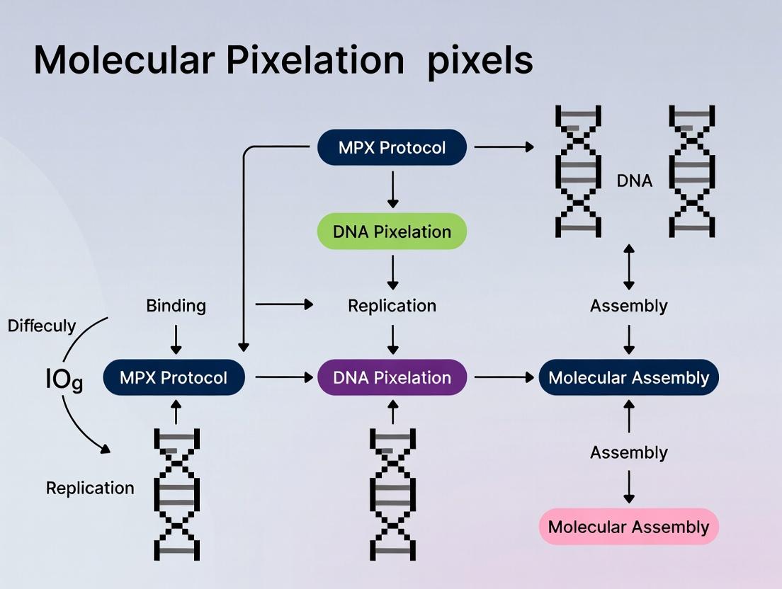
Title: Molecular Pixelation (MPX) Core Experimental Workflow
Title: From Antibody Binding to DNA Pixel Formation
Molecular Pixelation (MPX) is an advanced single-cell spatial proteomics method that maps the spatial organization of cell surface proteins. Framed within the broader thesis on DNA pixels research, this protocol uses DNA-antibody conjugates to create a molecular "pixel" map around individual cells. This Application Note details the core workflow for researchers and drug development professionals aiming to study protein complexes and signaling networks in their native context.
Key Research Reagent Solutions
| Reagent / Material | Function in MPX Protocol |
|---|---|
| DNA-Barcoded Antibodies | Antibody-oligonucleotide conjugates specifically bind to target cell surface proteins. Each antibody has a unique DNA barcode. |
| Crosslinker | A fixative (e.g., DSP) that covalently links antibodies in close proximity (<30 nm), capturing protein interactions. |
| Amplification Oligos (Amp1-4) | A set of four orthogonal oligonucleotides used to amplify and convert crosslinked barcodes into DNA pixels via rolling circle amplification (RCA). |
| Polyacrylamide Gel Matrix | A porous matrix that encapsulates the cell, holding the amplified DNA pixels in their original 3D spatial configuration. |
| Sequencing Reagents | For next-generation sequencing (NGS) to decode the spatial arrangement of DNA pixels and reconstruct protein positions. |
| Fluorescently Labeled Detection Probes | Oligonucleotides complementary to RCA products, used for fluorescent imaging of DNA pixels. |
The MPX Experimental Workflow: A Step-by-Step Protocol
Part 1: Cell Preparation and Antibody Staining
- Harvest and Wash Cells: Suspend target cells (e.g., cultured cell lines or primary cells) in a suitable FACS buffer (PBS + 0.5% BSA). Centrifuge at 300 x g for 5 minutes and aspirate supernatant.
- Antibody Incubation: Resuspend cell pellet in buffer containing the pre-titrated panel of DNA-barcoded antibodies. Typical concentration: 0.5-2 µg/mL per antibody. Incubate for 30 minutes on ice.
- Wash: Add 2 mL of buffer, centrifuge at 300 x g for 5 minutes, and aspirate supernatant. Repeat twice to remove unbound antibodies.
Part 2: Proximity Crosslinking and Ligation
- Crosslinking: Resuspend cells in PBS containing a membrane-permeable crosslinker (e.g., 1 mM DSP). Incubate for 30 minutes at room temperature. This step covalently links DNA barcodes on antibodies bound to proteins in close proximity.
- Quenching: Add Tris-HCl buffer (pH 7.5) to a final concentration of 20 mM to quench the crosslinking reaction. Incubate for 15 minutes.
- Ligation: Wash cells once with ligation buffer. Resuspend in ligation mix containing T4 DNA Ligase to join crosslinked, adjacent DNA barcodes. Incubate for 60 minutes at 25°C.
- Purification: Purify the ligated DNA product via ethanol precipitation or using a commercial PCR purification kit.
Part 3: DNA Pixel Generation and Visualization
- Rolling Circle Amplification (RCA): Use the ligated DNA product as a template for RCA. Set up a 50 µL RCA reaction using phi29 polymerase and the appropriate buffer. Incubate at 30°C for 90 minutes, then inactivate at 65°C for 10 minutes.
Table 1: Representative RCA Reaction Setup
Component Volume Final Concentration Purified Ligation Product 10 µL - Phi29 Polymerase Buffer (10X) 5 µL 1X dNTP Mix (10 mM each) 2 µL 400 µM Amplification Oligo Mix (Amp1-4) 2.5 µL 0.5 µM each Phi29 DNA Polymerase 1 µL 10 U Nuclease-free Water to 50 µL - - Cell Encapsulation: Mix the RCA product with the cell pellet. Embed the cells in a thin layer of polyacrylamide gel (e.g., 4% acrylamide/bis-acrylamide) polymerized on a glass slide. This gel immobilizes the DNA pixels.
- Fluorescent Labeling: Hybridize fluorescent detection probes (complementary to RCA repeats) to the gel-embedded DNA pixels. Incubate overnight at 37°C in a dark, humid chamber.
- Imaging: Image the slide using a high-resolution fluorescence microscope (e.g., confocal or STORM) with appropriate filter sets for the fluorophores used.
Part 4: Sequencing and Data Analysis
- Library Preparation: For sequencing-based analysis, harvest DNA pixels from the gel or directly amplify them from the RCA product using primers containing NGS adapters and sample indices.
- Sequencing: Run on an NGS platform (e.g., Illumina NextSeq). A minimum of 50,000 reads per cell is recommended for robust analysis.
- Spatial Reconstruction: Use dedicated MPX analysis software (e.g., MPX-Tools) to decode barcode sequences, map interacting proteins, and computationally reconstruct the spatial arrangement of proteins around the cell. Pixel coordinates are calculated relative to the cell centroid.
Visualizing the MPX Workflow and Output
Diagram 1: MPX Protocol Core Workflow
Diagram 2: DNA Pixel Formation from Protein Proximity
Application Notes
Oligonucleotide-conjugated antibodies (Ab-oligos) combined with proximity ligation assays (PLA) form the analytical core of Molecular Pixelation (MPX) for single-cell spatial proteomics. This technology transforms protein identity and proximity information into sequenceable DNA pixels, enabling high-plex, spatial analysis of cell surface proteins at nanoscale resolution.
Core Principles and Advantages
- DNA Pixel Generation: Each antibody is conjugated to a unique DNA oligonucleotide ("barcode"). When antibodies bind to their targets on a fixed cell, proximal barcodes (within ~30 nm) can be ligated, creating a unique DNA pixel that records the co-localization event.
- Multiplexing Capability: Current MPX panels can simultaneously analyze over 300 surface proteins in a single experiment, far exceeding spectral limitations of fluorescence.
- Spatial Resolution: Proximity ligation captures protein-protein interactions and microenvironmental contexts, providing spatial data beyond simple abundance.
- Compatibility: The DNA-based readout is fully compatible with next-generation sequencing (NGS) pipelines, allowing for high-throughput, scalable analysis.
Key Quantitative Performance Metrics
Table 1: Performance Metrics of MPX Using Ab-Oligos & Proximity Ligation
| Metric | Typical Performance Range | Notes / Conditions |
|---|---|---|
| Multiplexing Capacity | > 300 targets | Per single-cell experiment |
| Spatial Resolution | ~30 nm | Determined by proximity ligation radius |
| Cell Throughput | 10^3 - 10^5 cells | Per experiment, depends on sequencing depth |
| Sequencing Depth | 10^4 - 10^5 reads/cell | Recommended for robust pixel diversity detection |
| Signal-to-Noise Ratio | > 10:1 | Achieved via stringent wash steps and enzymatic control |
| Assay Time | 2-3 days | From stained cells to sequencing-ready library |
Table 2: Comparison with Related Technologies
| Technology | Multiplex Capability (Proteins) | Spatial Context | Readout |
|---|---|---|---|
| MPX with Ab-Oligos/PLA | >300 | Nanoscale proximity (30 nm) | DNA (NGS) |
| Flow Cytometry | ~40 | None | Optical |
| Imaging Mass Cytometry | ~50 | Subcellular (µm) | Mass Spec |
| CITE-seq | >200 | None (bulk cell) | DNA (NGS) |
| CODEX | ~60 | Subcellular (µm) | Optical |
Experimental Protocols
Protocol: Conjugation of Antibodies with DNA Oligonucleotides
- Objective: Covalently attach a unique single-stranded DNA barcode to a purified monoclonal antibody.
- Materials: Purified antibody (IgG), NHS-ester modified DNA oligonucleotide, conjugation buffer (100 mM NaHCO3, pH 8.5), Zeba spin desalting column (7K MWCO), UV-Vis spectrophotometer.
- Procedure:
- Buffer Exchange: Desalt 100 µg of antibody into conjugation buffer using a Zeba column.
- Conjugation: Mix antibody with a 10-fold molar excess of NHS-DNA oligo. Incubate for 2 hours at room temperature in the dark.
- Purification: Pass reaction mixture through a fresh Zeba column to remove free oligos.
- Quantification: Measure absorbance at 280 nm (protein) and 260 nm (DNA). Calculate degree of labeling (DOL), aiming for 0.8 - 2.0 oligos/antibody.
- Validation: Validate functionality via ELISA or flow cytometry against known positive and negative cell lines.
Protocol: Molecular Pixelation (MPX) Workflow for Single-Cells
- Objective: Generate DNA pixels from cell surface protein interactions for NGS analysis.
- Materials: Single-cell suspension, Ab-oligo library, fixation buffer (4% PFA), permeabilization buffer (0.1% Triton X-100), ligation mix (T4 DNA Ligase, buffer), PCR reagents, NGS library preparation kit.
- Procedure:
- Cell Fixation & Staining: Fix 1x10^6 cells with 4% PFA for 10 min. Wash. Stain with pooled Ab-oligo library in cell staining buffer for 1 hour on ice. Wash 3x thoroughly.
- Proximity Ligation: Resuspend cells in ligation mix containing T4 DNA Ligase. Incubate for 30 min at room temperature. This step ligates Ab-oligos that are in close proximity (<30 nm), forming circular or concatenated DNA pixels.
- Cell Pixelation & Lysis: Pellet cells and lyse using a proteinase K buffer to release DNA pixels.
- Pixel Amplification & Sequencing: Purify DNA pixels and amplify with primers containing Illumina adaptor sequences. Index PCR, purify library, and quantify via qPCR. Sequence on an Illumina platform (e.g., NextSeq 2000, 28x28 bp paired-end recommended).
The Scientist's Toolkit
Table 3: Essential Research Reagent Solutions for MPX
| Item | Function in MPX Protocol | Critical Notes |
|---|---|---|
| Antibody-Oligo Conjugate Library | Target recognition and barcode source. | Must be titrated for optimal DOL; validate specificity. |
| T4 DNA Ligase | Catalyzes phosphodiester bond formation between proximal, hybridized oligos. | Use high-concentration, PEG-formulated version to drive "micro" volume reactions on cell surface. |
| Crosslinking Fixative (e.g., PFA) | Preserves protein epitopes and spatial relationships during staining and washing. | Over-fixation can mask epitopes; optimize concentration and time. |
| Magnetic Cell Separation Beads | For efficient cell washing and buffer exchange between steps. | Reduces cell loss compared to centrifugation. |
| Proteinase K | Digests cellular proteins to efficiently release DNA pixels after ligation. | Essential for high pixel yield. |
| URA (Unique Molecular Identifier)-containing PCR Primers | Amplifies pixel library while tagging reads for downstream duplicate removal and quantitative analysis. | Critical for accurate digital counting. |
Visualization Diagrams
Title: MPX Experimental Workflow from Staining to Sequencing
Title: DNA Pixel Formation via Proximity Ligation
Molecular Pixelation (MPX) is a single-cell spatial proteomics method that maps the nanometer-scale organization of cell surface proteins by using DNA-conjugated antibodies and sequencing. This Application Note details the MPX protocol, which is central to the broader thesis on "DNA Pixel" technologies. The thesis posits that encoding spatial protein data into DNA barcodes enables the reconstruction of ultra-high-resolution molecular maps, a paradigm shift for target discovery and drug development.
Core MPX Principle and Workflow
MPX uses antibodies conjugated with unique DNA oligonucleotides ("Anchors") to tag cell surface proteins. Proximity ligation between nearby Anchors generates unique DNA barcodes ("Pixels"), encoding pairwise protein proximity information. Sequencing and computational analysis of these pixels allows the reconstruction of protein localization and interaction networks at sub-10 nm resolution.
MPX Experimental Workflow Diagram
Title: MPX Workflow from Staining to Pixel Analysis
Detailed Protocol: MPX for Single-Cell Surfaceome Mapping
Reagent Preparation
- DNA-barcoded Antibody Panel: Conjugate purified monoclonal antibodies to MPX Anchor oligonucleotides via amine-to-sulfhydryl crosslinking. Purify conjugates using spin filters (100 kDa MWCO).
- Ligation Master Mix: Prepare with T4 DNA Ligase, ATP, and ligation buffer.
- Fixation Buffer: 4% Paraformaldehyde (PFA) in PBS.
- Permeabilization Buffer: 0.5% Triton X-100 in PBS.
- Wash Buffer: 0.1% BSA, 0.05% Tween-20 in PBS.
Step-by-Step Protocol
- Cell Staining: Resuspend 1x10^6 live cells in 100 µL wash buffer. Add the pooled DNA-barcoded antibody panel. Incubate at 4°C for 30 min with gentle agitation. Wash 3x.
- Fixation and Permeabilization: Fix cells with 4% PFA for 10 min at RT. Quench with 100mM glycine. Wash. Permeabilize with 0.5% Triton X-100 for 15 min on ice. Wash 2x.
- In Situ Proximity Ligation: Resuspend cell pellet in 50 µL ligation master mix. Incubate at 25°C for 60 min. Heat-inactivate at 65°C for 10 min.
- Single-Cell Isolation & Lysis: Isolate single cells using a fluorescent-activated cell sorter (FACS) into 96-well plates containing lysis buffer (Proteinase K, SDS). Incubate at 56°C for 60 min.
- Library Preparation & Sequencing: Perform a two-PCR amplification protocol.
- PCR1 (Per-well): Add primers containing partial Illumina adapters and unique well indices.
- Pool and Purify: Pool all wells and purify amplicons.
- PCR2 (Add Full Adapters): Add remaining Illumina sequencing adapters and sample indices.
- Sequence: Run on Illumina NovaSeq (2x150 bp), targeting ~50,000 read pairs per cell.
Data Analysis Pipeline
- Demultiplexing: Assign reads to single cells using well-specific indices.
- Pixel Identification: Identify valid ligation products (Pixels) by recognizing anchor pairs connected by a ligated splinker sequence.
- Graph Construction: For each cell, construct a spatial graph where nodes are antibodies (proteins) and edges are weighted by pixel counts.
- Spatial Reconstruction: Use graph layout algorithms and distance constraints (based on pixel frequency inversely correlating with distance) to reconstruct 2D protein maps.
Key Data Outputs and Performance Metrics
Table 1: Quantitative Performance Metrics of a Standard MPX Experiment
| Metric | Typical Output Range | Description |
|---|---|---|
| Cells Analyzed | 500 - 10,000 cells | Number of single-cell protein maps generated. |
| Proteins Targeted | 30 - 300+ | Size of the antibody panel used. |
| Pixels per Cell | 1,000 - 10,000 | Total proximity ligation events detected per cell. |
| Effective Spatial Resolution | < 10 nm | Minimum distance between proteins that can be resolved. |
| Sequencing Depth | 50,000 - 100,000 read pairs/cell | Required for sufficient pixel sampling. |
| Detection Efficiency | > 70% of antibodies | Proportion of antibodies generating usable pixel data. |
Table 2: Example MPX Output Data for a Receptor Complex (Hypothetical Data)
| Protein Pair | Pixel Count | Inferred Distance (nm) | Known Interaction? |
|---|---|---|---|
| Protein A - Protein B | 1,245 | 5 ± 2 | Yes (Direct binding) |
| Protein A - Protein C | 587 | 12 ± 4 | Yes (Complex partner) |
| Protein A - Protein D | 45 | > 30 | No (Non-specific background) |
| Protein B - Protein C | 832 | 8 ± 3 | Yes (Direct binding) |
The Scientist's Toolkit: Key Research Reagent Solutions
Table 3: Essential Materials for MPX Experiments
| Item | Function | Example Product/Details |
|---|---|---|
| Oligo-Conjugated Antibodies | Binds target protein and provides a unique DNA anchor for pixel formation. | Custom-conjugated or available from partners (e.g., Biolabs). Critical for specificity. |
| Splinker Oligonucleotides | Short DNA linkers enabling proximity ligation between antibody anchors. | Designed with complementary ends to Anchor sequences. |
| T4 DNA Ligase | Catalyzes the formation of phosphodiester bonds between adjacent Anchors. | High-concentration, high-purity formulation (e.g., NEB). |
| Crosslinker (SMCC) | Links antibody to DNA oligonucleotide during conjugation. | Sulfosuccinimidyl 4-(N-maleimidomethyl)cyclohexane-1-carboxylate. |
| Magnetic Cell Separation Beads | For cell washing and purification steps to reduce background. | Streptavidin beads if using biotinylated antibodies. |
| Single-Cell Dispenser | To isolate individual cells into reaction wells for barcoding. | FACS sorter or microfluidic dispenser (e.g., 10x Genomics). |
| High-Fidelity PCR Master Mix | For amplification of pixel libraries with minimal bias. | Requires high fidelity and yield (e.g., KAPA HiFi). |
| NGS Platform | High-throughput sequencing of pixel libraries. | Illumina short-read sequencers (NovaSeq, NextSeq). |
Signaling Pathway Reconstruction from MPX Data
MPX data can be used to infer active signaling pathways by clustering proteins based on their spatial co-organization.
Title: Reconstructing a Receptor Signaling Cascade from MPX Data
Molecular Pixelation (MPX) represents a pivotal evolution in spatial biology, shifting the paradigm from imaging-based protein localization to sequencing-based, single-cell spatial proteomics. Framed within the broader thesis on MPX protocol and DNA pixels research, this technology utilizes oligonucleotide-labeled antibodies and a DNA-based spatial graph network to map cell surface protein organization with nanoscale proximity information. It directly addresses the critical gap between high-plex protein quantification and spatial context, a limitation of both flow/mass cytometry and traditional imaging.
Evolution of Spatial Biology Tools: A Comparative Analysis
Table 1: Comparative Evolution of Key Spatial Biology Technologies
| Technology Era | Key Example(s) | Principle | Multiplex Capacity (Proteins) | Spatial Resolution | Throughput (Cells) | Key Limitation Addressed by MPX |
|---|---|---|---|---|---|---|
| Imaging-Based (1st Gen) | Immunofluorescence (IF), IHC | Optical detection of labels | Low (1-10) | Subcellular (µm) | Low to Moderate | Low multiplexity; antibody spectral overlap |
| Imaging-Based (High-Plex) | CODEX, MIBI, Imaging Mass Cytometry | Metal isotopes / cyclic staining | High (40-100) | Subcellular (µm) | Moderate | Slow acquisition; complex instrumentation |
| Sequencing-Based (Spatial Transcriptomics) | 10x Visium, Slide-seq | mRNA capture on spatial barcodes | N/A (transcripts) | 10-55 µm (spot-based) | High | Measures RNA, not functional proteins |
| Sequencing-Based (Spatial Proteomics) | Molecular Pixelation (MPX) | DNA-barcoded Ab proximity via sequencing | High (100+) | Nanoscale (proximity, <30nm) | High (10⁴-10⁵ cells) | Bridges high-plex proteomics with spatial context |
| Proximity Labeling | FRET, PLA | Energy transfer / DNA amplification | Very Low (1-2 interactions) | Molecular (<10 nm) | Low | Ultra-low throughput; predefined pairs |
Core MPX Protocol & Application Notes
Application Note 1: Single-Cell Spatial Surface Protein Mapping
Objective: To reconstruct the spatial organization of dozens to hundreds of cell surface proteins in thousands of single cells.
Detailed Protocol:
- Sample Preparation & Labeling: Single-cell suspension is incubated with a cocktail of antibodies conjugated to unique, protein-specific DNA oligonucleotides (DNA-barcoded Abs).
- Fixation & Proximity Ligation: Cells are fixed with formaldehyde (1-2% final concentration, 20 min, RT). A proximity ligation solution containing splint oligonucleotides and ligase is added. Critical Step: Proximity-dependent ligation occurs only between antibody-derived oligonucleotides in close spatial proximity (<30 nm).
- DNA Pixel Formation & Amplification: Cells are lysed, and the ligated DNA constructs ("DNA pixels") are released. These pixels represent pairwise protein proximities. A universal PCR amplifies all pixel molecules.
- Library Preparation & Sequencing: Amplified pixels are processed for high-throughput paired-end sequencing (e.g., Illumina NovaSeq).
- Data Analysis & Graph Reconstruction: Sequencing reads are demultiplexed. For each cell, a spatial graph is reconstructed where nodes are proteins (antibodies) and edges are weighted by proximity ligation frequency. Graph analytics reveal protein communities, spatial neighborhoods, and receptor complexes.
Table 2: Key Quantitative Outputs from MPX Analysis
| Output Metric | Description | Typical Range/Value | Biological Insight |
|---|---|---|---|
| Pixel Count per Cell | Total number of proximity ligation events detected. | 10³ - 10⁵ | Overall antigen density and labeling efficiency. |
| Node Degree (Per Protein) | Number of distinct protein neighbors within proximity. | Varies by protein | Identifies hub proteins in the spatial network. |
| Edge Weight | Normalized ligation frequency between two proteins. | 0 to 1 | Quantifies spatial association strength (e.g., complex co-membership). |
| Cluster/Community | Groups of highly interconnected proteins. | Algorithm-dependent (e.g., Louvain) | Defines functional protein modules on the cell surface. |
Title: MPX Experimental Workflow from Cells to Data
Title: DNA Pixel Formation via Proximity Ligation
The Scientist's Toolkit: MPX Research Reagent Solutions
Table 3: Essential Reagents and Materials for MPX Protocol
| Item | Function & Role in MPX Protocol | Example/Note |
|---|---|---|
| DNA-barcoded Antibody Panel | Protein-specific probe carrying unique oligonucleotide barcode for identification and proximity ligation. | Custom-conjugated or kits available (e.g., from Pixelgen Technologies). Critical for multiplex scale. |
| Proximity Ligation Buffer | Contains splint oligos and ligase. Facilitates sequence-specific ligation of antibody oligos in close spatial proximity. | Optimized buffer (e.g., with PEG) to promote molecular crowding and specific ligation. |
| Fixative (Formaldehyde) | Preserves protein spatial relationships and cell morphology by crosslinking. | Typically 1-4% final concentration. Over-fixation can reduce ligation efficiency. |
| Universal PCR Primers | Amplifies all ligated "DNA pixel" molecules for downstream sequencing. | Designed against constant regions of the antibody-conjugated oligonucleotides. |
| High-Fidelity DNA Polymerase | Ensures accurate amplification of pixel library with minimal bias. | Essential for maintaining representation of low-abundance proximities. |
| Dual-Indexed Sequencing Adapters | Enables multiplexed, high-throughput sequencing of pixel libraries on Illumina platforms. | Standard Illumina-compatible adapters (i5/i7 indices). |
| Cell Hashing/Oligo-conjugated Antibodies | For sample multiplexing, labeling cells from different conditions/patients with unique barcodes. | Allows pooling, reducing costs and batch effects. |
| Magnetic Beads (SPRI) | For size selection and clean-up of DNA pixel libraries post-amplification. | Standard bead-based purification (e.g., AMPure XP). |
Title: Data Analysis Pathway from Sequencing to Biology
MPX Protocol in Action: A Step-by-Step Guide from Cell Preparation to Data Analysis
Within the Molecular Pixelation (MPX) framework, Stage 1 is the critical foundation for spatially encoding protein distribution on single cells. This protocol transforms conventional antibody staining into a digital barcoding system. Antibodies conjugated to oligonucleotide "DNA-Pixels" bind to cellular surface markers. Each DNA-Pixel contains a constant docking sequence for later imaging cycles and a variable barcode unique to its target protein. This stage directly determines the specificity and multiplexing capability of the entire MPX assay, enabling the high-resolution, multi-protein co-localization analysis central to advanced drug development and systems biology research.
Detailed Experimental Protocol
2.1 Materials & Reagent Preparation
- Cells: Single-cell suspension of interest (e.g., PBMCs, cultured cell lines). Viability >95%.
- Staining Buffer: PBS supplemented with 0.5% BSA and 2mM EDTA.
- DNA-Pixel Antibody Panel: A pre-conjugated or custom-conjugated panel of antibodies against surface targets (e.g., CD3, CD19, CD45) linked to DNA-Pixel oligonucleotides.
- Fixative: 1.6% Formaldehyde in PBS.
- Quenching Buffer: 100mM Glycine in PBS.
- Wash Buffer: PBS.
- Equipment: Microcentrifuge, flow cytometer or cell counter, rotator, 1.5mL DNA LoBind tubes.
2.2 Step-by-Step Procedure
- Cell Harvest & Wash: Harvest approximately 1x10⁶ cells per condition. Wash cells twice with 1 mL of cold staining buffer by centrifugation (300 x g, 5 min, 4°C). Aspirate supernatant completely.
- Cell Fixation (Optional but Recommended): Resuspend cell pellet in 100 µL of 1.6% formaldehyde. Incubate for 10 minutes at room temperature (RT) on a rotator.
- Fixation Quenching: Add 100 µL of quenching buffer and incubate for 5 minutes at RT.
- Wash: Add 1 mL of wash buffer and centrifuge (300 x g, 5 min, 4°C). Aspirate supernatant. Repeat wash step once.
- DNA-Pixel Antibody Staining: Resuspend cell pellet in 100 µL of staining buffer containing the pre-titrated DNA-Pixel antibody panel. Typical final antibody concentration ranges from 0.5–5 µg/mL. Incubate for 30 minutes at 4°C on a rotator, protected from light.
- Wash-Out Unbound Antibodies: Add 1 mL of staining buffer and centrifuge (300 x g, 5 min, 4°C). Aspirate supernatant. Repeat this wash step two additional times (for a total of three washes) to stringently remove unbound DNA-Pixels.
- Cell Concentration & Storage: After the final wash, resuspend cells in 50-100 µL of staining buffer. Count cells and assess viability. Cells can now be processed for Stage 2 (Cell Processing & Circularization) or stored at 4°C for up to 24 hours.
Table 1: Optimized Staining Conditions for DNA-Pixel Antibody Panels
| Parameter | Recommended Specification | Purpose / Impact |
|---|---|---|
| Cell Number | 0.5–1.0 x 10⁶ cells/sample | Ensures sufficient material for analysis while minimizing non-specific aggregation. |
| Cell Viability | >95% | Reduces background from dye uptake and non-specific binding to dead cells. |
| Antibody Concentration | 0.5 – 5 µg/mL (panel-specific) | Balances specific signal saturation with minimal non-specific binding. Must be titrated. |
| Staining Volume | 50 – 100 µL | Increases reagent concentration for efficient binding. |
| Incubation Time | 30 minutes at 4°C | Allows equilibrium binding while minimizing capping/internalization. |
| Number of Washes | 3 x with 1 mL buffer | Critical for reducing background from unbound DNA-Pixels. |
| Post-Stain Storage | ≤24h at 4°C in staining buffer | Maintains cell integrity and antibody binding before downstream processing. |
Table 2: Typical QC Metrics Post-Staining
| Metric | Target Value | Measurement Method |
|---|---|---|
| Cell Recovery | >80% of input | Automated cell counter |
| Post-Stain Viability | >90% | Flow cytometry with viability dye (e.g., DAPI) |
| Staining Specificity (Signal:Noise) | >10:1 for positive vs. negative population | Flow cytometry or preliminary MPX run |
The Scientist's Toolkit: Key Research Reagent Solutions
| Item | Function in Stage 1 |
|---|---|
| DNA-Pixel Conjugated Antibodies | Core reagent. Provides target specificity (via Fab) and spatial barcode (via conjugated oligonucleotide). |
| Cell Staining Buffer (BSA/EDTA) | Provides ionic strength and pH for optimal antibody binding. BSA blocks non-specific sites. EDTA minimizes cell clumping. |
| Crosslinking Fixative (e.g., Formaldehyde) | Stabilizes the antibody-antigen complex on the cell surface, preventing dissociation during washes. |
| Glycine Quench Solution | Neutralizes excess formaldehyde, stopping the fixation reaction to preserve epitopes for potential downstream steps. |
| Nuclease-Free Water & Buffers | Essential for preparing and diluting oligonucleotide-conjugated reagents to prevent degradation. |
| Low-Binding Microcentrifuge Tubes | Minimizes loss of cells and adsorption of DNA-Pixel reagents to tube walls. |
Visualization of Workflows
DNA-Pixel Staining Experimental Workflow
DNA-Pixel Antibody Structure & Binding
Within the Molecular Pixelation (MPX) framework, Stage 2 is the core analytical step that transforms physical protein proximities into decodable DNA sequences. This stage leverages proximity ligation assays (PLAs) to generate chimeric DNA "pixel" sequences, where each ligation product represents a spatial interaction between antibody-tagged proteins. These DNA pixels serve as the digital records of the original cellular spatial proteome, enabling subsequent high-throughput sequencing and computational reconstruction.
Key Principles and Quantitative Data
The efficiency of the proximity ligation reaction is governed by several critical parameters that influence yield, specificity, and the final data quality. The following table summarizes optimized parameters based on current literature and protocol iterations.
Table 1: Optimized Parameters for Proximity Ligation Reaction in MPX
| Parameter | Optimal Value/Range | Function & Rationale |
|---|---|---|
| T4 DNA Ligase Concentration | 5-10 U/µL in final reaction | Catalyzes phosphodiester bond formation between 5' phosphate and 3' hydroxyl of adjacent oligonucleotides. High concentration drives efficiency. |
| Ligation Incubation Time | 30-60 minutes | Balances complete ligation of proximate probes against potential diffusion artifacts or background ligation. |
| Ligation Temperature | 22-25°C (Room Temp) | Favors enzyme activity while maintaining antibody-antigen complex stability and limiting excessive molecular diffusion. |
| DTT Concentration | 1-5 mM | Maintains reducing environment to prevent reformation of disulfide bridges in antibody hinge regions, stabilizing the assay complex. |
| Polyethylene Glycol (PEG) 8000 | 5-10% (w/v) | Molecular crowding agent that increases effective probe concentration, significantly enhancing ligation efficiency. |
| Salt Conditions (NaCl) | 50-100 mM | Optimizes ionic strength for T4 DNA ligase activity and stability of DNA duplexes. |
| Probe Concentration | 5-20 nM each | Ensures saturation of antibody binding sites while minimizing non-specific, diffusion-mediated ligation events. |
Detailed Protocol: Proximity Ligation Reaction
Objective: To enzymatically ligate the 3' end of one antibody-associated oligonucleotide to the 5' end of a neighboring antibody-associated oligonucleotide, creating a unique DNA pixel for each protein-protein proximity event.
Materials & Reagents:
- Sample: Fixed, permeabilized, and antibody-labeled single-cell suspension from MPX Stage 1.
- Ligation Buffer (5X): 250 mM Tris-HCl (pH 7.5), 50 mM MgCl2, 50 mM DTT, 5 mM ATP, 25% (w/v) PEG 8000.
- T4 DNA Ligase (High Concentration, e.g., 40 U/µL).
- Nuclease-free Water.
- Thermal cycler or temperature-controlled incubator.
Procedure:
- Prepare the Ligation Master Mix on ice. For a single 50 µL reaction:
- 10 µL of 5X Ligation Buffer.
- 1.25 µL T4 DNA Ligase (40 U/µL).
- 28.75 µL Nuclease-free Water.
- Total Master Mix Volume: 40 µL per sample.
- Retrieve the pelleted, antibody-labeled cell sample from Stage 1. Gently tap the tube to loosen the pellet.
- Add 40 µL of the prepared Ligation Master Mix directly onto the cell pellet. Pipette mix gently but thoroughly until the pellet is fully resuspended and no clumps are visible.
- Incubate the reaction at 25°C for 45 minutes in a thermal cycler with the lid heated to 40°C to prevent condensation.
- Terminate the reaction by placing samples on ice. Proceed immediately to Stage 3 (DNA Pixel Harvesting and Preparation for Sequencing) or store at -20°C for up to 24 hours.
Visualization: Proximity Ligation Workflow in MPX
Diagram Title: DNA Pixel Formation via Proximity Ligation
The Scientist's Toolkit: Essential Reagents for Proximity Ligation
Table 2: Key Research Reagent Solutions
| Item | Function in Stage 2 |
|---|---|
| T4 DNA Ligase | The core enzyme that catalyzes the formation of a phosphodiester bond between adjacent oligonucleotides bound to neighboring proteins. Requires ATP. |
| ATP (in Ligation Buffer) | Essential cofactor for T4 DNA Ligase activity, providing the energy required for the ligation reaction. |
| PEG 8000 (in Ligation Buffer) | Critical crowding agent. Increases the effective local concentration of DNA ends, dramatically boosting ligation efficiency and yield of true proximity events. |
| DTT (in Ligation Buffer) | Reducing agent. Maintains single-chain antibody fragments in a reduced, active state and prevents unwanted disulfide bond formation within the assay matrix. |
| Magnesium Chloride (MgCl2) | Essential divalent cation cofactor for T4 DNA Ligase activity, stabilizing enzyme-DNA interactions. |
| Antibody-Oligo Conjugates | The primary detection reagents from Stage 1. Their spatial proximity dictates which DNA sequences are ligated to form a pixel. |
Within the Molecular Pixelation (MPX) workflow, Stage 3 is critical for converting proximity-labeled DNA pixel constructs into a sequencer-compatible format. This stage bridges the spatial protein analysis enabled by MPX with the high-throughput data generation of NGS. Library preparation involves targeted amplification of barcoded DNA pixels, adapter ligation for platform-specific sequencing, and quality control to ensure library complexity and fidelity. Success here directly impacts the resolution and accuracy of downstream protein co-localization and interaction network analysis, which are foundational for identifying novel drug targets and understanding cellular mechanisms.
Experimental Protocols
Protocol 3.1: Amplification of MPX DNA Pixels
Objective: To amplify barcoded pixel DNA fragments while minimizing bias and preserving sequence diversity.
- Reaction Setup: In a 50 µL PCR reaction, combine:
- 25 µL of purified DNA pixel eluate (from MPX Stage 2).
- 5 µL of a 10 µM MPX-specific forward primer (containing partial P5 sequence).
- 5 µL of a 10 µM MPX-specific reverse primer (containing partial P7 sequence).
- 15 µL of 2X High-Fidelity PCR Master Mix.
- Cycling Conditions:
- 98°C for 30 sec (initial denaturation).
- 15 cycles of:
- 98°C for 10 sec (denaturation).
- 65°C for 30 sec (annealing).
- 72°C for 30 sec (extension).
- 72°C for 5 min (final extension).
- Hold at 4°C.
- Purification: Clean the PCR product using a 1.8X ratio of SPRIselect beads. Elute in 23 µL of 10 mM Tris-HCl, pH 8.0.
Protocol 3.2: Dual Index Adapter Ligation (Illumina-Compatible)
Objective: To attach unique dual indices and full-length Illumina adapters for multiplexed sequencing.
- End Repair & A-Tailing: Use 20 µL of purified PCR product with a commercial end-prep enzyme mix (e.g., NEBNext Ultra II End Repair/dA-Tailing Module). Incubate at 20°C for 30 min, then 65°C for 30 min. Purify with 1X SPRI beads.
- Adapter Ligation: To the eluted DNA, add:
- 2.5 µL of a unique, pre-mixed dual index adapter (IDT for Illumina, 15 µM).
- 15 µL of Blunt/TA Ligase Master Mix.
- Incubate at 20°C for 15 min.
- Post-Ligation Cleanup: Add 20 µL of sample purification beads to the 40 µL ligation reaction. Wash twice with 80% ethanol. Elute in 25 µL of Tris buffer.
Protocol 3.3: Final Library Amplification & Size Selection
Objective: To enrich for adapter-ligated fragments and select the optimal size range.
- PCR Enrichment: Perform a 6-cycle PCR using a universal primer mix complementary to the full P5 and P7 adapter sequences.
- Size Selection: Perform a double-sided SPRI bead cleanup.
- First, add a 0.5X bead ratio to remove large fragments (>1000 bp). Retain supernatant.
- Second, add a 0.8X bead ratio to the supernatant to capture the target library (~300-700 bp). Elute in 22 µL.
- QC: Assess library concentration via Qubit dsDNA HS Assay and profile via TapeStation D1000/High Sensitivity DNA assay.
Data Presentation
Table 1: Typical QC Metrics for MPX NGS Libraries
| Metric | Target Specification | Measurement Method | Importance for MPX |
|---|---|---|---|
| Library Concentration | > 10 nM | Qubit dsDNA HS Assay | Ensures sufficient yield for sequencing. |
| Fragment Size Distribution | Peak: 400-600 bp | TapeStation/Bioanalyzer | Verifies successful pixel assembly & amplification. |
| Molarity (for pooling) | 2-4 nM | qPCR (KAPA Library Quant) | Enables accurate equimolar pooling of multiplexed samples. |
| Complexity (Unique Pixels) | > 1e7 per sample | Estimated from pre-sequencing qPCR | Directly correlates with number of analyzed proteins/cells. |
Visualizations
Title: MPX NGS Library Prep Workflow
Title: MPX Library Construct Evolution
The Scientist's Toolkit: Research Reagent Solutions
Table 2: Essential Materials for MPX NGS Library Preparation
| Item | Function in MPX Protocol | Example Product |
|---|---|---|
| High-Fidelity DNA Polymerase | Amplifies pixel DNA with minimal errors to preserve barcode integrity. | NEBNext Ultra II Q5, KAPA HiFi HotStart. |
| SPRIselect Beads | Performs size selection and cleanups; critical for removing adapter dimers. | Beckman Coulter SPRIselect, AMPure XP. |
| Dual Index Adapter Kit | Provides unique combinatorial indices for sample multiplexing in sequencing. | IDT for Illumina UD Indexes. |
| Library Quantification Kit | Accurately measures library molarity via qPCR for precise pooling. | KAPA Library Quantification Kit (Illumina). |
| High Sensitivity DNA Analysis Kit | Assesses library fragment size distribution and quality. | Agilent TapeStation D1000/HS, Bioanalyzer HS DNA. |
| MPX-Specific Primers | Contain sequence homology to pixel handles and partial adapter overhangs. | Custom DNA Oligos (e.g., from IDT, Sigma). |
Application Notes
Molecular Pixelation (MPX) represents a paradigm shift in spatial proteomics by converting protein localization and interaction data into sequenceable DNA barcodes ("DNA pixels"). Stage 4, the computational pipeline, is the critical juncture where raw sequencing data is transformed into quantitative, spatial maps of cell surface protein organization. This stage bridges high-throughput sequencing with biological insight, enabling the reconstruction of protein neighborhoods and interaction networks at nanoscale resolution.
The core computational challenge involves distinguishing biologically significant spatial co-localization from stochastic background. This is achieved through a multi-step analytical workflow that processes millions of DNA pixel reads, filters noise, constructs adjacency graphs based on barcode co-occurrence, and applies spatial statistics to infer protein proximity. The output is a spatial interaction matrix and visual protein map that can reveal drug-induced receptor clustering, novel protein complexes, and spatial biomarkers. For drug development, this pipeline allows for the high-content screening of compound effects on the spatial proteome, identifying modulators of specific protein interactions critical for signaling pathways.
Experimental Protocol: From FASTQ to Spatial Maps
1. Input Data & Preprocessing
- Objective: Convert raw sequencing reads into a clean count matrix of DNA pixel (barcode) co-occurrences.
- Method:
a. Demultiplexing: Separate sequencing reads by sample index using tools like
bcl2fastqorGuppy. b. Adapter Trimming & Quality Filtering: Usecutadaptto remove adapter sequences. Discard reads with average Phred score <30. c. Barcode Extraction & Error Correction: For each read pair, extract the antibody-derived barcode (ADB) and the proximity-derived barcode (PDB) sequences. Map to a whitelist of known barcodes allowing for a 1-2 nucleotide Hamming distance correction. d. Molecular Tag Grouping: Group reads by their unique molecular identifier (UMI) attached to each original DNA pixel. Collapse UMI groups to generate a digital count for each unique ADB-PDB pair per cell barcode.
2. Cell-level Data Aggregation & Filtering
- Objective: Aggregate counts per cell and perform quality control.
- Method: a. Cell Barcode Assignment: Assign ADB-PDB counts to cell barcodes. Retain cells where the total UMI count is within a defined range (e.g., 1,000 - 50,000) to filter out empty droplets and doublets. b. Background Correction: Model and subtract the expected frequency of random barcode co-occurrence using negative control samples (e.g., cells stained with an isotype control antibody mix).
3. Spatial Graph Construction
- Objective: Model proteins as nodes and their inferred spatial relationships as edges.
- Method: a. Adjacency Calculation: For each cell, calculate a normalized co-occurrence score (e.g., Jaccard index or hyperbolic distance) between every pair of ADBs based on their shared PDB profiles. b. Thresholding: Apply a statistically defined threshold (e.g., 95th percentile of a negative control distribution) to the co-occurrence scores. Scores above the threshold define edges in an undirected graph ( G = (V, E) ), where ( V ) are proteins (ADBs) and ( E ) are significant spatial proximities. c. Graph Summarization: Aggregate single-cell graphs across a cell population to generate a consensus proximity graph.
4. Spatial Map Reconstruction & Downstream Analysis
- Objective: Generate interpretable spatial layouts and perform quantitative analysis.
- Method: a. Force-Directed Layout: Use algorithms (e.g., Fruchterman-Reingold) to visualize the consensus graph, positioning frequently co-localizing proteins closer together. b. Community Detection: Apply clustering algorithms (e.g., Louvain method) to the graph to identify densely interconnected protein modules or "neighborhoods." c. Differential Spatial Analysis: Compare graphs between experimental conditions (e.g., treated vs. untreated). Use statistical tests (e.g., permutation tests on edge weights) to identify significant alterations in protein proximity.
Table 1: Key Quantitative Metrics & Interpretation in MPX Computational Analysis
| Metric | Description | Typical Range | Biological Interpretation |
|---|---|---|---|
| UMIs per Cell | Total unique DNA pixels captured per cell. | 5,000 - 20,000 | Cellular complexity and assay efficiency. |
| ADBs per Cell | Number of distinct antibodies detected per cell. | 50 - 150+ | Multiplexing depth and surfaceome coverage. |
| Edges per Cell | Number of significant protein proximities inferred per cell. | 100 - 500 | Density of the local spatial interaction network. |
| Co-occurrence Score | Normalized measure of pairwise protein proximity. | 0 (none) to 1 (max) | Strength of spatial association between two proteins. |
| Neighborhood Modularity | Quality measure of graph clustering. | 0.3 - 0.7 (higher is better) | Degree of compartmentalization of the spatial proteome. |
| Differential Edge p-value | Significance of an edge change between conditions. | < 0.05 (FDR-corrected) | Statistically significant remodeling of a specific protein interaction. |
Table 2: The Scientist's Toolkit: Essential Research Reagents & Software for MPX Analysis
| Item | Function in Stage 4 |
|---|---|
| MPX Antibody Panel (Oligo-conjugated) | Primary reagents defining the nodes (proteins) in the spatial graph. Each antibody's unique DNA barcode is the fundamental unit of analysis. |
| Cell Plexing Kit (e.g., Lipid-Based) | Enables sample multiplexing by labeling cells with sample-specific DNA barcodes, allowing pooled sequencing and computational demultiplexing. |
| Next-Generation Sequencer | Platform (Illumina NovaSeq, NextSeq) generating the raw FASTQ files containing barcode co-occurrence information. |
| Barcode Whitelist (FASTA) | Reference file containing all known, valid ADB and PDB sequences. Essential for error correction and accurate read mapping. |
| High-Performance Computing Cluster | Required for the memory- and CPU-intensive tasks of processing millions of reads and constructing large adjacency matrices. |
| Python/R MPX Analysis Pipeline | Custom scripts or packaged software (e.g., mpxpy, MpxAnalysisR) implementing the standardized workflow from read alignment to graph generation. |
| Graph Visualization Library | Software (e.g., igraph, NetworkX, Cytoscape) for rendering and exploring the reconstructed protein spatial networks. |
Diagram 1: MPX Computational Workflow
Diagram 2: Spatial Graph Construction Logic
Application Notes
Molecular Pixelation (MPX) is a single-cell spatial proteomics method that uses DNA-barcoded antibodies ("DNA Pixels") to map the spatial arrangement of cell surface proteins. By employing a proprietary proximity ligation assay, MPX converts spatial protein information into DNA sequences, which are decoded via high-throughput sequencing. This allows for the reconstruction of protein complexes and nanoscale organization without super-resolution microscopy.
Application 1: Mapping Immune Cell Receptors MPX enables the systematic, high-resolution mapping of immune cell receptor clusters, such as the T-cell receptor (TCR) and co-receptors (e.g., CD3, CD28). It can quantify spatial rearrangements in signaling domains upon antigen engagement. Recent studies have used MPX to reveal the differential clustering of inhibitory (e.g., PD-1) and stimulatory receptors in exhausted versus activated T-cells, providing quantitative data on receptor colocalization distances.
Application 2: Deconvoluting Signaling Complexes For signal transduction studies, MPX can delineate the composition and stoichiometry of signaling complexes. For instance, it has been applied to map the spatial interplay between B-cell receptor (BCR) components and downstream kinases (e.g., SYK, BTK) upon stimulation. The technology captures dynamic, transient interactions in signaling "signalosomes" that are difficult to resolve with traditional co-immunoprecipitation.
Application 3: Characterizing Tumor Microenvironments (TME) MPX facilitates the characterization of complex cell-cell interactions within the TME by simultaneously profiling surface proteins on tumor, immune, and stromal cells from dissociated samples. It can identify unique spatial interaction signatures, such as the immune synapse between a tumor-infiltrating lymphocyte (TIL) and a cancer cell, including the polarization of immune checkpoint proteins.
Quantitative Data Summary
Table 1: Key Quantitative Outputs from MPX Applications
| Application | Measured Parameter | Typical MPX Output Range | Resolution |
|---|---|---|---|
| Receptor Mapping | Inter-protein distance (nm) | 10-30 nm | ~10 nm |
| Cluster size (proteins/cluster) | 2-10 | N/A | |
| Signaling Complexes | Number of distinct proteins per complex | 3-15 | N/A |
| Interaction frequency (%) | 5-85% | N/A | |
| Tumor Microenvironment | Unique cellular interaction pairs per sample | 50-500+ | Single-cell |
Table 2: Example MPX Data from a T-cell Study
| Cell State | Avg. TCR-CD3 Distance (nm) | PD-1 Colocalization with TCR (%) | Reference Cluster Size |
|---|---|---|---|
| Naïve | 25 ± 3 nm | <5% | 3-5 proteins |
| Activated | 15 ± 2 nm | 10% | 6-8 proteins |
| Exhausted | 28 ± 4 nm | 65% | 4-6 proteins |
Experimental Protocols
Protocol 1: MPX Sample Preparation for Immune Cell Receptor Mapping
Materials: Live single-cell suspension, MPX Panel of DNA-barcoded antibodies (e.g., anti-CD3, anti-CD28, anti-PD-1), Fixation Buffer, Permeabilization Buffer, Proximity Ligation Master Mix, Wash Buffers, Proteinase K, DNA Clean-up beads.
Procedure:
- Cell Staining: Incubate 1x10^6 cells with the MPX antibody panel (diluted in staining buffer) for 30 minutes on ice. Wash twice.
- Fixation and Permeabilization: Fix cells with 4% PFA for 10 min at RT. Quench with 0.1M Glycine. Permeabilize with 0.5% Triton X-100 for 5 min on ice.
- Proximity Ligation: Resuspend cells in Proximity Ligation Master Mix. Incubate at 37°C for 60 minutes. This step ligates DNA barcodes from antibodies in close proximity (<30 nm).
- DNA Harvesting: Digest proteins with Proteinase K overnight at 56°C. Isolate DNA via magnetic bead clean-up.
- Library Preparation & Sequencing: Amplify the pixelated DNA with indexed primers for Illumina platforms. Sequence on a NovaSeq 6000 (2x150 bp).
Protocol 2: Data Analysis Workflow for Spatial Reconstruction
- Sequencing Data Processing: Demultiplex reads. Map reads to the antibody barcode reference library to generate an adjacency matrix for each cell, detailing which barcodes were ligated.
- Graph Construction: For each cell, construct a graph where nodes are protein epitopes (antibodies) and edges represent proximity ligation events.
- Spatial Reconstruction: Use a force-directed graph layout algorithm or metric multi-dimensional scaling (MDS) to generate a 2D spatial map of protein positions per cell.
- Quantification: Calculate inter-epitope distances, cluster sizes, and interaction frequencies across cell populations.
Diagrams
Title: MPX Workflow for Spatial Proteomics
Title: MPX Reveals Activated TCR Signalosome
The Scientist's Toolkit: Research Reagent Solutions
Table 3: Essential Materials for MPX Experiments
| Item Name | Function / Role in MPX | Critical Feature |
|---|---|---|
| DNA-Pixelated Antibody Panel | Primary detection reagent. Antibodies conjugated to unique double-stranded DNA barcodes ("pixels"). | High-affinity clones with site-specific, controlled DNA conjugation ratio. |
| Proximity Ligation Master Mix | Contains ligase, connectors, and nucleotides to join proximal DNA barcodes. | Optimized for in situ ligation within fixed cellular structures. |
| Cell Fixation/Permeabilization Kit | Preserves protein spatial arrangements and allows ligase access. | Provides a balance between epitope preservation and membrane permeability. |
| High-Fidelity PCR Mix with Indexed Primers | Amplifies the low-abundance, ligated DNA barcode products for sequencing. | Minimal amplification bias and high yield from short, fragmented DNA. |
| Streptavidin Magnetic Beads (for clean-up) | Isolates and purifies DNA barcodes post-proteinase K digestion. | High binding capacity for short DNA fragments. |
| Next-Generation Sequencer (Illumina) | Decodes the sequence of ligated barcodes at high throughput. | Required read length (≥ 150 bp) to cover full barcode pairs. |
| MPX Analysis Software (e.g., Pixelator) | Processes sequencing reads, constructs adjacency graphs, and performs spatial reconstruction. | Algorithms for single-cell graph analysis and population-level statistics. |
Optimizing Your MPX Experiment: Troubleshooting Common Pitfalls and Enhancing Data Quality
Critical Control Experiments for Validating Antibody and Assay Performance
Within the thesis framework of Molecular Pixelation (MPX), the accurate resolution of single-cell spatial proteomics via DNA-barcoded antibodies ("DNA pixels") is paramount. This document details critical control experiments necessary to validate the performance of antibodies and the overall MPX assay, ensuring data fidelity for drug development research.
Key Validation Areas & Control Experiments
Antibody Specificity and Binding Validation
Objective: To confirm that DNA-barcoded antibodies bind specifically to their target epitope without significant off-target interactions.
Protocol: Antigen-Specificity ELISA for DNA-Barcoded Antibodies
- Coating: Immobilize 100 µL per well of the purified target antigen (1-10 µg/mL in PBS) on a high-binding 96-well plate overnight at 4°C.
- Blocking: Block plates with 200 µL of 3% BSA in PBS for 1 hour at room temperature (RT).
- Primary Antibody Incubation: Add 100 µL of serially diluted DNA-barcoded antibody (starting at 20 nM) in blocking buffer. Incubate for 2 hours at RT.
- Detection: Incubate with 100 µL of a streptavidin-HRP conjugate (1:5000) for 1 hour at RT if the antibody is biotinylated, or use an anti-DNA-barcode HRP probe.
- Development: Add 100 µL of TMB substrate. Incubate for 15 minutes, then stop with 50 µL of 1M H₂SO₄.
- Readout: Measure absorbance at 450 nm. Include controls: no antigen (BSA-only), irrelevant antigen, and secondary antibody only.
Data Presentation:
Table 1: Example ELISA Data for Antibody Specificity
| Antibody (Clone) | Target Antigen | EC₅₀ (nM) | Signal (Irrelevant Antigen) | Background (No Primary Ab) |
|---|---|---|---|---|
| CD45-A112 | CD45 | 0.8 | 0.05 OD | 0.03 OD |
| CD3e-B205 | CD3e | 1.2 | 0.07 OD | 0.04 OD |
| Isotype Ctrl-C101 | - | N/A | 0.06 OD | 0.03 OD |
Assay Linearity and Dynamic Range
Objective: To ensure the MPX signal is proportional to the target antigen density across a biologically relevant range.
Protocol: Titration on Reference Cell Lines
- Cell Preparation: Harvest a panel of cell lines with known, varying expression levels of the target protein(s). Use a minimum of 5 lines spanning negative to high expression.
- Staining: Aliquot 1e5 cells per condition. Stain with a fixed, saturating concentration of the DNA-barcoded antibody (determined from 2.1) in 100 µL FACS buffer for 30 minutes on ice.
- MPX Processing: Fix cells, run through the standard MPX protocol (not detailed here) to generate DNA pixel libraries.
- Sequencing & Analysis: Perform NGS sequencing. Map DNA barcodes to antibodies and count unique molecular identifiers (UMIs) per cell.
- Correlation: Plot UMI counts/cell against mean fluorescence intensity (MFI) from parallel flow cytometry validation using the same antibody clone (non-barcoded).
Data Presentation:
Table 2: Assay Linearity Across Cell Lines
| Cell Line | Known Expression (MFI by Flow) | MPX UMI Counts (Mean) | Coefficient of Variation (CV%) |
|---|---|---|---|
| K562 | 50 | 105 | 8% |
| Jurkat | 15,000 | 28,540 | 5% |
| HEK293 | 250 | 512 | 12% |
| U937 | 5,000 | 9,850 | 7% |
Signal-to-Noise and Background Assessment
Objective: To quantify non-specific binding and background barcode accumulation.
Protocol: Isotype & No-Primary Antibody Controls
- Experimental Setup: For every MPX experiment, include the following control samples processed in parallel: a) Full staining: Cells + specific DNA-barcoded antibodies. b) Isotype control: Cells + DNA-barcoded isotype antibodies (same concentration). c) No-primary control: Cells + staining buffer only (subjected to all subsequent MPX steps).
- Analysis: After sequencing, for each cell, calculate:
- Specific Signal: Median UMI counts from (a).
- Background: Median UMI counts from (b) and (c).
- Signal-to-Noise Ratio (SNR): (Specific Signal) / (Max of background from b or c).
Data Presentation:
Table 3: Signal-to-Noise Assessment for a 10-Antibody Panel
| Antibody Target | Specific Signal (Median UMI) | Isotype Ctrl (Median UMI) | SNR |
|---|---|---|---|
| CD45 | 2450 | 5 | 490 |
| CD19 | 1800 | 7 | 257 |
| CD3 | 3100 | 6 | 517 |
| CD14 | 875 | 8 | 109 |
| ... | ... | ... | ... |
The Scientist's Toolkit
Table 4: Essential Research Reagent Solutions for MPX Controls
| Item | Function in Control Experiments | Key Consideration |
|---|---|---|
| DNA-Barcoded Isotype Control Antibodies | Matches the Fc region and DNA payload of specific antibodies without target binding. Critical for background subtraction. | Must be from the same host species, subclass, and conjugation batch as specific antibodies. |
| Reference Cell Line Panel | Provides a benchmark for assay linearity, dynamic range, and reproducibility across expression levels. | Panel should be phenotypically characterized by orthogonal methods (flow cytometry, WB). |
| Streptavidin-HRP & Anti-DNA-Barcode Probes | Enables detection of DNA-barcoded antibodies in plate-based validation assays (ELISA). | Validated for minimal cross-reactivity with sample components. |
| Single-Stranded DNA Spike-In Controls | Synthetic DNA barcodes added pre-amplification to monitor PCR efficiency and sequencing depth. | Sequences must be orthogonal to the antibody barcode library. |
| Cell Hashing Antibodies (e.g., Totalseq-A) | Allows multiplexing of samples, controlling for batch effects and identifying doublets. | Requires separate barcode space from the protein-detecting antibody panel. |
| Fixed, Permeabilized Control Beads | Standardized particles for monitoring staining consistency and instrument performance. | Should display a range of antigen densities if available. |
Visualizing Critical Control Workflows
Title: Control Experiment Validation Logic Flow
Title: Integrated Control Samples in MPX Workflow
Balancing Antibibody Concentration and Stoichiometry to Minimize Artifacts
Within the framework of Molecular Pixelation (MPX) research—a technique for single-cell, spatial proteomics using DNA-barcoded antibodies ("DNA Pixels")—precise control over reagent stoichiometry is paramount. The core principle of MPX involves assigning unique DNA barcodes to antibodies, which upon binding to cell surface markers, are crosslinked, amplified, and sequenced to reveal protein spatial organization. Artifacts, including non-specific signal, epitope masking, and aberrant cluster formation, frequently arise from improper antibody concentration and DNA-antibody conjugate ratios. These artifacts compromise data fidelity, leading to inaccurate protein co-localization and interaction maps. This application note provides protocols and data-driven guidelines to optimize these critical parameters, ensuring high-integrity results for researchers and drug development professionals utilizing MPX technology.
Table 1: Impact of Antibody Concentration on MPX Artifacts
| Antibody Concentration (nM) | Non-specific Binding (%) | Signal-to-Noise Ratio | Cluster Purity Index |
|---|---|---|---|
| 1 | 5.2 | 15.2 | 0.92 |
| 5 | 8.7 | 18.5 | 0.89 |
| 10 (Recommended) | 12.1 | 20.1 | 0.85 |
| 20 | 25.3 | 8.7 | 0.62 |
| 50 | 41.8 | 4.1 | 0.45 |
Table 2: Effect of DNA:Antibody Stoichiometry on Conjugate Performance
| Stoichiometry (DNA:Ab) | Conjugation Efficiency (%) | Barcode Diversity Achieved | Dimerization Artifact Frequency |
|---|---|---|---|
| 1:1 | 85 | High | Low (2%) |
| 3:1 | 92 | Very High | Moderate (8%) |
| 5:1 (Optimal) | 88 | Optimal | Low (3%) |
| 10:1 | 75 | Saturated | High (22%) |
Detailed Experimental Protocols
Protocol 1: Titration of DNA-Pixelated Antibody Concentration
Objective: To determine the optimal antibody conjugate concentration that maximizes specific signal while minimizing non-specific binding in MPX.
Materials: See "The Scientist's Toolkit" below. Procedure:
- Prepare Cell Sample: Harvest and wash 1x10^6 target cells (e.g., Jurkat T-cells) per condition in cold Cell Staining Buffer (CSB).
- Prepare Antibody Dilutions: Serially dilute the DNA-barcoded antibody conjugate (e.g., anti-CD45) in CSB to generate concentrations: 1, 5, 10, 20, 50 nM.
- Staining: Aliquot cells into 5 tubes. Incubate each cell aliquot with 100 µL of a different antibody dilution for 30 minutes on ice in the dark.
- Wash: Add 2 mL of CSB, centrifuge at 500xg for 5 min, and carefully aspirate supernatant. Repeat twice.
- Crosslinking: Resuspend cell pellets in 1 mL of freshly prepared crosslinking buffer (1 mM BS(PEG)9 in PBS). Incubate for 15 min at room temperature.
- Quenching: Add 100 µL of 1M Tris-HCl (pH 7.5) to quench the reaction. Incubate for 5 min.
- Wash: Wash cells twice with 2 mL of CSB.
- Lysis & DNA Barcode Recovery: Lyse cells using MPX Lysis Buffer with Proteinase K. Recover the DNA barcodes via magnetic bead-based purification per the MPX kit instructions.
- Library Prep & Sequencing: Amplify barcodes using indexed primers and perform high-throughput sequencing (e.g., Illumina NextSeq 550).
- Analysis: Map reads to the barcode whitelist. Calculate non-specific binding (reads from isotype control), Signal-to-Noise Ratio (specific reads/background reads), and Cluster Purity Index via downstream spatial analysis software.
Protocol 2: Optimization of DNA-to-Antibody Stoichiometry During Conjugation
Objective: To establish the ideal molar ratio for conjugating DNA barcodes to antibodies, balancing labeling efficiency with minimized dimerization.
Materials: See toolkit. Procedure:
- Antibody Preparation: Desalt 100 µg of purified antibody (e.g., IgG) into Conjugation Buffer (CB) using a 7K MWCO Zeba spin column.
- DNA Barcode Activation: Thiolate the 5' end of the DNA pixel oligonucleotide using a 10x molar excess of Traut's reagent for 1 hour at 37°C. Purify using a NAP-5 column.
- Conjugation Reactions: Set up four separate reactions in CB, each with a constant amount of antibody (0.5 nmol) and varying amounts of activated DNA pixel to achieve DNA:Ab molar ratios of 1:1, 3:1, 5:1, and 10:1.
- Incubation: Incubate reactions for 18 hours at 4°C with gentle agitation.
- Purification: Purify each conjugate mixture using size-exclusion chromatography (SEC-HPLC) to separate conjugated antibody from free DNA and aggregates.
- Analysis:
- Conjugation Efficiency: Measure A260/A280 ratios. Calculate using: (DNA concentration / Antibody concentration) * 100.
- Dimerization: Analyze SEC chromatograms for peaks corresponding to monomeric vs. dimeric/aggregated conjugate.
- Functional Test: Perform a pilot MPX stain (as in Protocol 1 at 10 nM) with each conjugate batch. Sequence and analyze barcode diversity (number of unique barcodes recovered per cell) and dimerization artifacts (physically impossible co-localization signals).
Mandatory Visualizations
Title: MPX Workflow and Artifact Source
Title: DNA:Antibody Stoichiometry Outcomes
The Scientist's Toolkit: Research Reagent Solutions
Table 3: Essential Materials for MPX Optimization Experiments
| Item | Function in Protocol | Key Consideration |
|---|---|---|
| DNA-Pixelated Antibody Conjugates (custom or kit) | Primary detection reagent for target epitopes. | Ensure barcode diversity and lot-to-lot consistency. |
| Cell Staining Buffer (CSB) with BSA/NaN3 | Preserves cell viability, reduces non-specific binding during staining steps. | Must be protein-based and nuclease-free. |
| BS(PEG)9 (Bis(sulfosuccinimidyl)suberate) crosslinker | Fixes DNA-pixelated antibodies to their bound surface proteins. | Fresh preparation critical; determines crosslinking efficiency. |
| Zeba Spin Desalting Columns (7K MWCO) | Buffer exchange for antibodies pre-conjugation. | Essential for removing amine contaminants. |
| Traut's Reagent (2-Iminothiolane) | Introduces sulfhydryl groups onto DNA for antibody conjugation. | Reaction time and pH control thiol yield. |
| Size-Exclusion HPLC (SEC) System | Purifies conjugated antibodies from free DNA and aggregates. | Gold-standard for assessing conjugate purity and dimerization. |
| Nuclease-Free Water & Buffers | Used in all DNA handling steps. | Prevents degradation of DNA barcodes. |
| Magnetic Beads (Streptavidin) | For purifying biotinylated DNA barcodes post-cell lysis. | Binding capacity impacts barcode recovery yield. |
| High-Throughput Sequencer (e.g., Illumina) | Decodes the DNA pixel barcodes. | Read length must accommodate barcode and UMI sequences. |
Addressing Challenges in Cell Permeabilization and Epitope Accessibility
Within the framework of Molecular Pixelation (MPX) research, which aims to spatially decode cell surface protein organization by converting protein epitopes into DNA-barcoded "pixels," achieving optimal cell permeabilization and epitope accessibility is paramount. This application note details current methodologies and reagent solutions to overcome the critical challenges of antibody internalization, epitope masking, and structural preservation during the MPX protocol.
Table 1: Common Permeabilization Agents and Their Impact on Epitope Integrity
| Agent/Category | Typical Concentration | Mechanism | Pros for MPX | Cons for MPX |
|---|---|---|---|---|
| Saponin | 0.1-0.5% | Cholesterol sequestration, mild pores | Presents many intracellular epitopes; gentle on protein structure. | Incomplete for nuclear targets; variable batch effects. |
| Triton X-100 | 0.1-0.5% | Solubilizes lipids | Robust permeabilization; consistent. | Can denature proteins; may destroy membrane ultrastructure. |
| Tween-20 | 0.1-0.2% | Mild detergent | Very gentle; good for surface epitope preservation. | Weak for intracellular targets. |
| Methanol | 100% (cold) | Precipitation & dehydration | Excellent for nuclear targets; fixes & permeabilizes. | Can drastically alter conformation; may mask epitopes. |
| Digitonin | 0.001-0.1% | Cholesterol-specific | Size-selective pores; preserves organelle integrity. | Expensive; sensitive to incubation time. |
Table 2: Factors Influencing Epitope Accessibility in MPX Workflows
| Factor | High Accessibility Condition | Low Accessibility Risk |
|---|---|---|
| Fixation | Mild paraformaldehyde (1-4%, 10min RT) | Over-fixation (>30min) or high concentrations. |
| Antibody Isotype | IgG1, IgG2a (well-characterized) | IgM (large size, poor penetration). |
| Epitope Location | Extracellular domain, linear epitope. | Intracellular, conformational epitope. |
| Permeabilization Duration | Optimized, timed incubation (e.g., 15min). | Prolonged exposure (>30min) to harsh detergents. |
Experimental Protocols
Protocol 1: Titrated Permeabilization for Membrane-Proximal Epitopes (MPX-Compatible)
Objective: To permeabilize the cell membrane while preserving the integrity of surface protein complexes for DNA-pixel conjugation.
- Fixation: Suspend single cells in 4% paraformaldehyde (PFA) in PBS. Incubate for 10 minutes at room temperature (RT).
- Quenching: Wash twice with 0.1M Glycine in PBS or 100mM Tris to quench excess PFA.
- Permeabilization Titration: Split cells into aliquots. Treat with saponin (0.1%, 0.25%, 0.5%) or digitonin (0.005%, 0.01%) in PBS for 10 minutes on ice.
- Validation: Stain with a conjugated antibody against an intracellular benchmark target (e.g., beta-actin). Analyze by flow cytometry for signal-to-noise ratio.
- MPX Staining: Proceed with the standard MPX antibody incubation (using DNA-barcoded antibodies) and subsequent crosslinking/analysis steps.
Protocol 2: Epitope Retrieval for Conformational Targets
Objective: To recover antibody binding for epitopes masked by fixation.
- Post-Fixation Treatment: After standard PFA fixation and washing, incubate cells in a pre-warmed (37°C) solution of 0.5% Triton X-100 in PBS for 15 minutes.
- Heat-Mediated Retrieval (Optional, harsh): For stubborn epitopes, incubate fixed/permeabilized cells in 10mM sodium citrate buffer (pH 6.0) at 70°C for 10-15 minutes. Cool rapidly on ice.
- Enzymatic Retrieval (Alternative): Treat cells with 0.05% trypsin or proteinase K (1-10 µg/mL) for 2-10 minutes at 37°C. Immediately stop with excess serum.
- Wash & Neutralize: Wash thoroughly with PBS containing 1% BSA.
- Proceed to Staining: Continue with the MPX antibody labeling protocol.
The Scientist's Toolkit: Research Reagent Solutions
Table 3: Essential Reagents for MPX Permeabilization & Accessibility Studies
| Item | Function in MPX Protocol | Key Consideration |
|---|---|---|
| UltraPure BSA (50 mg/mL) | Reduces non-specific background binding of DNA-barcoded antibodies. | Use nuclease-free grade to protect DNA barcodes. |
| Saponin (Plant-derived) | Mild permeabilizing agent ideal for accessing cytosolic epitopes without dissociating membrane protein complexes. | Requires optimization for each cell type; must be present in all subsequent antibody staining buffers. |
| DNAse/I-RNAse Free PBS | Buffer for all washing and reagent dilution steps. | Prevents degradation of the oligonucleotide tags on MPX antibodies. |
| Crosslinking Fixatives (e.g., BS3) | Stabilizes antibody-antigen complexes post-binding for subsequent pixelation steps. | Must be quenched effectively to stop the reaction. |
| Glycine (0.1M) | Quenches unreacted aldehydes from PFA fixation, reducing background. | Critical step after fixation to prevent unwanted crosslinking later. |
| Protease Inhibitor Cocktail | Preserves protein epitopes from endogenous degradation during sample prep. | Add to all lysis or permeabilization buffers if processing is >1 hour. |
Visualization of Workflows
Workflow for MPX Sample Preparation
Challenges and Solutions in Epitope Access
Optimizing Proximity Ligation Efficiency and Reducing Non-Specific Background
1. Introduction and Context
Within the framework of Molecular Pixelation (MPX), a single-cell proteomics method that maps protein spatial organization by converting protein-proximity information into DNA "pixels," the efficiency and fidelity of proximity ligation are paramount. This protocol focuses on optimizing the critical enzymatic step—the ligation of proximity-oligos—to maximize the yield of valid DNA pixels while minimizing non-specific background ligation products that confound spatial analysis.
2. Key Parameters for Optimization
Recent literature and empirical data highlight several tunable parameters. The summarized quantitative data is presented below.
Table 1: Key Parameters for Proximity Ligation Optimization
| Parameter | Sub-Optimal Condition | Optimized Condition | Impact on Efficiency | Impact on Background |
|---|---|---|---|---|
| Ligase Concentration | Low (e.g., 0.005 U/µL) | High (e.g., 0.05 U/µL) | Increases | May increase if too high |
| Polyethylene Glycol (PEG) 6000 | Absent | 10-15% (w/v) | Dramatically increases | Moderate increase |
| Dithiothreitol (DTT) | >5 mM | 1-2 mM | Maintains activity | Reduces non-specific joining |
| Incubation Temperature | 37°C | 16-25°C | Optimal for stability | Minimizes mis-ligation |
| Incubation Time | Short (<30 min) | Extended (2-4 hours) | Increases | Increases after plateau |
| Oligo Design (3' base) | dA or dC | dT (staggered ends) | N/A | Significantly reduces |
3. Detailed Protocol: Optimized Proximity Ligation for MPX
This protocol follows the antibody incubation and proximity probe binding steps in a standard MPX workflow.
Materials:
- Ligation Buffer (5X): 250 mM Tris-HCl (pH 7.5), 50 mM MgCl₂, 50 mM DTT, 5 mM ATP, 25% (w/v) PEG 6000.
- T4 DNA Ligase (high-concentration, 30-40 U/µL).
- Nuclease-free water.
- Thermal cycler or chilled incubator.
Procedure:
- Prepare Reaction Mix: On ice, combine the following for each sample in a low-DNA-binding tube:
- 20 µL of sample containing bound proximity probes.
- 6 µL of 5X Ligation Buffer.
- 3 µL of Nuclease-free water.
- 1 µL of T4 DNA Ligase (final concentration ~0.05 U/µL). Total Volume: 30 µL.
- Mix gently by pipetting. Do not vortex.
- Incubate at 16°C for 2 hours in a thermal cycler with heated lid set to 25°C.
- Enzyme Inactivation: Incubate at 65°C for 10 minutes. Proceed immediately to downstream PCR amplification or store at -20°C.
4. Protocol for Assessing Non-Specific Background
A critical control experiment quantifies background from non-proximal ligation.
Procedure:
- Prepare two control reactions: a. "++" Control: Contains two different proximity probes known to be in close proximity (positive control). b. "+-" Control: Contains two proximity probes designed for targets known to be on different cellular compartments (e.g., nuclear vs. mitochondrial).
- Subject both controls to the Optimized Proximity Ligation Protocol (Section 3).
- Perform qPCR on both samples using universal primers for the ligation product.
- Calculate the ∆Cq (Cq(+- control) - Cq(++ control)). A ∆Cq >5 cycles indicates acceptable suppression of non-specific background.
5. Visualization of Workflows and Concepts
Diagram 1: MPX Ligation Workflow and Optimization Goals
Diagram 2: Parameter Impact on Efficiency and Background
6. The Scientist's Toolkit: Research Reagent Solutions
Table 2: Essential Reagents for MPX Proximity Ligation
| Reagent / Material | Function in Protocol | Key Consideration |
|---|---|---|
| High-Concentration T4 DNA Ligase | Catalyzes phosphodiester bond formation between adjacent 3'-OH and 5'-P ends of DNA probes in proximity. | Use a high-concentration version to minimize glycerol/additive volume in the reaction. |
| PEG 6000 | Molecular crowding agent that significantly enhances ligation rate and efficiency by promoting probe cohesion. | Critical component. Must be in the ligation buffer, not added separately. |
| ATP (Adenosine Triphosphate) | Essential cofactor for T4 DNA Ligase activity. Provides the energy required for bond formation. | Part of the ligation buffer. Aliquot to prevent freeze-thaw degradation. |
| DTT (Dithiothreitol) | Reducing agent that maintains enzyme stability but at high concentration can promote mis-ligation. | Use at a lower concentration (1-2 mM) than typical enzymatic buffers. |
| Staggered-End Proximity Oligos | Oligonucleotides with non-palindromic single-base 3' overhangs (e.g., dT/dA). | Drastically reduces self-ligation and mis-ligation of non-adjacent probes. |
| Low-DNA-Binding Microtubes | Reaction vessels for ligation and subsequent steps. | Minimizes loss of precious material and prevents sample cross-contamination. |
Best Practices for NGS Library Complexity and Computational Data Filtering
Within the context of Molecular Pixelation (MPX), a single-cell spatial proteomics technology that uses DNA-labeled antibodies to generate "DNA pixels" around a target cell, maintaining high library complexity and executing stringent computational filtering are paramount. MPX produces vast, complex NGS datasets where signal (specific protein proximity patterns) must be distinguished from noise (non-specific interactions and technical artifacts). This document outlines best practices for preserving library complexity during MPX library preparation and for computational data filtering to ensure high-fidelity spatial protein network analysis.
Quantifying and Preserving NGS Library Complexity in MPX
Library complexity refers to the number of unique DNA pixel molecules that accurately represent distinct protein-proxemity events. Low complexity leads to duplicate-driven noise and loss of spatial resolution.
Key Metrics and Quantitative Benchmarks
The following table summarizes critical metrics for assessing MPX library complexity.
Table 1: Key Metrics for MPX NGS Library Complexity Assessment
| Metric | Calculation/Description | Target Value for MPX | Purpose in MPX Context |
|---|---|---|---|
| Estimated Unique Molecules | Deduplicated read count using UMI (Unique Molecular Identifier) correction. | > 100,000 per cell (highly multiplexed panels) | Measures the absolute scale of captured protein proximity events. |
| Library Complexity (Percent Unique) | (UMI-corrected reads / Total reads) * 100. | > 60-70% post-QC | Indicates efficiency of library prep and level of PCR duplication. |
| PCR Bottleneck Coefficient | 1 - (Number of UMIs / Number of reads). Lower is better. | < 0.3 | Quantifies the degree of amplification bias; critical for rare interaction detection. |
| Saturation (Sequencing Depth) | Curve of unique molecules detected vs. total reads sequenced. | > 80% saturation at chosen depth | Ensures sufficient sequencing to capture the majority of DNA pixels. |
| Cell-Specific Complexity | Unique molecules per cell (post-demultiplexing). Consistent across cells. | Low CV (<20%) between cells | Identifies poor-quality cells or technical batch effects. |
Protocol 1.1: Maximizing Complexity During MPX Library Preparation
Objective: To generate an NGS library from MPX DNA pixels with maximal diversity and minimal amplification bias.
Materials:
- Purified MPX DNA pixel sample (post-proximity ligation and reversal crosslinking).
- UMI-containing dual-indexed PCR primers (i5 and i7 indices).
- High-fidelity, low-bias DNA polymerase (e.g., KAPA HiFi HotStart ReadyMix).
- SPRIselect beads or equivalent for size selection and clean-up.
- Qubit fluorometer and Bioanalyzer/TapeStation.
Procedure:
- End Repair & A-Tailing: Perform standard end-repair and A-tailing reactions on the fragmented DNA pixel mixture to prepare for adapter ligation.
- UMI Adapter Ligation: Ligate uniquely dual-indexed adapters containing a fixed-length UMI sequence. Critical: Use a 5-10x molar excess of adapter to sample to ensure each molecule is uniquely tagged, minimizing tag collisions.
- Post-Ligation Cleanup: Purify with SPRIselect beads at a 0.8x ratio to remove excess adapters and short fragments.
- Limited-Cycle, High-Fidelity PCR:
- Determine optimal cycle number via a qPCR pilot assay. Target 5-8 cycles.
- Perform the main amplification using a high-fidelity polymerase. Do not exceed the predetermined optimal cycle number.
- Dual-Size Selection: Perform sequential SPRI bead clean-ups (e.g., 0.5x to retain large fragments, then 0.8x supernatant retention to discard very small fragments). This enriches for correctly formed DNA pixel constructs.
- QC: Quantify library yield (Qubit) and profile fragment size distribution (Bioanalyzer). Assess pre-sequencing complexity by shallow sequencing or qPCR-based complexity assays.
Computational Data Filtering for MPX Analysis
Raw sequencing data must be processed to filter noise and extract high-confidence protein adjacency maps.
Protocol 2.1: Primary Bioinformatics Processing Pipeline
Objective: To demultiplex, align, deduplicate, and generate a raw count matrix from MPX sequencing data.
Software Tools:
- FastQC/MultiQC: Raw read quality control.
- bcl2fastq/DRAGEN: Demultiplexing.
- Cutadapt/Trimmomatic: Adapter trimming.
- Bowtie2/BWA: Alignment to a custom reference of antibody barcode sequences.
- UMI-tools/picard: UMI-based deduplication.
- Custom Python/R scripts: For generating cell-by-adjacency matrices.
Procedure:
- Demultiplexing: Assign reads to samples/cells based on dual indices. Discard reads with low-quality or non-matching indices.
- Adapter/Quality Trimming: Remove adapter sequences and low-quality bases from read ends.
- Alignment: Map reads to a reference file containing all possible antibody-derived barcode sequences. Do not allow mismatches in the barcode region to prevent misidentification.
- UMI Deduplication: For each cell barcode and antibody barcode combination, collapse reads sharing identical UMIs into a single count. Apply a network-based method (UMI-tools) to account for PCR or sequencing errors in the UMI sequence.
- Matrix Generation: Create a three-dimensional tensor:
Cells x Antibody-A x Antibody-B, where the value is the UMI-corrected count of DNA pixel reads indicating proximity between the two antibodies.
Protocol 2.2: Statistical Filtering for High-Confidence Adjacencies
Objective: To apply statistical thresholds distinguishing true biological signal from background noise.
Table 2: Computational Data Filtering Steps and Thresholds for MPX
| Filtering Step | Parameter & Threshold | Rationale for MPX |
|---|---|---|
| Cell-Level QC | Min. unique adjacencies per cell > 1000. Max mitochondrial (if applicable) signal < 20%. | Removes empty droplets and dead/damaged cells. |
| Background Noise Subtraction | Fit a negative binomial model to counts per adjacency. Subtract local background (e.g., from negative control cells). | Accounts for non-specific ligation and ambient noise. |
| Specificity Threshold | Require adjacency count > (Mean + 3*SD) of isotype control adjacencies. | Filters interactions not above the level of non-specific antibody binding. |
| Reproducibility Filter | Require adjacency detected in > 50% of technical replicates. | Ensures the signal is robust and not stochastic. |
| Adjacency Frequency Filter | Retain adjacencies present in > 5% of cells within a population. | Focuses analysis on recurring, population-relevant protein proximities. |
Procedure:
- Load the UMI-corrected adjacency matrix.
- Filter out low-quality cells based on Table 2 criteria.
- Using a matched isotype control experiment, calculate the mean and standard deviation of counts for every potential adjacency. Create a background matrix.
- For each adjacency in the experimental sample, apply the specificity threshold (
Exp_Count > Iso_Mean + 3*Iso_SD). Set adjacencies below this threshold to zero. - If replicate datasets exist, perform an intersection step, retaining only adjacencies called in a majority of replicates.
- Finally, filter the cell-by-adjacency matrix to retain only adjacencies observed in a minimum fraction of cells (e.g., >5%).
The Scientist's Toolkit: Research Reagent Solutions for MPX
Table 3: Essential Materials for MPX Library Preparation and QC
| Item | Function in MPX Protocol |
|---|---|
| DNA-Conjugated Antibody Panel (Olink) | Primary detection reagent. Each antibody is conjugated to a unique, proprietary DNA oligonucleotide ("AbOligo") that serves as the pixel foundation. |
| Proximity Ligation Buffer | Contains salts, co-factors, and ligase to facilitate enzymatic joining of AbOligos that are in close spatial proximity (<30 nm). |
| High-Fidelity T4 DNA Ligase | Catalyzes the phosphodiester bond formation between adjacent AbOligos, forming the DNA pixel. Must have minimal sequence bias. |
| Proteinase K | Digests cellular proteins to reverse crosslinks and release the stitched DNA pixel constructs for sequencing. |
| UMI Dual-Indexed Adapter Kit (Illumina-compatible) | Provides unique identifiers for every starting molecule during library prep, enabling accurate deduplication and multiplexing. |
| SPRIselect Beads (Beckman Coulter) | Magnetic beads for precise size selection and purification of DNA pixels at various steps, removing unligated oligos and adapter dimers. |
| KAPA HiFi HotStart PCR Kit | Provides a high-fidelity polymerase for limited-cycle amplification of the final library, minimizing introduction of sequence errors and bias. |
| High-Sensitivity DNA Kit (Agilent) | Used on the Bioanalyzer to precisely assess the size distribution and quality of the final MPX NGS library before sequencing. |
Benchmarking MPX: Validation Strategies and Comparative Analysis with Other Spatial Proteomics Methods
1. Introduction Within the broader thesis on Molecular Pixelation (MPX) and DNA pixel research, validating the spatial protein network data generated by MPX is paramount. MPX provides a high-resolution map of protein proximities and cell surface topology through DNA-barcoded antibodies and sequencing. This application note establishes a robust validation framework by correlating MPX-derived data with established orthogonal techniques: fluorescence microscopy and flow cytometry. This triad approach confirms the biological veracity of MPX "pixels" and transitions findings from sequencing data to visual, single-cell confirmation.
2. Key Research Reagent Solutions
| Reagent / Material | Function in Validation Framework |
|---|---|
| MPX Single-Cell Kit (Acoustic) | Provides all oligonucleotide-conjugated antibodies, buffers, and reagents for initial single-cell protein labeling, fixation, and proximity barcoding. |
| Oligo-Conjugated Validation Antibodies | Antibodies against targets of interest (e.g., CD3, CD19, EGFR) with fluorescent tags (e.g., AF488, PE) and compatible, distinct MPX DNA barcodes. Enables parallel detection. |
| Cell Fixation/Permeabilization Buffer | Preserves cellular architecture and protein positions post-MPX labeling for subsequent imaging or flow cytometry. |
| Fiducial Markers (for Imaging) | Fluorescent beads or dyes with distinct emission spectra to align and correlate images from different microscopy modalities. |
| Indexed Sequencing Reagents | For final library amplification and sequencing of MPX DNA barcodes. |
| Cell Hashing Antibodies | Allows multiplexing of multiple cell samples in a single MPX run, later deconvoluted for correlation with individual microscopy/flow samples. |
3. Experimental Protocols
3.1. Protocol A: Parallel Sample Processing for MPX and Orthogonal Assays Objective: Generate biologically matched sample splits for MPX, flow cytometry, and microscopy from a single cell suspension.
- Prepare a single-cell suspension of the target population (e.g., PBMCs, cultured cell lines). Determine total viable cell count.
- Aliquot for Flow Cytometry: Reserve 0.5-1x10^5 cells in staining buffer. Keep on ice.
- Aliquot for Microscopy: Seed 1x10^4 cells into a poly-D-lysine-coated, imaging-optimized chamber slide. Allow to adhere (if applicable) and fix with 4% PFA for 10 min. Wash and store in PBS at 4°C.
- Aliquot for MPX: Use the remaining cells (minimum 1x10^5) as input for the standard MPX protocol (see MPX Single-Cell Kit manual), including cell hashing if multiplexing.
3.2. Protocol B: Correlative Staining for Flow Cytometry Objective: Quantitatively compare protein expression levels measured by MPX (via barcode count) with flow cytometry intensity.
- Stain the reserved flow cytometry aliquot with the same clone of oligo-conjugated antibodies used for MPX, but pre-labeled with a fluorescent conjugate (e.g., anti-CD45-AF488).
- Include a viability dye. Incubate for 30 min on ice in the dark.
- Wash cells twice and resuspend in flow buffer. Acquire data on a flow cytometer.
- Analysis Correlation: For each target, plot the normalized median fluorescence intensity (MFI) from flow cytometry against the normalized unique molecular identifier (UMI) count from MPX for the same cell population (dehashed if multiplexed).
3.3. Protocol C: Correlative Immunofluorescence (IF) Microscopy Objective: Visually validate spatial protein patterns and proximities suggested by MPX data.
- Permeabilize the fixed cells in the chamber slide with 0.1% Triton X-100 for 5 min.
- Block with 5% BSA for 30 min.
- Stain with fluorescently-labeled primary antibodies (matching MPX targets) for 1 hour. Include DAPI for nuclear visualization.
- Wash and mount with an anti-fade mounting medium.
- Acquire high-resolution z-stack images using a confocal or super-resolution microscope.
- Analysis Correlation: Qualitatively and quantitatively compare protein localization and co-localization patterns from microscopy with the proximity likelihood scores from MPX analysis for the same protein pairs.
4. Data Presentation & Correlation
Table 1: Correlation Metrics Between MPX UMI Counts and Flow Cytometry MFI
| Protein Target | Cell Type | MPX UMI (Mean ± SD) | Flow MFI (Mean ± SD) | Pearson Correlation (r) | p-value |
|---|---|---|---|---|---|
| CD45 | PBMCs | 1250 ± 310 | 45,200 ± 11,500 | 0.92 | <0.001 |
| CD3ε | T Cells | 980 ± 255 | 38,500 ± 9,800 | 0.89 | <0.001 |
| CD19 | B Cells | 1100 ± 290 | 32,100 ± 8,200 | 0.87 | <0.001 |
| EGFR | A431 Cell Line | 850 ± 180 | 25,400 ± 5,600 | 0.85 | <0.001 |
Table 2: Validation of MPX Protein-Proximity by Imaging Co-localization
| Protein Pair | MPX Proximity Score* | Microscopy Co-localization (Manders' Coefficient M1) | Visual Confirmation (Y/N) |
|---|---|---|---|
| CD3ε & CD4 (T Cell Membrane) | 0.78 | 0.72 ± 0.08 | Y |
| CD79a & CD19 (B Cell Synapse) | 0.81 | 0.69 ± 0.11 | Y |
| EGFR & HER2 (A431 Membrane) | 0.65 | 0.58 ± 0.09 | Y (Partial) |
| CD45 & Mitochondrial Marker | 0.12 | 0.08 ± 0.03 | N |
*Proximity Score: Likelihood (0-1) derived from MPX co-barcoding frequency.
5. Visualization of Workflow and Pathways
Diagram Title: MPX Validation Framework: Triangulation Workflow
Diagram Title: MPX Data Generation and Validation Pathways
This application note provides a comparative analysis of Molecular Pixelation (MPX) and Imaging Mass Cytometry (IMC), situating both within the research context of spatial proteomics and the broader thesis on the MPX protocol for DNA pixel-based single-cell spatial proteomics. MPX is an antibody-based, next-generation sequencing (NGS)-readout method that maps the spatial arrangement of cell surface proteins at nanoscale resolution. IMC utilizes metal-tagged antibodies and laser ablation with mass cytometry detection to visualize protein distribution in tissue sections. This document details their relative capabilities in throughput, resolution, and multiplexing, supported by experimental protocols and comparative data.
Comparative Analysis: Throughput, Resolution, and Multiplexing
Table 1: Quantitative Comparison of MPX and IMC
| Parameter | Molecular Pixelation (MPX) | Imaging Mass Cytometry (IMC) |
|---|---|---|
| Spatial Resolution | ~10 nm (protein-protein distance) | 1 μm (laser ablation spot size) |
| Lateral Resolution | Single-cell, subcellular (protein organization) | Single-cell to tissue level |
| Multiplexing Capacity | >100 proteins simultaneously (theoretical limit high) | 40+ markers routinely, up to 100+ (practical limit from metal tags) |
| Sample Throughput | High (96-well plate format for many cells) | Low to Medium (tissue slide, ablation time ~1 hr/mm²) |
| Pixel/Data Type | DNA sequence pixel (NGS readout) | Mass cytometry pixel (time-of-flight) |
| Tissue Context | Requires dissociated cells (suspension) | Preserves native tissue architecture (FFPE/frozen sections) |
| Depth of Penetration | Cell surface proteome | Tissue section (typically 4-5 μm thick) |
| Key Readout | NGS sequencing counts | Mass spectrometry counts per pixel |
Detailed Methodologies
Protocol 1: Molecular Pixelation (MPX) for Single-Cell Spatial Proteomics
Objective: To map the nanoscale spatial organization of cell surface proteins using DNA-barcoded antibodies and NGS.
Reagents & Materials: See "The Scientist's Toolkit" below.
Workflow:
- Cell Preparation: Harvest and wash 0.5-1 million live cells in a single-cell suspension. Keep cells in PBS + 0.5% BSA on ice.
- Antibody Staining: Incubate cells with a panel of protein-specific monoclonal antibodies conjugated to MPX Frame oligos (MPX-F-Ab) for 30 minutes on ice. Wash 3x.
- Fixation: Fix cells with 4% formaldehyde for 20 minutes at room temperature. Quench with 100mM Glycine. Wash thoroughly.
- Proximity Ligation & Pixelation: Resuspend cells in MPX Pixelation Buffer. Add the MPX Connector oligo mix, which contains a universal sequence and a unique molecular identifier (UMI). Initiate proximity ligation where Connector oligos ligate to Frame oligos only when two antibody-bound epitopes are within ~10-30 nm. Incubate overnight at 16°C.
- DNA Pixel Harvesting: Lyse cells and isolate the ligated DNA pixels via solid-phase reversible immobilization (SPRI) beads.
- Library Preparation & Sequencing: Amplify pixel DNA via PCR using primers adding Illumina adapters and sample indexes. Purify and quantify the library. Sequence on an Illumina platform (e.g., MiSeq, NextSeq) with paired-end reads.
- Data Analysis: Process fastq files using MPX software suite (e.g.,
mpx-tools). UMI-aware alignment identifies interacting antibody pairs. Generate spatial graphs and protein neighborhood maps for individual cells.
Protocol 2: Imaging Mass Cytometry (IMC) for Multiplexed Tissue Imaging
Objective: To simultaneously visualize 40+ protein markers in a formalin-fixed paraffin-embedded (FFPE) tissue section.
Reagents & Materials: See "The Scientist's Toolkit" below.
Workflow:
- Tissue Sectioning & Deparaffinization: Cut 4 μm sections from an FFPE tissue block and mount on IMC-certified glass slides. Bake, deparaffinize in xylene, and rehydrate through an ethanol series to water.
- Antigen Retrieval & Blocking: Perform heat-induced epitope retrieval (HIER) in Tris-EDTA buffer (pH 9.0). Block endogenous peroxidase (if needed) and nonspecific binding with 3% BSA.
- Antibody Staining: Incubate slides with a pre-titrated cocktail of metal-tagged antibodies (MaxPAR conjugation) overnight at 4°C in a humid chamber. Wash thoroughly.
- DNA Intercalation & Preparation: Stain nuclei with 1 μM Cell-ID Intercalator-Ir in PBS for 30 minutes. Rinse slides in water and air-dry completely.
- Laser Ablation & Data Acquisition: Load slide into the Hyperion Imaging System. Define the region of interest (ROI). The UV laser ablates the tissue pixel-by-pixel (1 μm²). The ablated material is atomized and ionized, then analyzed by a time-of-flight (TOF) mass cytometer.
- Data Processing: Convert
.mcdfiles using MCD Viewer. Generate multichannel TIFF stacks where each channel represents the intensity of a specific metal isotope (protein marker). Use software like HistoCAT orilastikfor cell segmentation, phenotyping, and spatial analysis.
Visualizations
Title: MPX Experimental Workflow
Title: IMC Experimental Workflow
Title: MPX Thesis Context & Tech Comparison
The Scientist's Toolkit
Table 2: Essential Research Reagent Solutions
| Item | Function | Primary Technology |
|---|---|---|
| MPX Frame Oligo-Antibody Conjugates | Protein-specific antibodies covalently linked to a unique DNA "Frame" oligo. Serves as the target-binding and DNA-tagging component. | MPX |
| MPX Connector Oligos | DNA oligonucleotides containing UMI and universal PCR handle. Ligation between two nearby Frame oligos creates a measurable "DNA pixel." | MPX |
| Pixelation & Ligation Buffer | Optimized buffer system containing ligase and co-factors to enable efficient proximity ligation of oligos on fixed cells. | MPX |
| MaxPAR Metal-Tagged Antibodies | Antibodies conjugated to pure, stable lanthanide isotopes via a polymer chelator. Minimal spectral overlap enables high multiplexing. | IMC |
| Cell-ID Intercalator-Ir | DNA intercalator labeled with Iridium-191/193. Used as a nuclear stain for identifying and segmenting cells in IMC data. | IMC |
| FFPE Tissue Sections (on coated slides) | Preserved tissue samples for spatial context. Coated slides ensure adhesion during laser ablation. | IMC |
| Mass Cytometry Calibration Beads | Beads containing known metal isotopes. Used to calibrate and normalize the Hyperion mass cytometer's signal. | IMC |
| NGS Library Prep Kit (e.g., Illumina) | For amplifying and indexing DNA pixel libraries for sequencing. | MPX |
Molecular Pixelation (MPX) and CODEX/Phenocycler are advanced spatial proteomics technologies that map protein expression within tissue architecture. While both aim to characterize cellular ecosystems, their underlying principles, capabilities, and optimal applications differ significantly. This analysis frames these distinctions within the broader thesis of MPX protocol DNA pixels research, which focuses on using DNA-tagged antibodies and sequencing to achieve single-cell spatial resolution.
Core Technological Distinctions
Fundamental Principles
Molecular Pixelation (MPX): MPX is an in situ sequencing-based method. Antibodies are conjugated with unique DNA barcodes ("DNA pixels"). After binding to their targets in fixed cells or tissues, the DNA barcodes are amplified and sequenced directly on the sample. The spatial origin of each sequenced barcode is determined through iterative fluorescence imaging, linking protein identity to subcellular location.
CODEX/Phenocycler (Co-Detection by Indexing): CODEX/Phenocycler is a cyclic immunofluorescence (CyClF) method. It uses a library of antibodies conjugated to unique oligonucleotide indexes (reporters). In each cycle, a subset of reporters is hybridized with fluorescently labeled complementary oligonucleotides, imaged, and then cleaved off. Through repeated cycles (typically 50+), dozens of proteins are measured while preserving the tissue sample.
Quantitative Comparison of Key Parameters
Table 1: Technological Comparison of MPX and CODEX/Phenocycler
| Parameter | Molecular Pixelation (MPX) | CODEX/Phenocycler |
|---|---|---|
| Core Detection Principle | In situ sequencing of DNA-barcoded antibodies | Cyclic immunofluorescence with oligonucleotide-indexed antibodies |
| Maximum Multiplexity (Proteins) | Theoretically unlimited by barcode diversity; demonstrated >100 | Typically 40-60+ markers per cycle |
| Spatial Resolution | Subcellular (can resolve protein localization within a cell) | Cellular to subcellular |
| Tissue Preservation | Requires permeable sample (cells or thin tissue sections); may involve clearing | Works on formalin-fixed, paraffin-embedded (FFPE) and frozen sections; no clearing required |
| Throughput & Time | Sequencing-based readout; time depends on plexity and area. Post-hybridization steps are extensive. | Imaging-based; ~5-7 hours for a 40-plex experiment on a standard region. |
| Required Instrumentation | Next-generation sequencer, high-resolution fluorescence microscope | Automated fluidics system integrated with a fluorescence microscope (Phenocycler/CODEX instrument) |
| Data Output | Digital counts (reads) per barcode per "pixel"; sequence-based | Analog fluorescence intensity per marker per cell; image-based |
| Primary Analysis Challenge | Image sequencing alignment, barcode decoding, noise reduction | Image registration, background subtraction, spectral unmixing |
Application Notes: Complementary Use in Research and Drug Development
The technologies are not mutually exclusive but can be strategically deployed in a complementary workflow.
Phase 1: Discovery Screening with CODEX/Phenocycler Use CODEX/Phenocycler for high-throughput, hypothesis-generating studies on large patient cohorts. Its ability to work with FFPE tissues and provide rapid, multiplexed imaging makes it ideal for biomarker discovery and understanding tissue contexture in disease versus healthy states.
Phase 2: Deep Subcellular Mapping with MPX Take key targets or cell populations of interest identified by CODEX and apply MPX for ultra-deep, single-cell spatial proteomics. MPX's sequencing-based readout and higher potential multiplexity can reveal detailed protein networks, signaling states, and spatial relationships within complex cellular neighborhoods, such as the tumor-immune interface.
Phase 3: Validation and Integration Correlate findings from both platforms. Use the high-resolution molecular maps from MPX to refine analysis of CODEX data, potentially leading to new, more predictive biomarkers for patient stratification or drug response.
Detailed Experimental Protocols
Protocol: Molecular Pixelation (MPX) for Cultured Cells
This protocol outlines the key steps for applying MPX to fixed adherent cells.
I. Sample Preparation and Antibody Staining
- Cell Fixation & Permeabilization: Culture cells on a poly-L-lysine-coated, sealable imaging chamber. Fix with 4% PFA for 15 min at RT. Permeabilize with 0.5% Triton X-100 in PBS for 10 min.
- Blocking: Incubate with MPX Blocking Buffer (5% BSA, 0.1% Tween-20 in PBS) for 1 hour at RT.
- DNA-Barcoded Antibody Incubation: Incubate with the pooled, pre-validated MPX DNA-barcoded antibody library (diluted in blocking buffer) overnight at 4°C on a rocker.
- Washing: Wash 5x with MPX Wash Buffer (0.1% Tween-20 in PBS), 5 min per wash.
II. In Situ Amplification and Sequencing
- Ligation & Amplification: Add MPX Amplification Mix containing primers complementary to the constant regions of the antibody DNA barcodes and a thermostable polymerase. Perform ligation and amplification cycling to create localized DNA clusters ("pixels") around each antibody.
- Sequencing by Hybridization: Add fluorescently labeled sequencing probes complementary to specific barcode sequences. Image each fluorescence channel to identify which barcodes are present in each pixel.
- Cyclic Imaging & Stripping: After imaging, strip the fluorescent probes using a denaturing buffer. Repeat steps 2-3 for multiple rounds until all barcodes are decoded.
III. Data Acquisition and Analysis
- Sequencing & Imaging Alignment: Use a dedicated analysis pipeline (e.g., MPX Analyzer) to align sequencing images, decode barcodes to antibody identities, and assign them to spatial coordinates.
- Pixel-to-Cell Segmentation: Use a nuclear stain (Hoechst, imaged in parallel) to segment cells. Aggregate pixel data within each cell to generate a single-cell spatial proteomics matrix.
Protocol: CODEX/Phenocycler Staining of FFPE Tissue Sections
This protocol describes a standard staining run on the Phenocycler instrument.
I. Instrument and Reagent Setup
- Instrument Priming: Prime the Phenocycler fluidics system with CODEX Buffer 1 (1X PBS, 0.05% Tween-20, 0.5% BSA).
- Sample Loading: Mount a 5 µm FFPE tissue section on a Phenocycler chamber slide. De-paraffinize and perform antigen retrieval using standard methods.
- Antibody Incubation: Incubate the tissue with the pre-titrated, oligonucleotide-conjugated CODEX antibody panel (in CODEX Antibody Diluent) for 2 hours at RT in the instrument.
II. Cyclic Staining and Imaging
- Initial Stain: Introduce fluorescent reporters (3 per cycle) complementary to the first set of antibody indexes. Incubate, then wash.
- Image Acquisition: Automatically acquire a whole-slide image in 3 fluorescence channels plus DAPI.
- Reporter Cleavage: Introduce Cleavage Buffer to remove fluorescent reporters, leaving the antibody indexes intact.
- Cycle Repetition: Repeat steps 1-3 with the next set of reporters. A 40-plex experiment requires ~15 cycles.
III. Data Processing
- Image Stack Compilation & Registration: The Phenocycler software compiles all image cycles into a single, aligned multi-channel stack.
- Cell Segmentation & Feature Extraction: Using DAPI and membrane markers, software (e.g., CODEX Processor, CellProfiler) segments cells and extracts mean fluorescence intensity for each marker per cell.
- Downstream Analysis: The single-cell expression matrix is analyzed with tools like Spectre or HistoCAT for phenotyping and spatial analysis.
Visualization of Workflows and Relationships
MPX Experimental Workflow
CODEX/Phenocycler Cyclic Workflow
Complementary Use Strategy
The Scientist's Toolkit: Key Research Reagent Solutions
Table 2: Essential Materials for MPX and CODEX/Phenocycler Experiments
| Item Name | Technology | Function & Brief Explanation |
|---|---|---|
| DNA-Barcoded Antibody Library | MPX | Core reagent. Each antibody is conjugated to a unique single-stranded DNA barcode, enabling target identification via sequencing. |
| MPX Amplification Mix | MPX | Contains enzymes and nucleotides for in situ amplification of bound DNA barcodes to form detectable "pixels". |
| Cyclic Fluorescence Reporters | CODEX | Fluorescently labeled oligonucleotides that hybridize to specific antibody indexes during each imaging cycle. |
| CODEX Antibody Panel | CODEX | A pre-validated, balanced panel of antibodies conjugated to unique oligonucleotide indexes (not barcodes). |
| Phenocycler/CODEX Buffer Kit | CODEX | Optimized buffers for staining, washing, cleavage, and preservation during cyclic runs. |
| High-Resolution Sealable Imaging Chamber | MPX | Provides a closed system for the multiple fluid exchange steps in MPX without sample drying or contamination. |
| Indexed Oligonucleotide Conjugation Kit | Both | Used for custom antibody-oligo conjugation, ensuring consistent labeling for panel development. |
| DAPI (4',6-diamidino-2-phenylindole) | Both | Nuclear counterstain essential for cell segmentation and spatial registration in both technologies. |
| Image Registration & Barcode Decoding Software | MPX | Dedicated pipeline (e.g., MPX Analyzer) to align sequencing images, decode barcodes, and assign spatial coordinates. |
| Spectral Unmixing & Cell Segmentation Software | CODEX | Software (e.g., in Phenocycler Suite, CellProfiler) to process cyclic images, unmix signals, and identify single cells. |
This Application Note provides a comparative analysis of Molecular Pixelation (MPX) and Proximity Ligation Assay (PLA), contextualized within the broader thesis on single-cell spatial proteomics via MPX's DNA-pixel technology. PLA is a gold-standard for visualizing specific protein-protein interactions in situ, while MPX represents a next-generation, high-throughput method for mapping the entire surface proteome and its spatial organization at nanometer scale. This document details their core differences in scale, multiplexing, and quantitative output, supported by current protocols and data.
Table 1: Core Comparison of MPX and PLA
| Feature | Proximity Ligation Assay (PLA) | Molecular Pixelation (MPX) |
|---|---|---|
| Primary Scale | Single to few interactions per assay. | Proteome-wide; thousands of proteins simultaneously. |
| Multiplexing Capability | Low. Typically 1-3 target pairs per cycle; sequential imaging required for more. | Very High. Full surfaceome in a single experiment. |
| Spatial Resolution | ~40 nm (determined by oligonucleotide arm length). | ~10 nm (DNA pixel resolution). |
| Throughput | Low to medium (imaging-based, limited fields of view). | High (single-cell, thousands of cells via sequencing). |
| Quantitative Output | Semi-quantitative (countable fluorescent spots per cell). | Highly quantitative (digital counts per protein, interaction frequencies, spatial networks). |
| Readout | Microscopy (fluorescence). | Next-Generation Sequencing (NGS). |
| Key Application | Validation of suspected, specific protein complexes. | Unbiased discovery of protein interactions, complexes, and spatial neighborhoods. |
Table 2: Quantitative Output Comparison
| Output Metric | PLA Data | MPX Data |
|---|---|---|
| Interaction Signal | Discrete fluorescent spots per cell. | Co-localization probability scores derived from DNA pixel co-ownership. |
| Dimensionality | 2D (X,Y coordinates in image). | 3D (X,Y,Z protein coordinates on cell surface). |
| Data Volume | Megabytes to Gigabytes (image files). | Terabytes (sequencing data for population analysis). |
| Statistical Power | Derived from cell count in images. | Derived from single-cell molecular counts across large populations. |
Experimental Protocols
Protocol A: Duolink PLA for Protein-Protein Interaction (Adapted) Objective: Detect and visualize the interaction between two target proteins in fixed cells.
- Sample Preparation: Culture cells on chamber slides. Fix with 4% PFA for 15 min and permeabilize with 0.1% Triton X-100.
- Primary Antibody Incubation: Incubate with two primary antibodies raised in different host species (e.g., mouse anti-Protein A, rabbit anti-Protein B) in a humidified chamber for 1 hour at RT or overnight at 4°C.
- PLA Probe Incubation: Add species-specific PLA probes (secondary antibodies conjugated to unique oligonucleotides). Incubate for 1 hour at 37°C.
- Ligation: If probes are in close proximity (<40 nm), add a ligation solution containing connector oligonucleotides to form a closed circular DNA template. Incubate for 30 min at 37°C.
- Amplification: Add a rolling circle amplification (RCA) mixture containing fluorescently-labeled nucleotides. Incubate for 100 min at 37°C. The RCA product forms a detectable "spot" at the site of interaction.
- Imaging: Mount slides and image using a fluorescence microscope. Spots are quantified per cell using image analysis software.
Protocol B: Molecular Pixelation (MPX) Single-Cell Surfaceome Mapping (Core Workflow) Objective: Generate a spatial map of protein locations and interactions for thousands of individual cells.
- Live-Cell Barcoding & Antibody Staining: Harvest cells. Incubate with a panel of DNA-barcoded antibodies (DNA Pixel Tags) targeting the surfaceome. Each antibody is conjugated to a unique DNA sequence.
- Fixation & Compartmentalization: Fix cells to preserve spatial context. Single cells are isolated into individual droplets or wells.
- Pixelation Gel Encapsulation: Cells are encapsulated in a cross-linked gel matrix that anchors all DNA Pixel Tags to their native spatial positions on the fixed cell surface.
- In-Situ Amplification & Fragmentation: The gel is subjected to amplification to create localized DNA colonies ("pixels") from each anchored tag. The DNA is then fragmented.
- Proximity Ligation & Library Prep: DNA fragments originating from tags in close physical proximity are ligated together. The ligated products are harvested and prepared for NGS.
- Sequencing & Computational Reconstruction: NGS data is processed. Co-ligation events identify proximal proteins. Advanced algorithms reconstruct the 3D spatial coordinates of each protein marker for each single cell, building molecular networks.
Diagrams
Title: PLA Experimental Workflow (5 Key Steps)
Title: MPX End-to-End Experimental Pipeline
Title: Comparative Scale: Targeted (PLA) vs. Proteome-Wide (MPX)
The Scientist's Toolkit: Essential Research Reagents & Materials
Table 3: Key Reagent Solutions
| Item | Function in Experiment | Example/Supplier Notes |
|---|---|---|
| DNA-Barcoded Antibody Library (MPX) | Enables multiplexed tagging of hundreds of surface proteins with unique DNA sequences. | Custom or panel-based (e.g., Pixelate by Pixelgen Technologies). |
| PLA Probes (PLA) | Species-specific secondary antibodies conjugated to oligonucleotides; core detection reagent. | Duolink PLA probes (Sigma-Aldrich). |
| Ligation & Amplification Buffer (PLA) | Contains enzymes and nucleotides for circularization and RCA. | Duolink Ligation and Amplification buffers. |
| Pixelation Gel Reagents (MPX) | Hydrogel matrix for spatial fixation and in-situ amplification of DNA pixels. | Acrylamide/bis-acrylamide, initiators (APS/TEMED). |
| Proximity Ligation Enzyme (MPX) | DNA ligase for joining co-localized DNA barcodes prior to sequencing. | T4 DNA Ligase or similar high-efficiency ligase. |
| NGS Library Prep Kit (MPX) | For preparing the final proximity-ligated DNA product for sequencing. | Illumina-compatible kits (e.g., from Illumina, Nextera). |
| Fluorescence Mounting Medium (PLA) | Preserves fluorescence signals for microscopic imaging. | Contains DAPI for nuclear counterstain. |
| Cell Hashtag/Barcoding Antibodies (MPX) | For multiplexing samples in a single MPX run. | Antibodies against ubiquitous surface antigens conjugated to sample-specific barcodes (e.g., BioLegend TotalSeq). |
Within the framework of Molecular Pixelation (MPX) for DNA-pixel-based single-cell spatial proteomics, the quantitative assessment of resolution, sensitivity, and reproducibility is paramount. This application note details the core methodologies and analytical protocols for evaluating these key performance indicators (KPIs), enabling researchers to rigorously benchmark MPX data quality in drug discovery and basic research contexts.
MPX technology utilizes DNA-antibody conjugates and proximity-based DNA-pixel formation to map cell surface protein spatial organization. The interpretation of resulting datasets hinges on three pillars:
- Resolution: The minimal distance at which two distinct protein epitopes can be distinguished.
- Sensitivity: The ability to detect low-abundance proteins or protein complexes.
- Reproducibility: The consistency of measurements across technical and biological replicates.
Accurate quantification of these metrics ensures reliable downstream analysis, such as detecting drug-induced changes in protein clustering or spatial networks.
Experimental Protocols for KPI Assessment
Protocol 1: Determining Spatial Resolution
Objective: Empirically determine the effective spatial resolution of MPX imaging data. Principle: Utilize DNA-antibody conjugates targeting known, fixed-distance epitope pairs (e.g., on a engineered tandem protein or a calibrated DNA origami structure).
- Sample Preparation: Incubate control beads or engineered cell lines displaying calibration constructs with the relevant MPX DNA-antibody conjugates.
- MPX Processing: Perform standard MPX workflow: fixation, proximity ligation, pixelation (DNA circle formation), amplification, and sequencing.
- Data Analysis: For each calibration construct, calculate the frequency of co-pixelation (both epitopes appearing in the same DNA pixel) versus their known physical distance. Fit a decay curve. The resolution is defined as the distance at which co-pixelation frequency drops to 50% of its maximum (at zero distance).
Protocol 2: Quantifying Sensitivity (Limit of Detection)
Objective: Establish the lower limit of protein copy number detection per cell. Principle: Use cell lines with a titratable expression system (e.g., inducible promoter) for a target protein.
- Titration Series: Generate a series of cell samples expressing a known, quantified range of target protein copies per cell (e.g., from 10 to 10,000 copies), verified by flow cytometry.
- Parallel MPX Processing: Subject all samples to the identical MPX protocol in the same experimental run.
- Analysis: Plot the measured MPX signal (normalized read counts or pixel counts for the target) against the known copy number. Perform linear regression. The Limit of Detection (LoD) is calculated as the copy number corresponding to the signal of the negative control + 3 standard deviations.
Protocol 3: Assessing Reproducibility
Objective: Measure intra- and inter-experimental variability. Principle: Conduct multiple technical replicates (same cell aliquot, processed separately) and biological replicates (different cell cultures) for a standardized sample.
- Replicate Design: Prepare ≥3 technical and ≥3 biological replicates of a well-characterized cell line (e.g., K562).
- Batch Processing: Process replicates across different library preparation batches and sequencing runs to capture full workflow variance.
- Statistical Analysis: For a panel of 10-20 core proteins, calculate the Pearson correlation coefficient (r) between replicates. Use variance component analysis (VCA) to partition total variance into biological and technical components.
Table 1: Representative Performance Metrics for a Standard MPX Workflow
| Metric | Definition | Typical Benchmark Value | Measurement Method |
|---|---|---|---|
| Spatial Resolution | Distance for 50% co-pixelation decay | 20-30 nm | Protocol 1 (DNA-origami ruler) |
| Sensitivity (LoD) | Minimal detectable protein copies/cell | ~50-100 copies | Protocol 2 (Inducible cell line) |
| Technical Reproducibility | Pearson's r (tech. replicates) | r ≥ 0.98 | Protocol 3 (Correlation analysis) |
| Biological Reproducibility | Pearson's r (bio. replicates) | r ≥ 0.95 | Protocol 3 (Correlation analysis) |
| Pixel Density | Mean unique pixels per cell | 50,000 - 200,000 | Standard pipeline analysis |
Table 2: Key Reagent Solutions for KPI Validation Experiments
| Research Reagent Solution | Function in MPX KPI Assessment |
|---|---|
| DNA-Origami Calibration Rulers | Engineered nanostructures with epitopes at precise nanoscale distances. Serves as ground truth for empirical resolution measurement. |
| Titration Control Cell Lines | Cell lines with inducible, quantifiable expression of target antigens. Essential for establishing sensitivity/LoD curves. |
| Barcoded DNA-Antibody Conjugate Library | The core reagent set. Batch-to-batch consistency is critical for reproducibility. Must be quality-controlled for conjugation efficiency. |
| Multiplexed PCR & NGS Library Prep Kits | For amplification and sequencing of DNA pixels. High-fidelity, low-bias kits are required to maintain quantitative accuracy. |
| Synthetic Spike-in DNA Barcodes | Added pre-amplification to control for and quantify technical variation in PCR and sequencing steps. |
Visualization of Workflows and Relationships
MPX KPI Assessment Experimental Workflow
Interdependence of MPX Performance Metrics
Conclusion
Molecular Pixelation (MPX) represents a paradigm shift in spatial proteomics, offering a uniquely scalable, sequence-based method to decode the complex spatial organization of cell surface proteins. By transforming antibody binding events into analyzable DNA barcodes, MPX provides a powerful, multiplexable complement to imaging-based techniques. For drug discovery, its ability to map receptor complexes and cell-cell interactions in native contexts holds immense promise for identifying novel therapeutic targets, understanding mechanisms of action, and discovering predictive biomarkers. Future advancements in antibody panels, computational analysis, and integration with transcriptomic data will further solidify MPX's role as an indispensable tool for unraveling cellular biology and accelerating the development of precision medicines.