Revolutionizing Immunology Research: A Complete Guide to PBMC Migration Assays in Vascularized Organ-on-a-Chip Platforms
This comprehensive guide explores the implementation and application of peripheral blood mononuclear cell (PBMC) migration assays within advanced vascularized organ-on-a-chip (OOC) systems.
Revolutionizing Immunology Research: A Complete Guide to PBMC Migration Assays in Vascularized Organ-on-a-Chip Platforms
Abstract
This comprehensive guide explores the implementation and application of peripheral blood mononuclear cell (PBMC) migration assays within advanced vascularized organ-on-a-chip (OOC) systems. Targeting researchers, scientists, and drug development professionals, we cover foundational principles of immune cell-vessel interactions, detailed step-by-step protocols for chip seeding and assay execution, and critical troubleshooting strategies for common challenges like endothelial barrier integrity and signal-to-noise optimization. We further examine validation benchmarks against traditional transwell assays and in vivo models, highlighting the superior physiological relevance of vascularized chips for studying inflammation, cancer metastasis, and drug screening. This article synthesizes current methodologies, recent advancements, and practical insights to empower the adoption of this transformative technology in immunology and translational research.
Understanding PBMC Migration in Vascularized Microphysiological Systems: Core Principles and Biological Relevance
Peripheral Blood Mononuclear Cells (PBMCs) are a critical subset of blood cells comprising lymphocytes (T cells, B cells, NK cells) and monocytes. They are central mediators of the adaptive and innate immune response. Their function and migratory behavior are pivotal in health, inflammation, infection, and autoimmune diseases. Within vascularized chip research, understanding PBMC migration provides a dynamic, human-relevant model for studying immune trafficking, endothelial interactions, and therapeutic interventions.
Quantitative Characterization of PBMC Subsets
The composition of PBMCs is variable and can indicate immune status. The following table summarizes typical distribution ranges in healthy human blood.
Table 1: Typical PBMC Subset Distribution in Healthy Human Peripheral Blood
| Cell Type | Median Frequency (% of PBMCs) | Key Surface Markers | Primary Function |
|---|---|---|---|
| T Lymphocytes | 60-70% | CD3+ | Adaptive immunity; cellular response |
| Helper T Cells (Th) | 40-50% (of T cells) | CD3+, CD4+ | Activate other immune cells; cytokine secretion |
| Cytotoxic T Cells (Tc) | 20-30% (of T cells) | CD3+, CD8+ | Direct killing of infected/cancerous cells |
| B Lymphocytes | 10-15% | CD19+, CD20+ | Antibody production; antigen presentation |
| Natural Killer (NK) Cells | 10-15% | CD56+, CD16+, CD3- | Cytotoxic activity against virus-infected/cancer cells |
| Monocytes | 10-20% | CD14+, CD16-/+ | Phagocytosis; differentiate into macrophages/DCs |
Protocols for PBMC Isolation and Functional Assays
Protocol 2.1: Standard PBMC Isolation via Density Gradient Centrifugation
Objective: To isolate viable PBMCs from whole blood. Materials: Sodium Heparin or EDTA blood collection tubes, Ficoll-Paque PLUS or equivalent, PBS (Ca2+/Mg2+ free), 0.4% Trypan Blue, cell culture medium (e.g., RPMI-1640 + 10% FBS). Procedure:
- Dilution: Dilute fresh whole blood 1:1 with PBS.
- Layering: Carefully layer 25 mL of diluted blood over 15 mL of Ficoll-Paque in a 50 mL conical tube.
- Centrifugation: Centrifuge at 400 × g for 30-35 minutes at 20°C with no brake.
- Harvesting: Using a pipette, aspirate the thin PBMC layer at the plasma-Ficoll interface into a new tube.
- Washing: Wash cells with 30 mL PBS. Centrifuge at 300 × g for 10 minutes. Repeat wash step.
- Red Blood Cell Lysis: (Optional) If RBC contamination is high, resuspend pellet in 5 mL RBC lysis buffer (e.g., ACK) for 5 min on ice. Stop with PBS and centrifuge.
- Resuspension & Counting: Resuspend in complete medium. Count using a hemocytometer with Trypan Blue to assess viability (>95% expected).
Protocol 2.2: PBMC Migration Assay in a Vascularized Microfluidic Chip
Objective: To quantify and visualize PBMC transendothelial migration under inflammatory conditions in a microphysiological system. Materials: Microfluidic chip with endothelialized lumen (e.g., OrganoPlate, MIMETAS), PBMCs, recombinant human TNF-α and/or chemokine (e.g., CCL2, CXCL12), live-cell imaging microscope, analysis software (e.g., ImageJ, MATLAB). Procedure:
- Chip Preparation: Culture human endothelial cells (e.g., HUVECs) in the central lumen channel of the chip to form a confluent, quiescent monolayer over 2-3 days.
- Inflammatory Activation: Introduce TNF-α (10-20 ng/mL) in medium into the vascular lumen and incubate for 4-6 hours to upregulate endothelial adhesion molecules (e.g., ICAM-1, VCAM-1).
- PBMC Preparation: Isolate PBMCs per Protocol 2.1. Label cells with a fluorescent dye (e.g., Calcein-AM, 1 µM) for 30 minutes. Wash and resuspend in migration assay medium at 1-2 × 10^6 cells/mL.
- Migration Setup: Perfuse the labeled PBMC suspension through the vascular lumen at a physiological shear stress (~1-4 dyn/cm²). In parallel, introduce a chemokine gradient into the adjacent tissue chamber (e.g., CCL2 at 100 ng/mL).
- Real-time Imaging: Place the chip on a live-cell imaging stage. Acquire time-lapse images (e.g., every 5-10 minutes for 12-24 hours) at the endothelial-tissue interface.
- Quantitative Analysis: Export image stacks. Quantify:
- Adherent Cells: PBMCs stationary for >30 seconds.
- Transmigrated Cells: PBMCs that have fully crossed the endothelial barrier into the tissue chamber.
- Migration Velocity & Path: Track individual cells in the tissue chamber.
Visualizing Signaling in PBMC-Endothelial Interactions
The migration process is governed by a cascade of adhesive and signaling events.
Diagram 1: PBMC adhesion & transmigration cascade.
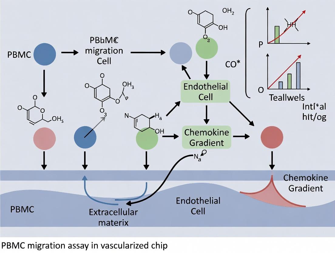
Experimental Workflow for Vascularized Chip Migration Studies
Diagram 2: Workflow for chip-based PBMC migration assay.
The Scientist's Toolkit: Essential Research Reagents & Materials
Table 2: Key Reagents and Materials for PBMC Migration Assays
| Item | Function/Description | Example Product/Catalog |
|---|---|---|
| Ficoll-Paque PLUS | Density gradient medium for isolating PBMCs from whole blood. | Cytiva, 17144003 |
| Recombinant Human TNF-α | Pro-inflammatory cytokine used to activate endothelial cells and upregulate adhesion molecules. | PeproTech, 300-01A |
| Recombinant Human CCL2 (MCP-1) | Key chemokine for monocyte recruitment; establishes chemotactic gradient. | R&D Systems, 279-MC |
| Calcein-AM (Fluorescent Dye) | Cell-permeant dye for live-cell fluorescent labeling of PBMCs. | Thermo Fisher, C3100MP |
| Anti-human CD3 / CD19 / CD14 / CD56 Antibodies | Flow cytometry antibodies for immunophenotyping PBMC subsets. | BioLegend, various |
| Organ-on-a-Chip Platform | Microfluidic device for 3D cell culture and vascularized model creation. | MIMETAS OrganoPlate |
| LIVE/DEAD Viability/Cytotoxicity Kit | Assay to determine cell viability before and after experiments. | Thermo Fisher, L3224 |
| Permeable Support Inserts (Transwell) | For standard 2D migration/chemotaxis assays (control experiments). | Corning, 3422 |
| Collagen I, Rat Tail | Extracellular matrix hydrogel for forming 3D tissue chambers in chips. | Corning, 354236 |
| Time-Lapse Imaging System | Microscope with environmental control for continuous live imaging. | Nikon BioStudio-T |
This application note details the molecular biology and experimental protocols for studying leukocyte extravasation—the multi-step adhesion cascade. Within the broader thesis on Peripheral Blood Mononuclear Cell (PBMC) migration assays in vascularized microfluidic chips, understanding this cascade is fundamental for modeling inflammatory diseases, immune cell trafficking, and evaluating therapeutic interventions in a physiologically relevant context.
The Multi-Step Adhesion Cascade: Key Steps & Molecular Players
The process involves sequential, checkpoint-driven interactions between leukocytes and the vascular endothelium.
Table 1: The Leukocyte Extravasation Cascade
| Step | Primary Function | Key Molecular Players (Examples) | Selectivity |
|---|---|---|---|
| 1. Tethering & Rolling | Initial contact and deceleration under shear flow. | Selectins (P, E, L), their glycoprotein ligands (e.g., PSGL-1). | Low-affinity, rapid on/off kinetics. |
| 2. Activation & Signaling | Inside-out signaling triggered by chemokines/cytokines. | Chemokine receptors (e.g., CXCR4, CCR7), Integrins (e.g., LFA-1, VLA-4) shift to high-affinity state. | G-protein coupled receptor signaling. |
| 3. Firm Adhesion | Stable arrest on endothelial surface. | Activated Integrins (LFA-1, VLA-4) bind to Ig-family CAMs (ICAM-1, VCAM-1). | High-affinity, shear-resistant bond. |
| 4. Crawling & Spreading | Locomotion to find optimal site for transmigration. | Integrins, ICAM-1, Actin cytoskeleton remodeling. | Polarized, adhesion-dependent. |
| 5. Transmigration (Diapedesis) | Crossing the endothelial barrier (paracellular or transcellular). | PECAM-1, JAMs, CD99, VE-cadherin. | Requires junctional rearrangement. |
Table 2: Quantitative Metrics in a Standardized PBMC Migration Assay (Example Data)
| Parameter | Typical Value (in Vascularized Chip) | Measurement Technique |
|---|---|---|
| Shear Stress in Capillary | 0.5 - 4 dyn/cm² | Computational fluid dynamics (CFD) or flow rate calibration. |
| Rolling Velocity | 5 - 50 µm/sec | Time-lapse microscopy, cell tracking software. |
| % of PBMCs Undergoing Firm Adhesion | 10-30% (stimulated) | Static analysis of arrested cells over total perfused. |
| Time to Firm Adhesion Post-Perfusion | 2 - 10 minutes | Time from initial contact to complete arrest. |
| Transmigration Efficiency | 5-20% (towards CXCL12 gradient) | Count of cells in collagen matrix vs. luminal cells. |
Detailed Protocol: PBMC Extravasation Assay in a Vascularized Chip
Protocol 1: Fabrication and Seeding of an Endothelialized Microfluidic Vessel
Objective: Create a 3D lumen lined with a confluent, cytokine-activated endothelium. Materials:
- Microfluidic chip (e.g., two-channel "OrganoPlate" or similar).
- Human Umbilical Vein Endothelial Cells (HUVECs) or induced pluripotent stem cell-derived ECs.
- Fibrinogen (10 mg/mL), Thrombin (10 U/mL), culture medium (EGM-2).
- Tumor Necrosis Factor-alpha (TNF-α) or Interleukin-1 beta (IL-1β). Procedure:
- Gel Channel Preparation: Mix fibrinogen (final 5 mg/mL) with cell suspension medium. Pipette into the gel channel. Add thrombin (final 2 U/mL) to initiate polymerization. Incubate at 37°C for 30 min.
- Lumen Formation: After gelation, inject HUVEC suspension (5x10^6 cells/mL) into the adjacent perfusion channel. Allow cells to attach for 15 min under static conditions.
- Culture & Activation: Connect channel to a perfusion system or rock the plate to establish gravity-driven flow. Culture for 3-5 days until a confluent endothelial tube forms.
- Inflammatory Stimulation: Prior to assay (16-24 hours), add TNF-α (10 ng/mL) to the perfusion medium to upregulate adhesion molecules (ICAM-1, VCAM-1, E-selectin).
Protocol 2: Isolation, Labeling, and Perfusion of PBMCs
Objective: Prepare fluorescently labeled human PBMCs for real-time tracking under flow. Materials:
- Human whole blood or leukopak.
- Ficoll-Paque PLUS density gradient medium.
- Fluorescent cell tracker (e.g., Calcein-AM, 1 µM; or CellTracker Green).
- Adhesion buffer (PBS + 0.1% HSA + Ca²⁺/Mg²⁺).
- Syringe pump or programmable perfusion system. Procedure:
- PBMC Isolation: Layer diluted blood over Ficoll-Paque. Centrifuge at 400 x g for 30 min (brake off). Collect PBMC interface, wash twice.
- Fluorescent Labeling: Resuspend PBMCs (1x10^7/mL) in serum-free medium containing Calcein-AM (1 µM). Incubate 30 min at 37°C. Wash twice with adhesion buffer.
- Perfusion Setup: Resuspend labeled PBMCs at 1x10^6 cells/mL in adhesion buffer. Load into a syringe connected to the chip inlet.
- Shear Calibration: Set syringe pump to achieve a wall shear stress of 1-2 dyn/cm² in the endothelialized channel (requires prior channel dimension calibration).
Protocol 3: Real-Time Imaging & Quantitative Analysis of the Adhesion Cascade
Objective: Capture and quantify each step of the cascade using time-lapse microscopy. Materials:
- Inverted fluorescent microscope with environmental chamber (37°C, 5% CO2).
- High-speed camera (for rolling).
- Time-lapse software (e.g., MetaMorph, ImageJ with plugins). Procedure:
- Data Acquisition: Mount the chip on the microscope stage. Begin perfusion of PBMCs. Acquire images.
- Rolling: 10 fps for 2 minutes at 10x objective. Capture near the channel inlet.
- Firm Adhesion: 1 frame every 30 sec for 20 minutes at 10x.
- Transmigration: 1 frame every 2 min for 4-12 hours using a 20x objective, acquiring z-stacks (5-10 µm steps).
- Quantitative Analysis:
- Rolling Velocity: Track the distance moved by individual cells between frames (>3 consecutive frames).
- Firm Adhesion: Count cells that remain stationary for >30 seconds.
- Transmigration Index: [(Cells in gel at tend) / (Total adherent cells at tstart)] * 100.
Signaling Pathways in Leukocyte Extravasation
The Scientist's Toolkit: Key Research Reagent Solutions
Table 3: Essential Materials for Leukocyte Extravasation Research
| Item | Function & Application | Example Product/Catalog Number |
|---|---|---|
| Functional Grade Monoclonal Antibodies | Block specific adhesion molecules to validate their role in each step (e.g., anti-PSGL-1 for rolling, anti-LFA-1 for adhesion). | Anti-human CD162 (PSGL-1) blocking antibody, clone KPL-1. |
| Recombinant Cytokines/Chemokines | Activate endothelium or create chemotactic gradients for leukocyte guidance. | Recombinant Human TNF-α, Recombinant Human CXCL12/SDF-1α. |
| Fluorescent Cell Trackers | Vital dyes for labeling PBMCs for live-cell, real-time microscopy. | CellTracker Green CMFDA, Calcein-AM. |
| Integrin Activation Reporter Antibodies | Detect high-affinity conformational states of integrins (e.g., LFA-1) on live cells. | mAb24 (reports active LFA-1), MEM-148. |
| Microfluidic Chip Platform | Provides 3D, perfusable, vascularized microenvironment for physiologically relevant assays. | Mimetas OrganoPlate (3-lane 40 or 96), Emulate Organ-Chip. |
| Live-Cell Imaging-Compatible Matrix | Hydrogel for 3D endothelial tubulogenesis and leukocyte transmigration. | Fibrinogen from human plasma, Collagen I (rat tail). |
| Shear Stress Calculator/Software | Calibrate flow rates to achieve physiologically relevant shear stresses in microchannels. | Calculator based on channel geometry (width, height) and fluid viscosity. |
Why Vascularized Chips? Overcoming the Limitations of 2D and Transwell Assays
The study of immune cell migration, particularly of Peripheral Blood Mononuclear Cells (PBMCs), is fundamental to understanding inflammation, immunity, and metastatic spread. Traditional methods like 2D monolayer cultures and Transwell assays have provided foundational insights but present significant limitations in recapitulating the physiological dynamics of the human vasculature. Vascularized microfluidic chips (Organ-on-a-Chip, OOC) emerge as a disruptive technology that overcomes these barriers by introducing fluid flow, 3D architecture, and endothelial barrier function. This application note details the advantages and protocols for implementing PBMC migration assays within vascularized chips, framed within a thesis on advancing mechanistic studies of extravasation.
Table 1: Comparative Analysis of Assay Platforms for PBMC Migration Studies
| Feature | 2D Monolayer Assay | Transwell/Boyden Chamber | Vascularized Chip (3D OOC) |
|---|---|---|---|
| Spatial Architecture | Flat, 2D | 2D compartments separated by a porous membrane | 3D lumen and tissue chamber; tubular vasculature |
| Fluid Flow & Shear Stress | None (static) | Optional, typically minimal | Physiological, programmable (0-10 dyn/cm²) |
| Endothelial Barrier | Poorly formed; no lumen | Formed on a flat filter | Polarized, lumen-forming; mature junctions |
| Extravasation Complexity | Adhesion only | Migration through pores into lower chamber | Full transendothelial migration into 3D matrix |
| Real-time Imaging | Excellent | Limited (endpoint typical) | High-resolution, live-cell tracking |
| Throughput | High | Medium-High | Low-Medium (increasing) |
| Data Output | Endpoint adhesion/morphology | Endpoint migrated cell count | Kinetics of rolling, adhesion, transmigration |
| Physiological Relevance | Low | Moderate | High |
| Typical Experimental Duration | 1-4 hours | 4-24 hours | 1-48 hours |
Data synthesized from recent literature (2023-2024) on immune- and vessel-on-a-chip models.
Key Experimental Protocols
Protocol 1: Fabrication and Seeding of a Basic Vascularized Chip for PBMC Migration
Objective: To create a microfluidic device containing a perfusable endothelial lumen embedded within a 3D extracellular matrix for PBMC perfusion and migration studies.
Materials: See "The Scientist's Toolkit" below.
Method:
- Chip Preparation: Place a sterile, commercially available or PDMS-glass microfluidic chip (e.g., two-channel design separated by a gel region) in a biosafety cabinet.
- Hydrogel Injection: Prepare a cold working solution of ECM hydrogel (e.g., 4 mg/mL collagen I). Pipette the solution into the central gel filling port until all channels are filled. Incubate at 37°C for 30 minutes to polymerize.
- Endothelial Channel Seeding: Reconstitute Human Umbilical Vein Endothelial Cells (HUVECs) or induced pluripotent stem cell-derived endothelial cells (iPSC-ECs) to 10-20 x 10⁶ cells/mL in complete EGM-2 medium. Using a pipette, introduce the cell suspension into one of the side channels (future "vascular" channel). Rotate the chip 90° every 20 minutes for 2 hours to allow even attachment to all sides of the channel, forming a monolayer.
- Lumen Formation & Maturation: Connect the chip to a programmable perfusion system via tubing. Begin perfusing EGM-2 medium at a low shear stress (0.5 dyn/cm²). Culture under flow for 48-72 hours to form a confluent, polarized endothelial tube with strong junctions (verify via ZO-1/VE-cadherin staining).
- PBMC Preparation & Stimulation (Optional): Isolate PBMCs from whole blood via density gradient centrifugation. For inflammation studies, pre-activate the endothelial lumen with TNF-α (10-20 ng/mL) or IL-1β for 6-24 hours via perfusion. Alternatively, PBMCs can be pre-stimulated.
- PBMC Perfusion & Assay: Resuspend fluorescently labeled (e.g., CellTracker) PBMCs in perfusion medium at 1-2 x 10⁶ cells/mL. Introduce the cell suspension into the endothelial channel via flow or a controlled bolus injection. Set a physiological shear stress (0.5-2 dyn/cm²). Begin real-time imaging.
Protocol 2: Quantitative Real-Time Analysis of PBMC Adhesion and Transmigration
Objective: To quantify the kinetics of PBMC rolling, firm adhesion, and transendothelial migration under physiological flow.
Method:
- Image Acquisition: Use an inverted confocal or high-content microscope with an environmental chamber (37°C, 5% CO₂). Acquire time-lapse images (e.g., every 30 seconds for 1 hour) at multiple positions along the vessel.
- Cell Tracking & Quantification: Use automated cell tracking software (e.g., TrackMate in Fiji/ImageJ, or commercial platforms).
- Rolling Cells: Defined as cells moving at a velocity significantly lower than the free-flow velocity (<50% of hydrodynamic velocity).
- Firmly Adherent Cells: Cells that remain stationary for >30 seconds under flow.
- Transmigrated Cells: Cells that have fully crossed the endothelial monolayer and are located within the 3D matrix. Use 3D reconstruction and z-stack analysis.
- Data Normalization: Express adherent cells as number/mm² of endothelial area. Express transmigrated cells as a percentage of total adherent cells or as absolute count per field.
- Endpoint Analysis: At the conclusion of the live assay, fix the chip (4% PFA) and immunostain for endothelial junctions (CD31) and nuclei (DAPI) to confirm transmigration events visually in 3D.
Visualizing Signaling and Workflows
Title: Signaling Pathway for PBMC Extravasation in Vascularized Chips
Title: Workflow for PBMC Migration Assay on Vascularized Chip
The Scientist's Toolkit: Research Reagent Solutions
| Item | Function & Importance in the Assay |
|---|---|
| Microfluidic Chip (2-Channel Design) | Provides the physical structure. The parallel channels separated by a gel region enable the creation of a perfusable vessel adjacent to a tissue compartment. |
| Collagen I, Rat Tail (High Concentration) | The most common ECM hydrogel for forming the 3D tissue matrix. It supports endothelial tube formation and provides a scaffold for immune cell migration. |
| Human Umbilical Vein Endothelial Cells (HUVECs) / iPSC-ECs | The source of vascular endothelium. iPSC-ECs offer donor-specific or disease-modeling potential. Essential for forming a biologically active barrier. |
| Endothelial Growth Medium-2 (EGM-2) | Specialized medium containing VEGF, FGF, and other factors critical for endothelial cell health, proliferation, and maintenance of barrier function under flow. |
| Programmable Perfusion Pump (Syringe or Peristaltic) | Generates physiological, pulsatile, or steady laminar flow. Critical for endothelial maturation and for presenting PBMCs to the vessel wall under shear stress. |
| Fluorescent Cell Tracker Dyes (e.g., CMFDA, CMTPX) | Vital for live-cell, real-time tracking of PBMCs without requiring fixation. Allows distinction from endothelial cells during kinetic analysis. |
| Recombinant Human TNF-α | Standard inflammatory cytokine used to activate endothelial cells, upregulating adhesion molecules (ICAM-1, VCAM-1) to induce an inflammatory phenotype. |
| Anti-CD31 / Anti-VE-Cadherin Antibodies | Used for immunofluorescence staining to visualize the endothelial junctions and confirm monolayer integrity post-assay. |
| Matrigel (Growth Factor Reduced) | Alternative/complement to collagen. Contains basement membrane proteins and can be mixed with collagen to create a more physiologically complex matrix. |
| Live-Cell Imaging Microscope (Confocal/Spinning Disk) | Equipped with environmental control. Essential for capturing high-resolution, multi-z-plane time-lapse data of the dynamic migration process. |
Application Notes
Vascularized chips are advanced in vitro microphysiological systems designed to recapitulate the structure and function of human vasculature within a controlled microenvironment. In the context of immune cell trafficking research, particularly for Peripheral Blood Mononuclear Cell (PBMC) migration assays, these chips provide a transformative platform to study dynamic processes like inflammation, cancer metastasis, and immune recruitment with high physiological relevance. The three core components—Endothelium, Perfusable Channels, and Stromal Cells—are indispensable for creating a functional and biomimetic system.
1. Endothelium: The confluent monolayer of endothelial cells (ECs) lining the perfusable channels is the primary interface for PBMC interaction. It is not merely a passive barrier; it is a dynamically responsive tissue. Under inflammatory cues (e.g., TNF-α, IL-1β), the endothelium upregulates adhesion molecules (e.g., ICAM-1, VCAM-1) and secretes chemokines (e.g., CXCL12, CCL2), establishing a chemotactic gradient essential for directed PBMC adhesion, rolling, and transmigration (paracellular or transcellular). The source of ECs (e.g., HUVECs, iPSC-derived ECs, or tissue-specific microvascular ECs) significantly influences the chip's phenotypic response.
2. Perfusable Channels: These three-dimensional microfluidic structures, typically fabricated from PDMS or hydrogels like collagen I or fibrin, provide the requisite architecture for physiologic fluid flow. Laminar shear stress generated by controlled perfusion (typically 1-10 dyn/cm²) is critical for endothelial cell polarization, barrier function maturation, and quiescence. For PBMC migration assays, a flow-based adhesion and transmigration protocol mimics the hemodynamic conditions of post-capillary venules, the primary site of leukocyte extravasation in vivo. The channel geometry (diameter, shape) directly influences flow profiles and cell-cell interaction probabilities.
3. Stromal Cells: Embedded within the extracellular matrix (ECM) surrounding the endothelialized channel, stromal cells (e.g., fibroblasts, pericytes, mesenchymal stromal cells) provide paracrine and juxtacrine signals that are vital for vascular stabilization, remodeling, and inflammatory signaling. They deposit and remodel the ECM, secrete basement membrane proteins, and release cytokines that can prime or modulate endothelial inflammatory responses. In tri-culture models, pericytes wrapping the endothelium significantly enhance barrier integrity (measured by reduced permeability) and provide more accurate signals for PBMC diapedesis.
Integration for PBMC Migration Assays: The interplay of these three components creates a model where PBMCs, introduced via the perfusate, can be quantitatively monitored as they undergo the multi-step cascade of capture, rolling, firm adhesion, and transmigration under defined flow conditions. The assay readouts include real-time imaging of fluorescently labeled PBMCs, quantification of transmigrated cells in the stromal compartment, and post-assay analysis of endothelial activation markers.
Protocols
Protocol 1: Fabrication and Seeding of a Basic Vascularized Chip
Objective: To create a collagen I-based microfluidic device containing a central endothelialized channel surrounded by a fibroblast-laden stroma.
Materials:
- PDMS microfluidic device (2 parallel channels, 1 mm wide, connected by a gel region).
- Rat tail Collagen I, high concentration (e.g., 8-10 mg/mL).
- Neutralization solution (NaOH, HEPES, and 10x PBS).
- Human Dermal Fibroblasts (HDFs).
- Human Umbilical Vein Endothelial Cells (HUVECs).
- Endothelial Growth Medium (EGM-2) and Fibroblast Growth Medium.
- CellTracker dyes (e.g., CMFDA for HUVECs, CMPTX for HDFs).
Procedure:
- Chip Preparation: Sterilize the PDMS device via UV ozone treatment for 30 minutes.
- Stromal Cell Embedding:
- Trypsinize and count HDFs. Resuspend in cold collagen I solution at 5x10⁶ cells/mL final density.
- Keep the collagen-cell mix on ice. Pipette the mixture into the central gel region of the device via side ports. Allow polymerization at 37°C for 30 min.
- Channel Hydration: After gelation, introduce fibroblast medium into the two adjacent side channels to hydrate the gel. Culture for 24-48 hours to allow fibroblast spreading.
- Endothelial Seeding:
- Trypsinize and count HUVECs. Resuspend in EGM-2 at 5x10⁶ cells/mL.
- Aspirate medium from one side channel and introduce the HUVEC suspension.
- Invert the device and incubate for 20 min to allow cell attachment to the upper channel wall.
- Repeat for the other channel. Return device to normal orientation, fill both channels with EGM-2, and culture under static conditions for 24-48 hours to achieve confluence.
- Perfusion Initiation: Connect the chip to a programmable syringe pump via tubing. Begin perfusion of EGM-2 at a low shear stress (0.5 dyn/cm²), gradually increasing to 2-4 dyn/cm² over 24 hours to condition the endothelium.
Protocol 2: PBMC Adhesion and Transmigration Assay under Flow
Objective: To quantify the TNF-α-induced migration of fluorescently labeled PBMCs across the chip's endothelial barrier.
Materials:
- Vascularized chip from Protocol 1 (HUVEC channel confluent under flow).
- Freshly isolated or cryopreserved human PBMCs.
- Recombinant Human TNF-α.
- Calcein-AM or CellTracker Green dye.
- Assay buffer: Hanks' Balanced Salt Solution (HBSS) with Ca²⁺/Mg²⁺ and 2% FBS.
- Live-cell imaging microscope with environmental chamber.
Procedure:
- Endothelial Activation: Replace EGM-2 in the endothelial channel with EGM-2 containing 10 ng/mL TNF-α. Perfuse for 6-8 hours to induce an inflammatory phenotype.
- PBMC Preparation: Isolate PBMCs via density gradient centrifugation. Label cells with 1 µM Calcein-AM in serum-free buffer for 30 min at 37°C. Wash and resuspend in assay buffer at 1x10⁶ cells/mL.
- Assay Setup: Mount the chip on the microscope stage. Switch the endothelial channel perfusate to assay buffer for 10 min to remove cytokines.
- Flow-Based Migration Assay:
- Introduce the PBMC suspension into the endothelial channel inlet reservoir.
- Program the syringe pump to achieve a wall shear stress of 1 dyn/cm².
- Start perfusion and immediately begin time-lapse imaging (e.g., 10x objective, 30-second intervals for 60-90 minutes) at multiple fields of view along the channel.
- Quantification:
- Adhesion: Count the number of firmly adherent (stationary for >10 seconds) PBMCs per field of view at the 30-minute time point.
- Transmigration: At the end of the assay (e.g., 90 min), acquire z-stack images. Cells that have moved into the 3D stromal compartment (below the focal plane of the endothelium) are counted as transmigrated. Use image analysis software (e.g., ImageJ) for automated cell counting where possible.
Data Presentation
Table 1: Comparative Performance Metrics of Vascularized Chip Components in PBMC Migration Studies
| Component & Variable | Typical Parameter Range | Impact on PBMC Migration (Key Readout) | Measurement Technique |
|---|---|---|---|
| Endothelium | |||
| Shear Stress | 0.5 - 4.0 dyn/cm² | Optimal adhesion at 1-2 dyn/cm²; Higher shear (>4) reduces binding. | Syringe pump flow rate calculation. |
| TNF-α Concentration | 1 - 20 ng/mL | Dose-dependent increase in adhesion/transmigration; 10 ng/mL is standard. | ELISA for secreted ICAM-1/VCAM-1. |
| Barrier Integrity (TEER) | 20 - 60 Ω*cm² (chip-adapted) | Inverse correlation with baseline transmigration. | Trans-endothelial Electrical Resistance (TEER) measurement. |
| Perfusable Channel | |||
| Channel Diameter/Width | 100 - 500 µm | Smaller diameters increase PBMC-endothelium interaction frequency. | Microscopy & design specifications. |
| Matrix Stiffness (Collagen I) | 2 - 6 mg/mL | Softer gels (2 mg/mL) may promote higher transmigration. | Rheometry. |
| Stromal Cells | |||
| Fibroblast Density in Matrix | 1 - 10 x 10⁶ cells/mL | Higher density (5-10 x 10⁶/mL) enhances chemokine secretion & migration. | Pre-seeding cell counting. |
| Pericyte Co-culture | 1:1 to 1:5 (EC:Pericyte) | Reduces baseline permeability; modulates inflammatory response. | Immunofluorescence for NG2/αSMA. |
Visualizations
Title: Signaling Pathway for PBMC Migration in Vascularized Chips
Title: Workflow for PBMC Migration Assay on Vascularized Chip
The Scientist's Toolkit: Research Reagent Solutions
| Item | Function & Application in Vascularized Chip/ PBMC Assay |
|---|---|
| Collagen I, High Concentration (Rat Tail) | The most common hydrogel for constructing the 3D stromal compartment; provides a physiologically relevant ECM for stromal cell embedding and PBMC migration. |
| Microfluidic PDMS Chips | The physical platform. Devices with defined channel architectures (e.g., from Emulate, AIM Biotech, or in-house fabricated) enable precise fluid control and compartmentalization. |
| Programmable Syringe Pump | Generates physiologically relevant, continuous laminar flow to condition the endothelium and perform PBMC adhesion/transmigration assays under shear stress. |
| Live-Cell Fluorescent Dyes (e.g., Calcein-AM, CellTracker) | Vital for real-time, label-free visualization and tracking of specific cell types (e.g., PBMCs, endothelium) during dynamic migration experiments. |
| Recombinant Human Cytokines (TNF-α, IL-1β) | Used to induce a reproducible inflammatory phenotype in the endothelium, upregulating adhesion molecules and creating a chemotactic gradient for PBMCs. |
| Fluorescence-Compatible Inverted Microscope | Equipped with an environmental chamber (CO₂, temperature control) for acquiring high-quality, time-lapse imaging data throughout the duration of the assay. |
| Image Analysis Software (e.g., ImageJ/FIJI, Imaris) | Essential for post-acquisition quantification of key metrics: number of adherent/transmigrated PBMCs, cell velocity, and endothelial barrier integrity. |
Application Notes
This document details the application of PBMC migration assays within vascularized organ-on-chip (OOC) platforms to model and interrogate key disease paradigms. These microphysiological systems provide a critical bridge between conventional in vitro studies and in vivo complexity, enabling precise dissection of cellular migration within a vascular context.
Inflammation Modeling
Vascularized chips enable the study of acute and chronic inflammation by modeling the multi-step adhesion and transmigration cascade of immune cells. Primary human PBMCs or isolated leukocyte subsets are introduced into the vascular channel. Their migration toward gradients of inflammatory chemokines (e.g., IL-8, MCP-1) present in the adjacent tissue chamber is quantified. These models are pivotal for studying conditions like atherosclerosis and cytokine storm syndromes.
Key Quantitative Metrics for Inflammation Studies:
| Metric | Typical Readout | Measurement Technique |
|---|---|---|
| Adhesion Density | 50-200 cells/mm² (under TNF-α stimulation) | Phase-contrast/fluorescence imaging |
| Transmigration Rate | 5-25% of perfused PBMCs (chemokine-dependent) | Confocal Z-stack analysis |
| Velocity on Endothelium | 5-15 µm/min | Time-lapse tracking |
| Activation Marker (e.g., CD11b) | 2-5 fold increase (MFI) | On-chip fixation & immunostaining |
Immuno-oncology (I-O) Applications
These assays are used to evaluate T-cell and NK-cell trafficking toward tumor compartments. Tumor spheroids or monolayers are cultured in the tissue chamber, often with cancer-associated fibroblasts. Autologous or allogeneic PBMCs, including engineered CAR-T cells, are perfused through the vascular lumen. Real-time monitoring of immune cell extravasation, tumor infiltration, and cytotoxic activity is performed. This platform is instrumental for screening bispecific antibodies, oncolytic viruses, and adoptive cell therapies.
Key Quantitative Metrics for Immuno-oncology Studies:
| Metric | Typical Readout | Measurement Technique |
|---|---|---|
| Tumor-infiltrating Lymphocytes (TILs) | 10-50% of perfused CD8+ T cells | Deep tissue imaging analysis |
| Tumor Killing (% cytotoxicity) | 20-60% over 72-96h | Live/dead staining, caspase-3 activity |
| Immune Cell Velocity in Tumor | 2-10 µm/min | Multiplexed time-lapse |
| Cytokine Secretion (e.g., IFN-γ) | pg/mL range, chip effluent | Multiplex ELISA/MSD |
Autoimmune Disease Modeling
To model autoimmunity, autologous PBMCs are perfused over an endothelium activated by disease-relevant cytokines (e.g., Type I IFN). The tissue chamber may contain stromal cells (e.g., synovial fibroblasts for RA, astrocytes for MS) or relevant autoantigens. This setup quantifies pathological migration and tissue invasion, and can test the efficacy of leukocyte-targeting therapeutics (e.g., integrin inhibitors).
Key Quantitative Metrics for Autoimmune Studies:
| Metric | Typical Readout | Measurement Technique |
|---|---|---|
| Pathogenic Th17/Tfh Migration | 1.5-4 fold increase vs. control | Flow cytometry of retrieved cells |
| Endothelial Barrier Disruption | 20-50% decrease in TEER | Trans-endothelial electrical resistance |
| Autoantibody Deposition | Semi-quantitative intensity score | On-chip immunostaining |
| Matrix Degradation | Release of fragments (e.g., C2C) | Fluorescent probe cleavage |
Experimental Protocols
Protocol 1: Standard PBMC Migration Assay in a Vascularized Dual-Channel Chip
Objective: To quantify chemotactic PBMC migration across a vascular endothelium into a 3D tissue matrix.
Materials: Dual-channel microfluidic chip (e.g., from Emulate, AIM Biotech, or MIMETAS), primary human umbilical vein endothelial cells (HUVECs), primary human PBMCs (healthy or donor-matched), fibrin or collagen I matrix, chemokine of interest (e.g., CXCL12 at 100 ng/mL), live-cell imaging microscope.
Procedure:
- Chip Preparation & Endothelialization: Sterilize the chip. Seed HUVECs at high density (e.g., 10x10⁶ cells/mL) into the vascular channel. Culture under flow (0.02-0.05 mL/hr) for 2-3 days to form a confluent, mature monolayer. Confirm by VE-cadherin staining.
- Tissue Chamber Preparation: Mix the chemokine into a fibrinogen solution (e.g., 5 mg/mL). Combine with thrombin and immediately inject into the adjacent tissue chamber. Allow polymerization for 30 min at 37°C.
- PBMC Preparation: Isolate PBMCs via density gradient centrifugation. Label with a cell tracker dye (e.g., Calcein AM, 1 µM) for 30 min. Resuspend in assay medium at 1-2x10⁶ cells/mL.
- Assay Initiation: Perfuse labeled PBMCs through the vascular channel at a physiological shear stress (0.5-1.0 dyn/cm²) for 1 hour. Switch to cell-free medium to remove non-adherent cells.
- Migration Phase: Maintain the chip under continuous flow for 6-48 hours. The chemokine gradient from the tissue chamber drives transmigration.
- Quantification: At endpoint, fix and immunostain for CD31 (endothelium) and DAPI (nuclei). Acquire confocal Z-stacks at multiple positions. Calculate:
- Adhesion: Labeled cells in contact with the apical endothelial surface.
- Transmigration: Labeled cells located beneath the endothelium and within the matrix.
Protocol 2: Tumor-Killing Assay with CAR-T Cells
Objective: To evaluate the infiltration and cytotoxic efficacy of engineered CAR-T cells against a patient-derived tumor spheroid.
Materials: Vascularized chip, patient-derived tumor cells, autologous CAR-T and non-transduced (NT) T-cells from PBMCs, viability dyes, cytokines for T-cell maintenance.
Procedure:
- Tumor Spheroid Formation: Generate tumor spheroids (~150 µm diameter) via hanging drop or ultra-low attachment plates. Load one spheroid into the tissue chamber pre-filled with a 3D matrix.
- Endothelialization & Chip Culture: Seed and mature endothelium in the vascular channel as in Protocol 1. Culture chip for 24-48h to allow tumor-endothelium crosstalk.
- T-cell Perfusion: Harvest expanded CAR-T and NT control T-cells. Label with distinct fluorescent dyes (e.g., CellTrace Violet/CFSE). Perfuse at a physiological concentration (e.g., 1x10⁶ cells/mL) for 2 hours.
- Co-culture & Monitoring: Switch to low-flow maintenance medium. Acquire time-lapse images every 30 minutes for 72-96 hours to track T-cell extravasation, spheroid contact, and tumor morphology.
- Endpoint Analysis: Stain with a live/dead marker (e.g., propidium iodide). Quantify:
- % Tumor Cytotoxicity: (1 - (Final viable tumor area/Initial viable tumor area)) x 100.
- T-cell Infiltration Index: Number of T-cells within the spheroid perimeter / total number of extravasated T-cells.
Signaling Pathways & Workflows
Title: Inflammatory Signaling & PBMC Migration Cascade
Title: Immuno-oncology Chip Assay Workflow
Title: Autoimmune Disease Modeling in a Vascularized Chip
The Scientist's Toolkit: Key Research Reagent Solutions
| Item | Function/Application in PBMC Migration Assays |
|---|---|
| Primary Human PBMCs (Fresh or Cryopreserved) | The primary immune cell source. Donor-matched cells are critical for autoimmune and immuno-oncology studies. |
| Recombinant Human Chemokines/Cytokines (e.g., CXCL12, CCL2, TNF-α, IFN-γ) | To establish chemotactic gradients or pre-condition the endothelium/tissue to model specific disease states. |
| Fluorescent Cell Linker Dyes (e.g., Calcein AM, CFSE, CellTrace Violet) | For stable, non-transferable labeling of PBMC populations to enable live tracking and quantification. |
| Blocking/Antagonistic Antibodies (e.g., anti-α4β1, anti-ICAM-1, anti-CXCR4) | To inhibit specific adhesion or signaling pathways and validate mechanistic involvement. |
| 3D Hydrogel Kits (e.g., Fibrin, Collagen I, Matrigel) | Provide the physiologically relevant extracellular matrix in the tissue chamber for cell migration and embedding. |
| Live/Dead Viability/Cytotoxicity Assay Kits | Essential for endpoint quantification of tumor or stromal cell killing in immuno-oncology and autoimmune models. |
| On-Chip Fixation & Permeabilization Buffer | Formulated for microfluidic chambers to enable high-quality immunostaining without disrupting the delicate 3D structure. |
| Specific Endothelial Cell Media (with shear-stress supplements) | To promote the formation and long-term maintenance of a robust, quiescent endothelial monolayer under flow. |
Step-by-Step Protocol: Establishing and Running a PBMC Migration Assay on a Vascularized Chip
Application Notes
Within vascularized chip research, particularly for Peripheral Blood Mononuclear Cell (PBMC) migration assays, the selection between commercial platforms and custom-fabricated chips is pivotal. The choice directly impacts experimental reproducibility, biological relevance, scalability, and resource allocation. This decision must be anchored in the specific requirements of modeling the vascular endothelium, establishing chemokine gradients, and quantifying leukocyte transmigration.
Core Considerations for PBMC Migration Assays
- Barrier Integrity & Characterization: The chip must support the formation of a confluent, adherent endothelial monolayer. Commercial chips often provide validated protocols, while custom chips offer flexibility in channel geometry to modulate shear stress.
- Gradient Generation & Control: Precise, stable chemokine gradients (e.g., CXCL12) are essential. Commercial systems frequently integrate programmable pumps. Custom microfluidics require external pump systems or rely on passive gradient generators.
- Real-time Imaging Compatibility: The material must be optically clear (e.g., PDMS, glass) for high-resolution, live-cell microscopy of PBMC adhesion and diapedesis.
- Sample Accessibility & Throughput: Commercial platforms may offer multi-channel formats. Custom chips can be designed for specific multiplexing needs but often require manual operation.
Table 1: Platform Comparison for Vascularized PBMC Migration Assays
| Feature | Commercial Platforms (e.g., Emulate, MIMETAS, AIM Biotech) | Custom-Fabricated Chips (PDMS-based, 3D-printed) |
|---|---|---|
| Development Time | Low (Days to weeks for protocol adaptation) | High (Months for design, fabrication, validation) |
| Unit Cost per Chip | High ($50 - $500+) | Very Low ($1 - $10, excluding capital equipment) |
| Throughput | Medium-High (Often 4-96 chips per run) | Low-Medium (Typically 1-12 devices per run) |
| Biological Complexity | Medium (Optimized for common cell types) | High (Fully customizable for co-cultures, stromal layers) |
| Fluidic Control | Integrated/Simplified (Often proprietary controllers) | Flexible but User-Assembled (Requires syringe/perpumps) |
| Assay Reproducibility | High (Standardized manufacturing) | Variable (Dependent on in-lab fabrication skill) |
| Optical Properties | Excellent (Standardized materials) | Excellent (PDMS/Glass), Variable (Other polymers) |
| ECM/Scaffold Options | Sometimes Constrained (Pre-coated/format-specific) | Unlimited (User-defined coating, hydrogel integration) |
| Best Suited For | Screening applications, standardized assays, labs prioritizing rapid start-up. | Mechanistic studies, novel geometries, labs with engineering expertise. |
Experimental Protocols
Protocol 1: PBMC Migration Assay in a Commercial Vascularized Chip Platform
This protocol is adapted for a generic 3-lane commercial plate (e.g., AIM Biotech DAX-1 chip).
Objective: To quantify cytokine-stimulated PBMC transmigration across a Human Umbilical Vein Endothelial Cell (HUVEC) monolayer.
Materials: See "The Scientist's Toolkit" below.
Procedure:
- Chip Preparation: Sterilize the chip under UV light for 15 minutes. Hydrate all channels with 70µL of sterile DPBS for 1 hour at 37°C.
- Gel Channel Seeding: Aspirate DPBS from the gel channel. Inject 2µL of ECM gel (e.g., collagen I, 4 mg/mL) into the central gel channel. Incubate at 37°C for 30 minutes to polymerize.
- Endothelial Seeding: Introduce a suspension of HUVECs (2x10^6 cells/mL) in endothelial growth medium into the two adjacent fluid channels. Allow cells to attach by inverting the chip for 20 minutes, then incubate normally for 24-48 hours to form a confluent monolayer.
- PBMC Preparation & Staining: Isolate PBMCs from whole blood via density gradient centrifugation. Resuspend in migration assay medium (RPMI + 0.5% BSA). Label cells with a fluorescent dye (e.g., Calcein AM, 1 µM) for 30 minutes at 37°C.
- Gradient Establishment & Assay: Replace medium in the "source" fluid channel with assay medium containing chemokine (e.g., 100 ng/mL CXCL12). Replace medium in the opposite "sink" channel with assay medium alone. Introduce stained PBMCs (1x10^6 cells/mL) into the "source" channel.
- Image Acquisition & Quantification: Place chip in a live-cell imaging system. Acquire time-lapse images (e.g., every 5 minutes for 4-24 hours) at the gel-fluid channel interface. Use image analysis software (e.g., ImageJ, MetaMorph) to count fluorescent cells that have migrated into the gel channel.
Protocol 2: Fabrication and Use of a Custom PDMS Dual-Channel Migration Chip
Objective: To fabricate a custom chip for generating a stable chemokine gradient and assaying PBMC migration under controlled shear.
Materials: SU-8 photoresist, Silicon wafer, PDMS and curing agent, Plasma cleaner, Polyethylene tubing (0.5 mm ID), Syringe pumps, Reagents as in Protocol 1.
Procedure: Part A: Chip Fabrication (Soft Lithography)
- Master Mold Creation: Spin-coat SU-8 photoresist onto a silicon wafer. Use a photomask with the design of two parallel channels (e.g., 1 mm wide x 100 µm high) connected by a series of micropillars (to act as a physical barrier supporting the endothelial monolayer). Perform UV exposure and development to create the relief master.
- PDMS Replica Molding: Mix PDMS elastomer and curing agent (10:1 ratio), degas, pour onto the master, and cure at 65°C for 2 hours. Peel off the cured PDMS slab.
- Bonding: Punch inlet/outlet ports. Treat the PDMS slab and a glass slide with oxygen plasma for 45 seconds. Bring surfaces into contact immediately to form an irreversible seal.
Part B: Migration Assay Setup
- Chip Priming & Coating: Treat channels with 1% Pluronic F-127 for 10 minutes to prevent non-specific adhesion. Coat both channels with 50 µg/mL fibronectin in DPBS for 1 hour at 37°C.
- Endothelial Seeding & Culture: Seed HUVECs into one channel at high density. After 4 hours of attachment, connect the chip to a syringe pump and perfuse endothelial medium at a low shear stress (0.5 - 2 dyne/cm²) for 24-48 hours to form a mature, aligned monolayer.
- Gradient Generation: Connect the endothelial channel inlet to a reservoir of assay medium with chemokine. Connect the opposite channel (representing the interstitial compartment) to a reservoir of assay medium alone. Use syringe pumps in withdrawal mode to establish slow, parallel flow in both channels, creating a stable diffusion gradient across the micropillar barrier.
- PBMC Perfusion & Imaging: Introduce fluorescently labeled PBMCs into the endothelial channel via the pump. Initiate time-lapse imaging as in Protocol 1. Quantify cells adhering to and migrating across the endothelial barrier.
Visualizations
Diagram 1: PBMC Transmigration Signaling in a Vascular Chip
Diagram 2: Experimental Workflow for Chip-Based Migration Assay
The Scientist's Toolkit
Table 2: Essential Research Reagents & Materials for PBMC Migration Assays
| Item | Function & Rationale |
|---|---|
| Primary Human Endothelial Cells (HUVECs, HMVECs) | Form the vascular barrier. Primary cells best replicate in vivo physiology and adhesion molecule expression. |
| Isolated Human PBMCs | The motile leukocyte population for studying transmigration. Should be used fresh for optimal viability and response. |
| Chemokines (CXCL12, CCL2, CCL5) | Establish the chemotactic gradient. Critical for directing specific leukocyte subsets. |
| Extracellular Matrix (Collagen I, Fibrin) | Provides a 3D scaffold in the "interstitial" or gel channel, supporting migrated cell infiltration and analysis. |
| Fluorescent Cell Trackers (Calcein AM, CFSE) | Vital dyes to label PBMCs for live-cell, real-time tracking without immediate cytotoxicity. |
| Adhesion Molecule Antibodies (anti-ICAM-1, anti-VCAM-1) | For validating endothelial activation state via immunofluorescence or flow cytometry. |
| Low-Protein Adhesion Assay Medium (RPMI + 0.5% BSA) | Reduces non-specific cell sticking. Serum-free conditions allow precise cytokine control. |
| Syringe Pumps (for custom systems) | Provide precise, steady flow rates to establish stable gradients and apply physiological shear stress. |
| Live-Cell Imaging Microscope | Equipped with environmental control (37°C, 5% CO₂) for kinetic data collection over extended durations. |
Within the broader thesis investigating PBMC migration assays in vascularized organ-on-chip (OoC) systems, the establishment of a quiescent, mature, and functionally intact endothelial layer is the critical foundational step. This protocol details the isolation, seeding, and maturation of primary human endothelial cells (ECs) within a microfluidic chip to create a physiological barrier capable of supporting leukocyte extravasation studies. The resulting endothelium must express appropriate adhesion molecules, form stable junctions, and exhibit low permeability.
Key Research Reagent Solutions
The following materials are essential for the successful execution of this protocol.
| Item Name | Supplier (Example) | Catalog # (Example) | Function & Rationale |
|---|---|---|---|
| Primary Human Umbilical Vein Endothelial Cells (HUVECs) | Lonza | C2519A | Gold-standard primary ECs for vascular modeling. |
| Endothelial Cell Growth Medium-2 (EGM-2) BulletKit | Lonza | CC-3162 | Serum-containing medium with VEGF, FGF, and other supplements for proliferation. |
| Endothelial Basal Medium-2 (EBM-2) | Lonza | CC-3156 | Serum-free basal medium for maturation and assay phases. |
| Microfluidic 3-lane Organ-Chip (μ-Slide VI 0.4) | ibidi | 80606 | Pre-coated, sterile chip with three parallel channels for EC seeding. |
| Fibronectin, Human Plasma | Corning | 354008 | Extracellular matrix protein for coating; promotes EC adhesion and spreading. |
| VE-Cadherin (CD144) Antibody | Santa Cruz Biotechnology | sc-9989 | Immunostaining for visualization of adherens junctions. |
| ZO-1 Tight Junction Protein Antibody | Invitrogen | 33-9100 | Immunostaining for visualization of tight junctions. |
| 4',6-Diamidino-2-Phenylindole (DAPI) | Sigma-Aldrich | D9542 | Nuclear counterstain for fluorescence microscopy. |
| Dextran, Tetramethylrhodamine, 70 kDa | Invitrogen | D1818 | Fluorescent tracer for quantifying endothelial monolayer permeability. |
Detailed Protocol: Seeding and Maturation
Chip Preparation and Coating
- Sterilization: Place the sterile microfluidic chip (e.g., ibidi μ-Slide) under a UV lamp in a laminar flow hood for 15 minutes.
- Coating Solution: Prepare a 50 µg/mL solution of human fibronectin in sterile phosphate-buffered saline (PBS).
- Channel Coating: Using sterile techniques, pipette 40 µL of the fibronectin solution into the central channel of the chip. Ensure the liquid fills the channel completely without introducing bubbles.
- Incubation: Place the chip in a humidified chamber (e.g., a Petri dish with a wet paper towel) and incubate at 37°C for 1 hour.
- Washing: Carefully aspirate the fibronectin solution. Rinse the channel three times with 50 µL of sterile PBS.
Endothelial Cell Seeding
- Cell Preparation: Thaw or harvest primary HUVECs (passage 3-5). Centrifuge and resuspend in complete EGM-2 medium to a final density of 8 x 10^6 cells/mL.
- Seeding: Introduce 40 µL of the cell suspension into the coated central channel. Pipette gently to avoid bubble formation.
- Attachment Phase: Place the chip in the cell culture incubator (37°C, 5% CO₂) for 20 minutes to allow initial cell attachment.
- Perfusion Initiation: After 20 minutes, carefully connect the chip to a perfusion system (e.g., ibidi pump system) or manually add medium to the reservoirs. Begin perfusion with EGM-2 at a low shear stress of 0.5 dyn/cm² for the first 24 hours.
Monolayer Maturation Under Flow
- Medium Transition: After 24 hours, switch the perfusion medium from growth-factor-rich EGM-2 to a maturation medium consisting of EBM-2 supplemented with 1% FBS and 50 ng/mL of TNF-α for 24 hours to prime the endothelium. For the final 48 hours, switch to EBM-2 with 1% FBS only.
- Shear Stress Ramp: Gradually increase the shear stress from 0.5 dyn/cm² to a physiological level of 5-10 dyn/cm² over 72 hours to promote junctional stabilization and quiescence.
- Duration: Total maturation time under flow is 96 hours (4 days) post-seeding.
Quality Control Assays & Data
Immunofluorescence for Junctional Markers
Protocol:
- Fixation: After maturation, perfuse the channel with 4% paraformaldehyde (PFA) for 15 minutes at room temperature.
- Permeabilization & Blocking: Perfuse with 0.1% Triton X-100 in PBS for 10 min, then with 3% BSA in PBS for 1 hour.
- Staining: Incubate with primary antibodies (VE-Cadherin, ZO-1) diluted in 1% BSA/PBS overnight at 4°C. Wash and incubate with appropriate fluorescent secondary antibodies and DAPI for 2 hours at room temperature.
- Imaging: Acquire confocal z-stacks.
Expected Quantitative Outcome:
| Junctional Marker | Expected Localization | Qualitative Metric | Success Criterion |
|---|---|---|---|
| VE-Cadherin (Adherens) | Continuous linear pattern at cell borders | % of perimeter with continuous staining | >90% |
| ZO-1 (Tight) | Continuous/overlapping punctate pattern at borders | Absence of large gaps | No gaps >5 µm |
Permeability Assay
Protocol:
- Tracer Introduction: After maturation, switch the perfusion medium to EBM-2 containing 70 kDa Tetramethylrhodamine-labeled dextran at 25 µM.
- Image Acquisition: Using time-lapse fluorescence microscopy, immediately start imaging the channel and the adjacent extracellular matrix (side channels) every 2 minutes for 60 minutes.
- Quantification: Measure fluorescence intensity (FI) in the central (Ichannel) and side chambers (Imatrix) over time. Calculate the apparent permeability coefficient (Papp) using the formula: Papp = (dImatrix/dt) * (Vmatrix / (A * Ichannel)), where Vmatrix is the side chamber volume, A is the monolayer surface area, and dI_matrix/dt is the initial slope of intensity increase.
Benchmark Data:
| Shear During Maturation (dyn/cm²) | Mean P_app (cm/s) ± SD | n | Reference (from recent search) |
|---|---|---|---|
| Static (0) | (3.2 ± 0.8) x 10⁻⁶ | 6 | Adapted from recent OoC studies |
| 5 | (1.1 ± 0.3) x 10⁻⁶ | 6 | Target for functional layer |
| 10 | (0.9 ± 0.2) x 10⁻⁶ | 6 | Optimal physiological flow |
Signaling Pathway for Endothelial Maturation Under Flow
Diagram Title: Flow-Mediated vs. Inflammatory Signaling in Endothelial Maturation
Experimental Workflow for Protocol Part 1
Diagram Title: Workflow for Seeding and Maturing Endothelial Layer
This protocol details the critical preparatory steps for investigating Peripheral Blood Mononuclear Cell (PBMC) migration within engineered vascularized microfluidic chips. This work forms an essential component of a broader thesis focused on modeling immune cell trafficking and endothelial interactions in physiological and pathological conditions, such as inflammation and cancer metastasis. Precise isolation, labeling, and loading of PBMCs are foundational for generating reproducible and biologically relevant data in these advanced in vitro systems.
PBMC Isolation from Whole Blood
The standard method for PBMC isolation is density gradient centrifugation using Ficoll-Paque.
Detailed Protocol: Density Gradient Centrifugation
Materials:
- Fresh human whole blood (anti-coagulated with EDTA, heparin, or citrate).
- Sterile Phosphate-Buffered Saline (PBS), pH 7.4.
- Ficoll-Paque PLUS or similar density gradient medium (density: 1.077 g/mL).
- Centrifuge tubes (e.g., 15 mL or 50 mL conical tubes).
- Tabletop centrifuge with swinging bucket rotor and brake-off capability.
- Serological pipettes and pipette controller.
- Cell culture medium (e.g., RPMI-1640 + 10% FBS).
Procedure:
- Dilution: Dilute the whole blood 1:1 with sterile PBS or a balanced salt solution.
- Layering: Carefully layer the diluted blood sample over the Ficoll-Paque medium in a centrifuge tube. Maintain a clear interface. A typical ratio is 3-4 mL of Ficoll to 6-8 mL of diluted blood in a 15 mL tube.
- Centrifugation: Centrifuge at 400-500 x g for 30-35 minutes at room temperature (20°C) with the brake OFF. This allows for the formation of distinct layers.
- Harvesting: After centrifugation, four layers will be visible. Carefully aspirate the upper plasma layer. Using a sterile pipette, collect the opaque PBMC interface layer (mononuclear cells) without disturbing the underlying granulocyte/Ficoll layer. Transfer to a new 15 mL tube.
- Washing: Fill the tube with wash buffer (PBS or culture medium) to dilute residual Ficoll. Centrifuge at 300-350 x g for 10 minutes with the brake ON. Carefully decant the supernatant.
- Red Blood Cell Lysis (Optional): If the pellet is contaminated with RBCs, resuspend in 1-2 mL of RBC lysis buffer (e.g., ammonium-chloride-potassium buffer) for 5-10 minutes at room temperature. Stop the reaction by adding excess PBS and centrifuge as in step 5.
- Final Wash & Count: Resuspend the PBMC pellet in 10 mL of complete culture medium or assay buffer. Centrifuge at 250 x g for 5-10 minutes. Resuspend the final pellet in an appropriate volume. Count cells using a hemocytometer or automated cell counter with trypan blue to assess viability.
Table 1: Expected Outcomes from Density Gradient Centrifugation
| Parameter | Typical Yield from Healthy Donor | Acceptable Range | Key Influencing Factors |
|---|---|---|---|
| PBMCs per mL blood | 1-2 x 10^6 cells | 0.5 - 3.0 x 10^6 cells | Donor health, age, hematocrit |
| Lymphocyte Purity | 70-90% | >70% | Gradient quality, careful harvesting |
| Monocyte Purity | 10-30% | N/A | Donor variability |
| Cell Viability (Trypan Blue) | >95% | >90% | Processing speed, sterile technique |
| Granulocyte Contamination | <5% | <10% | Centrifuge brake setting, interface disturbance |
| Platelet Contamination | Variable (can be high) | Can be reduced | Additional low-speed centrifugation washes (150 x g, 10 min) |
PBMC Labeling Strategies for Chip-Based Tracking
Fluorescent labeling is essential for real-time visualization and quantification of PBMC migration and adhesion in vascularized chips.
Detailed Protocol: Membrane Dye Labeling (e.g., PKH26, CellTracker)
Materials:
- Isolated PBMCs.
- Fluorescent cell linker dye kit (e.g., PKH26, PKH67, CellTracker CMFDA/CMTMR).
- Diluent Buffer (supplied with dye).
- Serum-free culture medium or PBS.
- Complete culture medium with 10% FBS (acts as a "stop" reagent).
- Centrifuge tubes.
Procedure for PKH Dyes (General Principle):
- Prepare Cells: Wash PBMCs twice in serum-free medium or the dye diluent. After the final wash, aspirate supernatant completely.
- Prepare Dye Solution: Prepare the dye working solution in diluent immediately before use. Concentration is critical (e.g., 2-10 µM for PKH26; refer to datasheet).
- Labeling: Resuspend the cell pellet in 1 mL of the dye solution. Incubate at room temperature for 3-5 minutes with gentle agitation. Important: Do not exceed recommended time.
- Stop Reaction: Add an equal volume of complete medium with FBS to bind excess dye. Incubate for 1 minute.
- Wash: Add a large volume (e.g., 10 mL) of complete medium. Centrifuge at 350 x g for 5 minutes. Repeat wash 2-3 times until supernatant is clear.
- Resuspend & Validate: Resuspend labeled PBMCs in assay-specific buffer. Check labeling efficiency and brightness using fluorescence microscopy or flow cytometry before loading into the chip.
Table 2: Comparison of Fluorescent Labeling Strategies for PBMC Migration Assays
| Dye Name | Excitation/Emission (nm) | Labeling Mechanism | Typical Working Conc. | Stability (Proliferation Dilution) | Best For |
|---|---|---|---|---|---|
| PKH26 | 551/567 | Membrane intercalation | 2-10 µM | Long-term (weeks), dilutes upon division | Long-term tracking, adhesion studies |
| PKH67 | 490/502 | Membrane intercalation | 2-10 µM | Long-term (weeks), dilutes upon division | Long-term tracking, high signal |
| CellTracker Green (CMFDA) | 492/517 | Cytoplasmic, reacts with glutathione | 0.1-10 µM | Moderate (days), transfers to daughter cells | Medium-term migration, viability indicator |
| Calcein-AM | 494/517 | Cytoplasmic esterase activity | 0.1-1 µM | Short-term (hours) | Viability & short-term migration |
| CFSE | 492/517 | Cytoplasmic, amine-reactive | 0.5-10 µM | Very long-term, dilutes upon division | Proliferation tracking & long-term migration |
| Hoechst 33342 | 350/461 | Nuclear DNA binding | 1-10 µg/mL | Stable while cell intact | Identification/co-staining, not primary tracking |
PBMC Loading Strategies into Vascularized Chips
Effective loading minimizes shear stress, ensures even distribution, and mimics physiological entry.
Detailed Protocol: Controlled Static Loading for Adhesion/Migration Assay
This protocol assumes a vascularized chip with an endothelialized channel adjacent to a tissue or collagen gel chamber.
Materials:
- Labeled PBMCs.
- Vascularized microfluidic chip.
- Syringe pumps and tubing (for perfusion loading).
- Assay buffer (e.g., cell culture medium +/- chemokines).
- Pipettes and tips.
Procedure:
- Chip Preparation: Ensure the endothelial channel is pre-perfused and conditioned with medium. If studying adhesion under flow, precondition with assay buffer at the desired shear stress.
- Cell Preparation: Concentrate labeled PBMCs to a high density (e.g., 5-10 x 10^6 cells/mL) in assay buffer. Keep on ice.
- Static Loading (For Initial Seeding/Adhesion):
- Stop any flow in the chip.
- Carefully pipette the PBMC suspension into the inlet reservoir of the endothelial channel. For chips with access ports, inject ~20-50 µL of cell suspension directly into the channel inlet.
- Gently tap the chip to distribute cells and minimize air bubbles.
- Incubate the chip statically (37°C, 5% CO2) for 15-30 minutes to allow cells to settle and make initial contact with the endothelium.
- Initiation of Flow/Migration Conditions:
- After the static adhesion period, carefully connect the chip inlet to a syringe pump containing fresh assay buffer (with or without a chemoattractant gradient).
- Initiate a very low, physiological shear stress (e.g., 0.5 - 2 dyn/cm²). This washes away non-adherent cells and establishes conditions for transmigration if a gradient is present.
- Begin real-time imaging to track labeled PBMC adhesion and migration.
The Scientist's Toolkit: Research Reagent Solutions
Table 3: Essential Materials for PBMC Isolation, Labeling, and Chip Loading
| Item | Function/Application | Example Product/Brand |
|---|---|---|
| Ficoll-Paque PLUS | Density gradient medium for isolating PBMCs from whole blood. | Cytiva Ficoll-Paque PLUS |
| DPBS (1X), no calcium, no magnesium | Washing and diluting cells during isolation and labeling. | Gibco DPBS |
| RPMI-1640 Medium | Base medium for culturing and maintaining PBMCs. | Corning RPMI-1640 |
| Fetal Bovine Serum (FBS) | Serum supplement for cell culture medium; also used to quench dye reactions. | Characterized FBS, various suppliers |
| PKH26 Red Fluorescent Cell Linker Kit | For stable, long-term fluorescent labeling of PBMC membranes. | Sigma-Aldrich PKH26GL |
| CellTracker Green CMFDA Dye | For cytoplasmic fluorescent labeling that transfers to daughter cells. | Thermo Fisher Scientific C2925 |
| Trypan Blue Solution (0.4%) | Viability stain for counting live/dead cells after isolation. | Gibco Trypan Blue Stain |
| Collagenase Type IV (for chip harvest) | Enzymatic digestion to harvest cells from microfluidic chips for endpoint analysis. | Worthington Collagenase Type IV |
| Recombinant Human MCP-1/CCL2 | Key chemokine to establish a gradient for monocyte migration in chips. | PeproTech 300-04 |
| SDF-1α/CXCL12 | Key chemokine for lymphocyte migration and homing studies. | R&D Systems 350-NS |
Visualization Diagrams
Title: PBMC Isolation by Density Gradient Centrifugation
Title: Decision Flow for Selecting a PBMC Fluorescent Label
Title: PBMC Loading and Migration Setup in Vascularized Chip
Within the broader thesis on modeling immune-vascular interactions using PBMC migration assays in vascularized microfluidic chips, the precise application of chemotactic and inflammatory stimuli is critical. This protocol details the preparation, application, and optimization of key mediators like TNF-α and CXCL12 to establish controlled, biologically relevant gradients for studying PBMC adhesion and trans-endothelial migration.
Research Reagent Solutions Toolkit
| Reagent/Material | Function/Application in Protocol |
|---|---|
| Recombinant Human TNF-α | Pro-inflammatory cytokine used to activate endothelial cells, upregulating adhesion molecules (e.g., ICAM-1, VCAM-1). |
| Recombinant Human CXCL12 (SDF-1α) | Canonical chemokine establishing a chemotactic gradient for CXCR4+ immune cells (e.g., monocytes, lymphocytes). |
| Serum-free Cell Culture Medium (e.g., RPMI-1640) | Basal medium for preparing chemoattractant working solutions, minimizing serum protein interference. |
| Vascularized Microfluidic Chip (e.g., 2-channel design) | Device featuring a 3D endothelialized vessel lumen adjacent to a collagen matrix compartment for gradient formation. |
| Precision Syringe Pumps & Tubing | For controlled medium perfusion in the vascular lumen and static loading in the matrix channel. |
| Fluorescent Cell Tracer (e.g., Calcein AM) | Labels PBMCs for real-time, quantitative tracking of migratory behavior. |
Quantitative Data: Optimizing Stimuli Concentrations
Table 1: Standard Working Concentrations and Key Effects of Applied Stimuli.
| Stimulus | Typical Concentration Range | Primary Target | Key Physiological Effect in Assay | Incubation Time Pre-Migration |
|---|---|---|---|---|
| TNF-α (for EC activation) | 10 – 100 ng/mL | Human Umbilical Vein Endothelial Cells (HUVECs) | Upregulates adhesion molecules; primes vessel for PBMC recruitment. | 4 – 6 hours |
| CXCL12 (for chemotactic gradient) | 50 – 300 ng/mL | PBMCs (via CXCR4 receptor) | Drives directed chemotaxis and trans-endothelial migration into matrix. | Loaded concurrently with PBMCs. |
Table 2: Impact of TNF-α Pre-treatment on HUVEC Activation (Representative Flow Cytometry Data).
| TNF-α Concentration (ng/mL) | % Increase in ICAM-1 Expression (MFI) vs. Control | % Increase in PBMC Adhesion (Static Assay) |
|---|---|---|
| 0 (Control) | 0% | 0% |
| 10 | 320% | 150% |
| 50 | 580% | 280% |
| 100 | 650% | 310% |
Detailed Experimental Protocol
Part A: Endothelial Activation with TNF-α
- Preparation: Reconstitute recombinant human TNF-α to a high-concentration stock (e.g., 10 µg/mL) in sterile, PBS containing 0.1% BSA. Aliquot and store at -80°C.
- Chip Preparation: Following the establishment of a confluent HUVEC monolayer in the vascular channel of the chip under perfusion, switch to a serum-free medium perfusion for 1 hour.
- Application: Dilute TNF-α stock in serum-free perfusion medium to the working concentration (e.g., 50 ng/mL). Completely replace the vascular channel medium with the TNF-α-containing medium.
- Incubation: Perfuse the TNF-α medium through the vascular lumen at a low, physiological shear stress (0.5 - 1.0 dyn/cm²) for 4-6 hours at 37°C, 5% CO₂.
Part B: Establishing a CXCL12 Chemotactic Gradient
- Preparation: Reconstitute recombinant human CXCL12 in sterile PBS with 0.1% BSA. Prepare a working solution in serum-free medium (e.g., 200 ng/mL).
- Gradient Setup: In a standard two-channel chip design:
- Vascular Lumen: Maintain TNF-α-activated endothelium under perfusion with fresh, serum-free medium (NO CXCL12).
- Collagen Matrix Channel: Gently aspirate the medium from the side port of the matrix channel and carefully backfill it with the CXCL12 working solution. Leave this channel under static, no-flow conditions.
- Gradient Formation: Allow the chip to incubate undisturbed for 30-60 minutes. Diffusion of CXCL12 from the matrix channel into the central collagen gel establishes a stable, linear chemotactic gradient toward the matrix compartment.
Part C: Initiating PBMC Migration Assay
- PBMC Preparation: Isolate PBMCs via density gradient centrifugation. Label cells with 1 µM Calcein AM in suspension for 20 minutes at 37°C.
- Cell Introduction: Resuspend labeled PBMCs (1-2 x 10⁶ cells/mL) in serum-free medium. Stop vascular perfusion and introduce the PBMC suspension into the inlet of the vascular lumen.
- Assay Start: Allow PBMCs to settle and interact with the activated endothelium for 10 minutes. Restart perfusion at physiological shear to remove non-adherent cells. This marks time T=0 for the migration assay.
- Imaging & Quantification: Use time-lapse confocal microscopy to track Calcein-AM-labeled PBMCs. Quantify parameters: (1) Number of firmly adherent cells per FOV, (2) Percentage of adherent cells undergoing trans-endothelial migration, (3) Migration velocity and directionality within the collagen matrix.
Visualizations
TNF-α Activation of Endothelium for PBMC Adhesion
CXCL12 Gradient-Driven PBMC Migration in Chip
Application Notes
Real-time imaging of PBMC behavior in vascularized microfluidic chips is a cornerstone technique for studying immune cell trafficking under physiological shear flow. This application note details the integration of live-cell microscopy with vascular chips to quantify the critical dynamic steps of the adhesion cascade: tethering/rolling, firm adhesion, and transendothelial migration (TEM). Key metrics derived from these assays provide quantitative insights into endothelial activation, ligand-receptor interactions, and the efficacy of therapeutic modulators in drug development pipelines.
Protocols
Protocol 1: Microfluidic Vascular Chip Preparation and PBMC Perfusion
Objective: To establish a confluent endothelial monolayer under physiological shear and perfuse isolated PBMCs for real-time adhesion/migration assays.
- Chip Priming & Seeding: Sterilize the microfluidic chip (e.g., two-channel "OrganoPlate" or similar) with 70% ethanol for 15 min. Rinse with PBS. Coat the gel channel with 10 µL of collagen I (3 mg/mL) and incubate at 37°C for 1 hr. Aspirate residual collagen. Introduce a suspension of Human Umbilical Vein Endothelial Cells (HUVECs, passage 3-6) at 10x10⁶ cells/mL into the perfusion channel. Allow cells to adhere for 20 min without flow, then connect chips to a perfusion system.
- Shear Conditioning: Place chips on a stage-top incubator (37°C, 5% CO₂). Initiate continuous flow of complete endothelial growth medium (EGM-2) at a low shear stress of 0.5 dyne/cm² using a programmable syringe pump. Gradually increase shear to 4-5 dyne/cm² over 48 hours to form a mature, confluent, and aligned monolayer.
- Inflammatory Stimulation (Optional): To model inflamed endothelium, perfuse the channel with EGM-2 containing 10 ng/mL TNF-α for 4-6 hours prior to the assay.
- PBMC Preparation & Perfusion: Isolate PBMCs from whole blood via density gradient centrifugation (Ficoll-Paque). Resuspend PBMCs at 1x10⁶ cells/mL in assay buffer (HBSS + 2% FBS + 10 mM HEPES). Load PBMC suspension into a separate syringe. Switch the inlet from medium to the PBMC suspension and initiate perfusion at a defined venous shear stress (0.5-2 dyne/cm²) to commence the real-time imaging assay.
Protocol 2: Real-Time Image Acquisition for the Adhesion Cascade
Objective: To capture high-temporal-resolution image sequences for quantifying rolling, adhesion, and transmigration events.
- Microscope Setup: Use an inverted epifluorescence or spinning-disk confocal microscope equipped with a environmental chamber (37°C, 5% CO₂), a high-sensitivity EMCCD or sCMOS camera, and a 10x or 20x air objective (or 20x/40x water immersion for higher resolution).
- Cell Labeling: Label PBMCs with a cytoplasmic dye (e.g., Calcein AM, 1 µM) for 30 min at 37°C. For transmigration studies, stain endothelial cells with a membrane dye (e.g., CellMask Deep Red, 5 µg/mL) for 10 min.
- Acquisition Parameters: Set up multi-position imaging for multiple chips/channels. For rolling/adhesion: Acquire phase-contrast and green fluorescence images at a high frame rate (1 frame every 100-500 ms) for 10-20 minutes. For transmigration: Acquire z-stacks (5-10 µm range, 2 µm steps) in both fluorescence channels every 60 seconds for 2-4 hours.
- Shear Control Synchronization: Synchronize the microscope acquisition software with the syringe pump controller to record the precise onset of PBMC perfusion and any changes in shear rate.
Protocol 3: Quantitative Image Analysis of Key Metrics
Objective: To extract quantitative metrics from time-lapse data using automated tracking software.
- Data Pre-processing: Apply flat-field correction and background subtraction to all image sequences.
- Rolling Velocity & Adhesion Analysis:
- Import the high-frame-rate sequence into tracking software (e.g., TrackMate in Fiji, or commercial packages like Imaris).
- Detect PBMCs and generate tracks. Filter tracks by duration (>2 sec).
- Rolling Cells: Define cells with a velocity between 5 and 50 µm/sec. Calculate the mean rolling velocity per field of view.
- Firmly Adherent Cells: Define cells with instantaneous velocity < 2 µm/sec for >30 seconds. Count these cells at a fixed time point (e.g., 10 min post-perfusion) per unit area.
- Transmigration Quantification:
- Use the 4D (x,y,z,t) data set from the transmigration experiment.
- Create a 3D surface rendering of the endothelial channel using the membrane dye signal.
- Use the "spots" function in Imaris or a similar 3D tracker to identify all Calcein-stained PBMCs. Classify spots as "luminal" (above endothelial surface) or "transmigrated" (below surface).
- Calculate the Transmigration Index: (Number of transmigrated cells / Total number of adherent cells) x 100% at each time point.
Data Tables
Table 1: Key Quantitative Metrics for PBMC Behavior under Shear Flow
| Metric | Definition | Typical Value (Resting Endothelium) | Typical Value (TNF-α Activated Endothelium) | Significance |
|---|---|---|---|---|
| Rolling Flux Fraction | (# cells rolling / # cells entering FOV) * 100% | < 5% | 15-30% | Measures initial tethering/selection-mediated interactions. |
| Mean Rolling Velocity | Average velocity of cells classified as rolling (µm/sec) | > 50 µm/sec | 10-30 µm/sec | Indicates bond dynamics with selectins (e.g., PSGL-1/P-selectin). |
| Number of Firm Adherent Cells | Cells stationary (<2 µm/sec) for >30 sec per mm² | 10-50 cells/mm² | 200-1000 cells/mm² | Quantifies integrin-mediated firm arrest (e.g., VLA-4/VCAM-1). |
| Transmigration Index | (% of adherent cells that complete diapedesis) at 2 hours | < 10% | 40-70% | Final step, indicates chemokine signaling and junctional remodeling efficiency. |
Table 2: Comparison of Microscopy Modalities for Vascular Chip Imaging
| Modality | Temporal Resolution | Spatial Resolution (xy) | Pros | Cons | Best For |
|---|---|---|---|---|---|
| Widefield Epifluorescence | Very High (10-100 ms) | ~250 nm | High speed, low phototoxicity, simple setup. | Low contrast, out-of-focus light, no 3D. | High-speed 2D tracking of rolling/adhesion. |
| Spinning-Disk Confocal | High (100-500 ms) | ~180 nm | Good optical sectioning, moderate speed, good for live cells. | Lower light throughput than widefield. | 3D transmigration assays with moderate time resolution. |
| TIRF | Very High (10-100 ms) | ~100 nm | Excellent surface specificity, very low background. | Images only ~100 nm from coverslip (2D). | Studying molecular events at the adhesion plane. |
Diagrams
Title: Workflow for Real-Time PBMC Migration Assay
Title: Molecular Steps in the Leukocyte Adhesion Cascade
The Scientist's Toolkit
Table 3: Essential Research Reagent Solutions for PBMC Migration Assays
| Item | Function & Role in Assay |
|---|---|
| Microfluidic Vascular Chips (e.g., from Emulate, AIM Biotech, Mimetas) | Provides a 3D, perfusable, physiologically relevant microenvironment with an endothelial lumen and optional stromal co-culture. Enables precise control of shear stress. |
| Primary Human Endothelial Cells (HUVEC, HMVEC) | Forms the vascular lumen. Responds to inflammatory cues (TNF-α, IL-1β) by upregulating adhesion molecules (E/P-selectin, VCAM-1, ICAM-1). |
| Ficoll-Paque Premium | Density gradient medium for the isolation of high-viability, uncontaminated PBMCs from human blood. |
| Recombinant Human TNF-α | Gold-standard cytokine for activating endothelial cells to an inflammatory phenotype, inducing adhesion molecule expression. |
| Calcein AM | Cell-permeant, non-fluorescent dye converted to green-fluorescent Calcein by intracellular esterases. Vital stain for live-cell tracking of PBMCs. |
| CellMask Deep Red Plasma Membrane Stain | Far-red fluorescent dye that stains endothelial cell membranes, enabling clear 3D segmentation of the vascular lumen for transmigration analysis. |
| Functional Blocking Antibodies (e.g., anti-PSGL-1, anti-VLA-4, anti-ICAM-1) | Tools to perturb specific molecular interactions to validate mechanism and quantify their contribution to each step of the cascade. |
| Syringe Pump with Low-Flow Capability (e.g., from Cetoni, Chemyx) | Precisely controls perfusion flow rates to generate physiological and pathological shear stress ranges (0.1 - 10 dyne/cm²) in the microfluidic chip. |
1. Introduction Within the broader thesis investigating PBMC migration dynamics in vascularized microfluidic chips, the post-assay retrieval of specific cell populations is a critical, non-trivial step. Successful downstream single-cell analysis via flow cytometry or transcriptomics hinges on efficient, high-viability recovery protocols that maintain cell state integrity. This application note details optimized methodologies for retrieving adherent and non-adherent cells from complex chip architectures for subsequent characterization.
2. Key Considerations for Cell Retrieval from Vascularized Chips
- Microenvironment Preservation: The retrieval process must minimize artificial activation or stress-induced gene expression changes.
- Spatial Specificity: Protocols must enable selective retrieval from distinct chip compartments (e.g., vascular channel vs. stromal matrix).
- Yield and Viability: Target recovery >70% with viability >90% for reliable downstream data.
- Compatibility: Methods must be compatible with staining buffers (flow cytometry) or lysis buffers (single-cell RNA-seq).
Table 1: Comparison of Cell Retrieval Methods for Different Chip Compartments
| Chip Compartment | Target Cell Type | Primary Retrieval Method | Average Yield (%) | Average Viability (%) | Key Consideration |
|---|---|---|---|---|---|
| Vascular Channel (Perfused) | Non-adherent PBMCs | Direct Collection via Outflow | 85-95 | 95-98 | Use enzyme-free buffer; collect on ice. |
| Vascular Channel (Lined with Endothelium) | Adherent Endothelial Cells | Enzymatic Detachment (e.g., Accutase) | 75-85 | 90-95 | Optimize exposure time to preserve surface markers. |
| Stromal / Extracellular Matrix (ECM) Region | Migrated PBMCs or Stromal Cells | Combined Enzymatic/Mechanical (Collagenase D + Pipetting) | 60-75 | 85-90 | Matrix density dictates enzyme concentration and time. |
| Interface / Luminal Surface | Adherent or Transmigrated Cells | Sequential Rinse + Mild Enzymatic Step | 70-80 | >90 | Crucial for collecting migrated immune cells. |
3. Detailed Experimental Protocols
Protocol 3.1: Retrieval of Non-Adherent Cells from Vascular Channels
- Objective: Collect non-adherent PBMCs from chip perfusion lines for flow cytometry analysis of surface markers.
- Materials: Collection tube (pre-chilled), Syringe with stopcock, Basal cell culture medium (ice-cold), Phosphate-Buffered Saline (PBS, ice-cold).
- Procedure:
- Stop the perfusion system.
- Flush the vascular channel with 2-3 chip volumes of ice-cold PBS using a slow, steady syringe flow (5-10 µL/min) to displace medium.
- Immediately flush with 2 chip volumes of ice-cold basal medium to collect cells. Direct effluent into a pre-chilled collection tube.
- Centrifuge cells at 300 x g for 5 min at 4°C. Proceed to staining for flow cytometry or cell sorting for transcriptomics.
Protocol 3.2: Retrieval of Adherent and Migrated Cells from ECM and Channels
- Objective: Recover endothelial cells and migrated PBMCs from the chip for single-cell RNA sequencing.
- Materials: Appropriate dissociation enzyme (e.g., Accutase, TrypLE), Collagenase D (for dense matrices), DNase I, Quenching medium (FBS-supplemented), Cell strainer (40 µm).
- Procedure:
- Perfusion & Rinse: Flush all channels with 37°C PBS to remove debris.
- Enzymatic Introduction: Introduce pre-warmed dissociation enzyme into all chip inlets. Incubate chip at 37°C for 5-15 min (optimize visually).
- Mechanical Agitation: Gently pipette fluid back-and-forth within channels. For matrix regions, use wider-bore tips.
- Collection & Quenching: Collect all effluent and quench immediately with 10% FBS medium.
- Filtration & Wash: Pass suspension through a 40 µm strainer. Centrifuge at 300 x g for 5 min. Resuspend in PBS + 0.04% BSA for scRNA-seq library preparation.
4. Visualization of Workflows
Title: Workflow for Cell Retrieval and Downstream Analysis
5. The Scientist's Toolkit: Essential Reagents & Materials
Table 2: Key Research Reagent Solutions for Post-Assay Cell Retrieval
| Item | Function & Rationale |
|---|---|
| Accutase / TrypLE | Gentle, enzyme-based cell dissociation reagents. Preserve cell surface epitopes better than traditional trypsin, critical for flow cytometry. |
| Collagenase D | Matrix-specific enzyme for degrading collagen-based hydrogels (e.g., Type I collagen) to recover embedded/migrated cells. |
| DNase I | Added during digestion to cleave DNA released from damaged cells, reducing clumping and improving single-cell yield. |
| DPBS (Ca²⁺/Mg²⁺-free) | Used for flushing and as a base for enzyme solutions. Lack of divalent cations promotes cell detachment and inhibits cell adhesion. |
| 0.04% Bovine Serum Albumin (BSA) in PBS | Standard suspension buffer for single-cell applications. Prevents non-specific cell adhesion and loss. |
| Viability Dye (e.g., 7-AAD, Propidium Iodide) | For flow cytometry, to exclude dead cells from analysis post-retrieval. |
| Single-Cell 3' Reagent Kit (v3.1) | Standardized commercial kit (e.g., 10x Genomics) for generating barcoded scRNA-seq libraries from retrieved cell suspensions. |
| 40 µm Cell Strainer | Essential for removing debris and cell aggregates to obtain a single-cell suspension for scRNA-seq or flow cytometry. |
Solving Common Challenges: A Troubleshooting Guide for Robust and Reproducible PBMC Migration Data
This document provides protocols and analytical frameworks for investigating poor endothelial barrier formation and integrity within the context of PBMC migration assays in organ-on-chip (OOC) vascular models. Deficiencies in barrier function compromise physiological relevance and experimental outcomes in modeling immune cell extravasation.
Quantitative Data on Barrier Integrity Metrics
Table 1: Key Quantitative Metrics for Assessing Endothelial Barrier Integrity
| Metric | Normal/Robust Barrier Range | Compromised/Poor Barrier Indicator | Primary Measurement Technique |
|---|---|---|---|
| Transendothelial Electrical Resistance (TEER) | 30-100 Ω·cm² (chip-specific) | < 15 Ω·cm² or sudden drop | Real-time TEER measurement (e.g., EVOM2) |
| Apparent Permeability (Papp) | ~1-5 x 10⁻⁶ cm/s (for 10 kDa FITC-Dextran) | > 10 x 10⁻⁶ cm/s for same tracer | Fluorescent tracer flux assay |
| Immunostaining Intensity & Localization | Continuous, sharp VE-cadherin/ZO-1 borders | Faint, discontinuous, or internalized staining | Confocal microscopy, line scan analysis |
| PBMC Transmigration Rate (under flow) | 1-5% of added cells (chemokine-dependent) | >15% (non-specific) or near-zero (dysfunctional) | Fluorescent cell counting in abluminal compartment |
| Actin Cytoskeleton Organization | Predominantly cortical actin bands | Prominent stress fibers & central actin bundles | Phalloidin staining, morphological analysis |
Core Experimental Protocols
Protocol 2.1: Establishing a Human Microvascular Endothelial Cell (HMVEC) Barrier in a Dual-Channel Vascular Chip
Objective: Seed and mature a confluent endothelial monolayer in the vascular channel.
- Chip Preparation: Coat the vascular channel with 50 µg/mL human fibronectin in PBS for 1 hour at 37°C or overnight at 4°C.
- Cell Seeding: Trypsinize HMVECs (e.g., HUVEC or primary microvascular cells). Resuspend at 5-10 x 10⁶ cells/mL in complete EGM-2MV medium. Introduce 10-20 µL of cell suspension into the vascular inlet. Invert the chip to promote attachment to the coated membrane. Incubate for 20 minutes.
- Perfusion Culture: Connect the chip to a perfusion system. Apply a low, continuous flow (0.02-0.05 mL/hour) for 6-12 hours, then increase to a physiological shear stress (1-5 dyn/cm²) for a minimum of 48 hours to promote barrier maturation.
- Barrier Validation: Monitor TEER daily. Proceed to experiments only when TEER stabilizes within the expected range (see Table 1).
Protocol 2.2: Integrated Assessment of Barrier Integrity and PBMC Migration
Objective: Quantify barrier function and its direct impact on immune cell transmigration in response to inflammatory stimuli.
- Pre-Stimulation (Optional): Introduce a pro-inflammatory cytokine (e.g., 10 ng/mL TNF-α) into the vascular channel for 4-24 hours to model inflammatory activation.
- Barrier Integrity Check: Measure TEER. In a parallel chip, perform a permeability assay: add 0.1 mg/mL FITC-labeled 10 kDa dextran to the vascular channel; sample from the interstitial/abluminal channel every 20 minutes for 2 hours; calculate Papp.
- PBMC Preparation & Labeling: Isolate PBMCs from whole blood via density gradient centrifugation. Label cells with 1-5 µM CellTracker Green CMFDA for 30 minutes at 37°C.
- Migration Assay: Introduce labeled PBMCs (1-2 x 10⁶ cells/mL) into the vascular inlet. To induce directed migration, add a chemoattractant (e.g., 100 ng/mL CXCL12) to the interstitial channel. Run assay under physiological flow for 4-24 hours.
- Quantification: Fix and immunostain for endothelial junctions (VE-cadherin/CD144). Use confocal z-stacks to count migrated (abluminal) PBMCs and analyze junction morphology.
Signaling Pathways & Experimental Workflow
Diagram Title: Signaling in Barrier Dysfunction & Integrated Chip Workflow
The Scientist's Toolkit: Research Reagent Solutions
Table 2: Essential Materials for Endothelial Barrier Research in Vascular Chips
| Item | Function/Application | Example Product/Catalog |
|---|---|---|
| Dual-Channel Microfluidic Chip | Provides luminal & abluminal compartments separated by a porous membrane for co-culture & transmigration. | Emulate µ-Slide VI 0.4, MIMETAS OrganoPlate, or custom PDMS chips. |
| Primary Human Microvascular Endothelial Cells (HMVECs) | Physiologically relevant cell source for forming the vascular barrier. | Lonza HMVEC-dermal neonatal (CC-2543) or -lung (CC-2527). |
| Endothelial Growth Medium (EGM-2MV) | Specialty medium containing required growth factors (VEGF, FGF) for endothelial health. | Lonza EGM-2 MV BulletKit (CC-3202). |
| Transendothelial Electrical Resistance (TEER) Instrument | Non-invasive, real-time quantification of barrier integrity. | World Precision Instruments EVOM2 with STX2 electrodes. |
| Fluorescent Tracers (e.g., FITC-Dextran) | Molecules of defined size used to calculate apparent permeability (Papp). | Sigma-Aldrich FD10S (10 kDa FITC-Dextran). |
| Anti-VE-cadherin (CD144) Antibody | Key marker for visualizing adherens junctions via immunostaining. | Santa Cruz Biotechnology sc-9989 (F-8) or Cell Signaling 2158. |
| Recombinant Human TNF-α | Gold-standard pro-inflammatory cytokine to experimentally induce barrier dysfunction. | PeproTech 300-01A. |
| CellTracker Fluorescent Probes | Cytoplasmic dyes for stable, non-transferable labeling of live PBMCs for migration tracking. | Thermo Fisher Scientific C2925 (CMFDA, Green). |
| Perfusion Pump (Syringe or Peristaltic) | Provides controlled, physiological shear stress to endothelial cells. | Harvard Apparatus PHD ULTRA or Elveflow OB1 MK3+. |
Within the broader thesis on PBMC migration assays in vascularized chips, consistent and robust migration is the critical functional readout for studying immune cell dynamics, endothelial barrier function, and therapeutic efficacy. Low or inconsistent migration rates compromise data integrity, leading to unreliable conclusions about immune-vascular interactions, a core pillar of organ-on-chip immunology research. This Application Note identifies primary failure points and provides optimized, standardized protocols to achieve reproducible, physiologically relevant PBMC migration.
Common Causes & Quantitative Analysis
A synthesis of current literature and troubleshooting guides identifies key variables impacting PBMC migration efficiency. The following table summarizes the major factors and their typical impact ranges.
Table 1: Primary Factors Affecting PBMC Migration Consistency
| Factor Category | Specific Parameter | Typical Problem Range | Optimized Range | Impact Level |
|---|---|---|---|---|
| PBMC Viability & State | Post-thaw viability | < 90% | ≥ 95% | High |
| Resting period post-thaw | < 2 hours | 16-24 hours (overnight) | High | |
| Activation status (CD69+%) | High baseline (>10%) | Low baseline (<5%) | Critical | |
| Endothelial Monolayer | Confluence at assay start | < 95% | 100% (verified microscopically) | Critical |
| TEER Value (if applicable) | Inconsistent/Low | Stable, platform-specific baseline | High | |
| Chemokine Expression | Low/Uneven | Uniform, induced (e.g., TNF-α) | High | |
| Assay Conditions | Chemoattractant Gradient | Steep/Unstable | Stable, defined gradient | Critical |
| Medium Composition | Serum-free or high serum | 0.5-2% FBS (chemotaxis medium) | Medium | |
| Assay Duration | Too short/long | 4-6 hours (for trans-endothelial) | Medium | |
| Chip & Matrix | ECM Coating | Incomplete/fragile | Uniform (e.g., Collagen IV, Fibronectin) | High |
| Microchannel Geometry | Too long/wide | 100-500 µm length, 3-10 µm pores | Medium |
Optimized Experimental Protocols
Protocol 1: Standardized PBMC Preparation for Migration Assays
Objective: To obtain highly viable, quiescent PBMCs responsive to chemotactic cues.
- Thawing: Rapidly thaw cryopreserved PBMC vial in 37°C water bath. Immediately transfer dropwise to 10 mL pre-warmed complete RPMI (with 10% FBS).
- Wash: Centrifuge at 300 x g for 10 minutes. Aspirate supernatant.
- Resting: Resuspend pellet gently in 10 mL complete RPMI. Count and assess viability (target ≥95% via Trypan Blue). Transfer to a T25 flask at a density of 1-2 x 10^6 cells/mL.
- Incubate: Place flask upright in a 37°C, 5% CO2 incubator for 16-24 hours (overnight). This step is critical for recovery from activation stress.
- Harvest: Gently collect non-adherent cells (monocytes may adhere). Centrifuge, resuspend in chemotaxis medium (RPMI + 0.5% HSA or 1% FBS). Keep at 37°C until use.
Protocol 2: Robust Endothelial Monolayer Formation in Vascularized Chips
Objective: To form a consistent, confluent, and responsive endothelial barrier.
- Chip Preparation: Sterilize microfluidic chip (e.g., two-channel design) with 70% ethanol (30 min), then UV for 20 min.
- ECM Coating: Introduce 50-100 µg/mL Collagen IV or 10 µg/mL Fibronectin in PBS into the vascular channel. Incubate ≥2 hours at 37°C or overnight at 4°C.
- Wash & Seed: Aspirate coating solution. Wash channel with PBS. Prepare endothelial cells (HUVEC or iPSC-EC) at 5-10 x 10^6 cells/mL in EGM-2 medium. Seed the vascular channel slowly, avoiding bubbles.
- Monolayer Formation: Place chip in incubator for 15 min to allow attachment, then flip and incubate another 15 min to coat opposite wall. Continue static culture for 4-6 hours, then connect to perfusion if desired. Culture for 48-72 hours until fully confluent.
- QC Check: Verify confluence via phase-contrast microscopy. Optionally, measure Transepithelial/Endothelial Electrical Resistance (TEER) if electrodes are integrated. Pre-activation: To study induced migration, perfuse the vascular channel with 10 ng/mL TNF-α in EGM-2 for 6-8 hours prior to assay.
Protocol 3: Controlled PBMC Migration Assay Execution
Objective: To perform a standardized migration assay with a stable chemokine gradient.
- Gradient Establishment: Replace medium in the tissue/interstitial channel (chemoattractant chamber) with chemotaxis medium containing the desired chemoattractant (e.g., 100 ng/mL CXCL12/SDF-1α or CCL2/MCP-1).
- PBMC Introduction: Wash vascular channel with chemotaxis medium. Introduce prepared PBMCs (1-5 x 10^6 cells/mL in chemotaxis medium) into the vascular channel inlet. Allow cells to settle for 15-30 min.
- Assay Run: Establish a slow, continuous flow (0.1-0.5 µL/min) of fresh chemotaxis medium (without chemoattractant) through the vascular channel to maintain gradient integrity. Run assay for 4-6 hours in the incubator.
- Fixation & Analysis: At endpoint, gently perfuse both channels with 4% PFA for 20 min at RT to fix. Wash with PBS.
- Quantification: Image multiple fields in the tissue channel. Manually count or use analysis software (e.g., ImageJ) to quantify migrated cells (e.g., DAPI+/CD45+).
- Calculation: Report as Migration Index = (Number of migrated cells in test condition) / (Number migrated in negative control).
Visualization of Signaling and Workflow
Title: Key Signaling Steps in PBMC Trans-endothelial Migration
Title: Optimized PBMC Migration Assay Workflow
The Scientist's Toolkit: Research Reagent Solutions
Table 2: Essential Materials for Reliable PBMC Migration Assays
| Item | Function & Rationale | Example/Note |
|---|---|---|
| Cryopreserved PBMCs | Standardized, donor-characterized starting material. Reduces variability from fresh isolation. | Ensure high pre-freeze viability (>98%). Characterized for major subsets. |
| Complete RPMI-1640 | Base medium for PBMC thawing and resting. Contains essential nutrients and glutamine. | Supplement with 10% FBS and 1% Pen/Strep for resting phase. |
| Chemotaxis Medium | Low-protein medium for assay execution. Minimizes non-specific chemotaxis. | RPMI-1640 + 0.5% Human Serum Albumin (HSA) or 1% FBS. |
| Recombinant Chemokines | Establish defined, potent chemoattractant gradients (e.g., CXCL12, CCL2, CCL5). | Use carrier-free, endotoxin-free variants. Prepare fresh aliquots. |
| Collagen IV / Fibronectin | ECM proteins for coating vascular channel. Promotes endothelial adhesion and maturation. | Collagen IV at 50-100 µg/mL is standard for HUVECs. |
| Tumor Necrosis Factor-alpha (TNF-α) | Inflammatory cytokine to activate endothelium, upregulating adhesion molecules and chemokines. | Use at 10-20 ng/mL for 6-8h pre-assay to induce a pro-migratory phenotype. |
| Microfluidic Chips | Physiologically relevant platform with endothelialized channels and porous membrane. | Choose designs with 3-10 µm pores and integrated ports for easy fluid handling. |
| Live-Cell Imaging System | For real-time tracking of migration kinetics and endpoint quantification. | System with environmental control (37°C, 5% CO2) is ideal. |
Within the broader thesis investigating PBMC migration in vascularized microfluidic chips, a critical technical challenge is the mitigation of high background noise and non-specific adhesion. These artifacts compromise data fidelity by obscuring true, chemokine-driven leukocyte migration and adhesion events. This application note details the sources of these issues and presents optimized protocols to enhance signal-to-noise ratio in chip-based assays.
Non-specific adhesion of PBMCs, particularly monocytes, to chip materials (e.g., PDMS) or unactivated endothelial monolayers generates high background. This is exacerbated by protein adsorption, shear stress variations, and unintended activation of cells during isolation or loading.
Table 1: Primary Contributors to Background in PBMC Migration Assays
| Contributor | Impact on Background | Quantitative Effect (Typical Range) |
|---|---|---|
| PDMS Hydrophobicity | Protein adsorption & cell adhesion | Can increase non-specific binding by 40-60% |
| Incomplete Endothelial Junction Formation | PBMC seepage underneath endothelium | Background "migration" up to 30% of input cells |
| Serum Proteins in Media | Non-specific adhesion via integrins | Can double baseline adhesion counts |
| Residual PBMC Activation (from isolation) | Upregulation of adhesion molecules (e.g., LFA-1) | Increases non-specific binding by 50-200% |
| Inadequate Blocking | Non-specific protein binding sites | Background signal increase of 70-100% |
Optimized Protocols
Protocol 1: Pre-Treatment of Microfluidic Chips to Minimize Non-Specific Adhesion
Objective: To render chip surfaces protein- and cell-resistant prior to endothelial seeding.
- Post-Fabrication Treatment: For PDMS chips, perform a sequential treatment:
- Oxygen plasma (100 W, 1 min) to hydroxylate surface.
- Immediate immersion in 0.01% (w/v) aqueous poly(L-lysine)-graft-poly(ethylene glycol) (PLL-g-PEG) for 1 hour at RT.
- Rinse 3x with sterile, endotoxin-free PBS.
- Alternative for Collagen/Gelatin Gels: Incorporate 1% (w/v) bovine serum albumin (BSA) into the gel matrix during polymerization.
- Universal Blocking: Prior to cell seeding, perfuse the chip with 5% (w/v) fatty-acid-free BSA in assay medium for 2 hours at 37°C.
Protocol 2: PBMC Preparation & Quiet State Induction
Objective: To reduce pre-activation of isolated PBMCs, lowering baseline adhesion.
- Isolation: Use a low-endotoxin, polysucrose-based density gradient medium (e.g., Ficoll-Paque PREMIUM). Perform all centrifugation steps with brakes OFF.
- Resting: Resuspend isolated PBMCs in "quiet state" medium (RPMI-1640 + 0.5% human serum albumin + 1 mM Mg2+). Do not use FBS.
- Incubation: Rest cells for 90 minutes in a non-tissue culture treated plate at 37°C, 5% CO2. This allows unstimulated cells to revert to a low-adhesion state.
- Labeling: Use a far-red cytoplasmic dye (e.g., CellTracker Deep Red) at ≤ 1 µM for 20 min. Avoid antibodies for labeling if possible.
Protocol 3: Assay Execution with Controlled Shear
Objective: To implement a standardized washing and assay protocol that removes unbound cells without inducing shear-activated adhesion.
- Endothelial Activation: Stimulate the vascular channel with a precise chemokine (e.g., CXCL12 at 100 ng/mL) or TNF-α (10 ng/mL for 4-6h) for directed studies.
- PBMC Perfusion: Introduce the rested, labeled PBMC suspension (1x10^6 cells/mL) at a low, defined shear stress (0.5 dyn/cm2) for 10 minutes.
- Differential Washing:
- Phase 1 (Gentle): Perfuse with warm assay medium at 0.5 dyn/cm2 for 5 min.
- Phase 2 (Definitive): Increase shear to 2.0 dyn/cm2 for 2 min to dislodge loosely bound cells.
- Imaging & Analysis: Acquire images at the endothelial plane. Use a rolling background subtraction (radius >50 µm) and set a stringent threshold for adhesion (minimum particle size >25 µm2, circularity >0.7).
Table 2: Key Reagent Solutions for Low-Noise PBMC Migration Assays
| Reagent / Material | Function & Rationale | Recommended Product/Specification |
|---|---|---|
| PLL(20)-g[3.5]-PEG(2) | Creates a non-fouling, protein-resistant surface on charged substrates. | SuSoS AG or Sigma-Aldrich |
| Fatty-Acid-Free BSA | Blocking agent; fatty-acid-free reduces unintended cell activation. | Sigma-Aldrich A7030 |
| Human Serum Albumin (HSA) | Provides essential proteins without exogenous cytokines found in FBS. | Gemini Bio, ≥99% pure |
| Low-Endotoxin Ficoll | PBMC isolation; minimal endotoxin reduces monocyte activation. | Cytiva Ficoll-Paque PREMIUM |
| CellTracker Deep Red | Far-red cytoplasmic dye; minimizes channel crosstalk, stable for >24h. | Thermo Fisher C34565 |
| Recombinant Human CXCL12 | Definitive chemokine for CXCR4+ lymphocyte migration studies. | PeproTech, carrier-free |
Signaling Pathways in Specific vs. Non-Specific Adhesion
Understanding the molecular drivers distinguishes signal from noise.
Integrated Workflow for Low-Background Assay
A step-by-step visual protocol integrating all optimization steps.
Implementing these surface passivation, cell handling, and fluidic control protocols can reduce non-specific adhesion by 60-80%, dramatically improving the sensitivity and reliability of PBMC migration studies in vascularized chips. This is paramount for accurately modeling inflammatory recruitment and screening therapeutic modulators within the thesis framework.
Within the broader thesis on PBMC (Peripheral Blood Mononuclear Cell) migration assays in vascularized organ-on-chip platforms, precise modeling of hemodynamic forces is paramount. Physiological shear stress is a critical regulator of endothelial cell phenotype, dictating the expression of adhesion molecules (e.g., ICAM-1, VCAM-1) that mediate PBMC rolling, adhesion, and transmigration. Accurately calculating and applying relevant shear stress ranges is therefore foundational to obtaining biologically relevant migration data for drug development applications.
Physiological Shear Stress Ranges & Computational Parameters
Shear stress (τ, dyn/cm² or Pa) is calculated using the formula for laminar flow in a rectangular channel: τ = (6μQ)/(wh²), where μ is dynamic viscosity (Pa·s), Q is volumetric flow rate (m³/s), w is channel width (m), and h is channel height (m). For physiological relevance, different vascular beds must be considered.
Table 1: Physiological Shear Stress Ranges and Corresponding Calculation Parameters
| Vessel Type | Shear Stress Range (dyn/cm²) | Shear Stress (Pa) | Typical Flow Rate (μL/min) for a 500μm x 100μm Channel | Key Pathophysiological Relevance |
|---|---|---|---|---|
| Large Arteries | 10 - 20 | 1.0 - 2.0 | 555 - 1110 | Atheroprone vs. atheroprotective |
| Venules (Post-capillary) | 1 - 6 | 0.1 - 0.6 | 55.5 - 333 | Primary site for PBMC extravasation |
| Capillaries | 5 - 30 | 0.5 - 3.0 | 278 - 1665 | High variation due to network |
| Inflamed Endothelium* | < 1 to > 30 | <0.1 to >3.0 | Variable | Dysregulated adhesion molecule expression |
Note: Under inflammatory conditions, local shear can be dramatically altered.
Protocol: Calibrating and Applying Physiological Shear in a Vascularized Chip for PBMC Migration Assay
Materials & Equipment
- Organ-on-chip device with a top endothelialized channel and a bottom stromal compartment.
- Programmable syringe pump or pressure-driven flow controller.
- In-line bubble trap and pressure sensor (optional but recommended).
- Cell culture medium (e.g., EGM-2 for HUVECs).
- PBMCs isolated via Ficoll-Paque density gradient.
- Acquisition: Phase-contrast or fluorescent time-lapse microscope.
- Software: Image analysis (e.g., ImageJ) and computational fluid dynamics (CFD) software (e.g., COMSOL, ANSYS) or analytical calculators.
Procedure
Part A: Pre-experiment Calculation and Calibration
- Define Target Shear: Based on your research question (e.g., modeling venular inflammation), select the target shear stress from Table 1 (e.g., 2 dyn/cm²).
- Measure Channel Geometry: Precisely measure the width (w) and height (h) of the device's vascular channel using microscopy or manufacturer specs.
- Calculate Required Flow Rate (Q):
- Use the formula: Q = (τ * w * h²) / (6μ)
- Assume a dynamic viscosity (μ) of ~0.007 Poise (0.0007 Pa·s) for cell culture medium at 37°C. For a 500 μm x 100 μm channel targeting 2 dyn/cm² (0.2 Pa):
- Q = (0.2 * 500e-6 * (100e-6)²) / (6 * 0.0007) ≈ 2.38e-10 m³/s ≈ 14.3 μL/min.
- CFD Validation (Optional but Recommended): Create a simplified 2D or 3D model of your channel geometry in CFD software. Apply the calculated Q as an inlet boundary condition and solve for the wall shear stress distribution to confirm uniformity and target value.
- Flow System Priming: Sterilize tubing, connect to the chip, and prime the entire system with medium, ensuring no bubbles are present in the vascular channel.
Part B: On-Chip Endothelial Conditioning and PBMC Perfusion
- Shear Conditioning: Seed human endothelial cells (e.g., HUVECs or iPSC-ECs) in the vascular channel and allow to form a confluent monolayer under static conditions for 24-48 hours.
- Apply Calculated Flow: Initiate flow using the pump at the calculated Q. Condition the endothelium under this defined, physiological shear for a minimum of 24 hours to induce appropriate mechanoadaptation.
- Inflammatory Stimulation (If Applicable): To model inflammation, introduce a cytokine (e.g., TNF-α at 10 ng/mL) into the flow medium for 4-6 hours prior to PBMC perfusion. This upregulates adhesion molecules in a shear-dependent manner.
- PBMC Migration Assay:
- Resuspend fluorescently labeled PBMCs in fresh flow medium at a concentration of 0.5-1 x 10⁶ cells/mL.
- Switch the inflow reservoir to the PBMC suspension and perfuse at the same pre-calculated Q for 30-60 minutes.
- Switch back to cell-free medium and continue flow for the desired assay duration (e.g., 2-24h).
- Use time-lapse microscopy to quantify PBMC adhesion (cells/area) in the vascular channel and transmigration (cells in the stromal compartment).
The Scientist's Toolkit: Key Research Reagent Solutions
Table 2: Essential Materials for Shear-Stress Controlled PBMC Migration Assays
| Item | Function & Relevance |
|---|---|
| Laminar Flow Syringe Pump | Provides precise, pulseless volumetric flow rate control essential for generating stable, calculable shear stress. |
| Microfluidic Chips with Defined Geometry | Channels with precise, uniform height and width are critical for accurate shear stress calculation and application. |
| Dynamic Viscosity Standard (~0.007 Poise) | Culture medium additive (e.g., dextran) to adjust viscosity to physiological levels (blood plasma ~1.2 cP), ensuring correct shear force translation. |
| Recombinant Human TNF-α / Cytokines | Standardized inflammatory stimulant to induce endothelial activation and adhesion molecule expression under flow. |
| Fluorescent Cell Linker Kits (e.g., CFSE, Calcein AM) | For stable, non-toxic labeling of PBMCs to enable quantitative tracking of adhesion and transmigration under flow. |
| Anti-human CD31 / ICAM-1 Antibodies | For post-assay immunofluorescence to correlate PBMC migration events with endothelial adhesion molecule density. |
| Computational Fluid Dynamics (CFD) Software | Enables modeling of complex channel geometries, validation of shear calculations, and identification of stagnation points. |
Visualizations
Diagram 1: Shear-Mediated Signaling in Endothelial-PBMC Interactions
Diagram 2: Experimental Workflow for Shear-Controlled PBMC Assay
Within the broader thesis investigating PBMC migration in vascularized microfluidic chips, the selection of endothelial cell (EC) sources is a critical variable. The choice between primary human endothelial cells (e.g., HUVEC, HMVEC) and induced pluripotent stem cell-derived endothelial cells (iPSC-ECs) fundamentally influences assay outcomes, donor variability, and translational relevance. Concurrently, the donor-specific characteristics of Peripheral Blood Mononuclear Cells (PBMCs) introduce another layer of biological variability that must be systematically controlled or characterized.
Primary ECs offer physiological maturity and established functional markers but are limited by donor availability, finite lifespan, and inherent inter-donor variability. iPSC-ECs provide an unlimited, genetically tractable source from specific donors or disease backgrounds but may exhibit immaturity or batch-to-batch differentiation inconsistencies. When co-cultured with PBMCs from matched or mismatched donors, the combinatorial effects on adhesion molecule expression, chemokine secretion, and resultant leukocyte transmigration can significantly impact the reproducibility and interpretation of vascular inflammation assays.
The following application notes and protocols are designed to guide researchers in making informed cell source decisions and executing robust, comparable experiments.
| Parameter | Primary ECs (e.g., HUVEC) | iPSC-Derived ECs (iPSC-ECs) |
|---|---|---|
| Source | Tissue biopsies (umbilical vein, lung dermis). | Reprogrammed somatic cells (fibroblasts, PBMCs). |
| Proliferation Capacity | Limited (≤ 15 population doublings). | Essentially unlimited via iPSC expansion. |
| Donor Availability | Limited, often pooled donors. | Unlimited, can derive from specific donors. |
| Genetic Manipulation | Difficult, low efficiency. | Highly amenable (CRISPR on iPSCs). |
| Functional Markers (Typical % Positive) | CD31 (>99%), vWF (>95%), VE-Cadherin (>98%). | CD31 (>85%), vWF (variable, 60-90%), VE-Cadherin (>80%). |
| Key Signaling Pathways | Robust VEGF/VEGFR2, Notch, Ang-1/Tie2. | Present but may be dysregulated; Wnt/β-catenin critical for differentiation. |
| Basal Permeability (Typical) | Low, stable junctions. | Higher, junctions can be less mature. |
| Response to Inflammatory Stimuli (TNF-α) | Strong, consistent upregulation of ICAM-1, VCAM-1. | Can be blunted or variable. |
| Cost per Experiment | Moderate to High. | High initial differentiation, lower long-term. |
| Throughput Potential | Lower due to sourcing limits. | Higher for large-scale studies. |
Table 2: Impact of PBMC Donor Variability on Migration Assay Metrics
| Donor Variable | Measurable Impact on PBMC Migration | Typical Coefficient of Variation (CV) |
|---|---|---|
| Age | ↑ Age can correlate with ↑ basal inflammatory state. | 15-25% in transmigration rate across age decades. |
| Health Status | Chronic conditions (e.g., diabetes) prime PBMCs. | Can cause >30% deviation from healthy baseline. |
| Genetic Background | SNPs in adhesion/chemokine receptor genes (e.g., CCR5). | 20-40% variation in chemotactic response. |
| Circadian Rhythm | Time of blood draw influences immune cell subsets. | 10-20% fluctuation in lymphocyte migration. |
| Immune Cell Subset Ratios | Varying CD4+/CD8+/Monocyte ratios alter total flux. | Major source of variability (CV 25-50%). |
Experimental Protocols
Protocol 1: Differentiation and Maturation of iPSC-ECs for Chip Seeding
Objective: Generate a consistent population of functional endothelial cells from iPSCs for microfluidic vessel culture.
Materials: iPSC line, mTeSR Plus medium, Recombinant Human BMP4, CHIR99021 (GSK3 inhibitor), Recombinant Human VEGF, Recombinant Human bFGF, CD31 MicroBeads, Endothelial Growth Medium-2 (EGM-2).
Procedure:
- Mesoderm Induction (Day 0-3): Culture iPSCs to 80% confluence. Switch to RPMI/B27 supplemented with 30 ng/mL BMP4 and 6 µM CHIR99021.
- Endothelial Progenitor Specification (Day 3-5): Change to RPMI/B27 with 100 ng/mL VEGF and 10 ng/mL bFGF.
- EC Expansion (Day 5-7): Dissociate cells and replate on collagen IV-coated dishes in EGM-2 + VEGF/bFGF.
- Purity Isolation (Day 7+): Harvest cells. Perform magnetic-activated cell sorting (MACS) using anti-CD31 MicroBeads.
- Maturation (Day 7-10): Culture sorted iPSC-ECs in EGM-2 on collagen IV. For chip seeding, dissociate and resuspend at 5-10 x 10⁶ cells/mL.
Protocol 2: Parallel PBMC Migration Assay on Primary vs. iPSC-EC Monolayers
Objective: Quantify and compare donor PBMC migration under flow across different EC sources under inflammatory stimulation.
Materials: Microfluidic chip (e.g., two-channel "vessel" chip), Ibidi pump system, Primary ECs (HUVEC), iPSC-ECs (from Protocol 1), PBMCs from 3+ donors, TNF-α, Histamine, Live-cell imaging setup, Calcein-AM.
Procedure:
- EC Monolayer Formation: Seed primary ECs or iPSC-ECs into the chip's collagen I gel-adjacent channel at high density. Perfuse with EGM-2 for 48h to form a confluent lumen.
- Inflammatory Activation: Treat EC lumen with 10 ng/mL TNF-α for 6 hours.
- PBMC Preparation: Isolate PBMCs via Ficoll density gradient from donors. Label with 2 µM Calcein-AM for 30 min.
- Migration Assay: Perfuse labeled PBMCs (1 x 10⁶/mL) through the EC lumen at 0.5 dyn/cm². Simultaneously perfuse the adjacent stromal channel with 100 nM Histamine (chemoattractant).
- Data Acquisition: Capture time-lapse images every 2 min for 90 min at 10x magnification.
- Quantification: Using ImageJ, quantify: a) Number of adherent PBMCs per FOV at 30 min, b) Number of transmigrated PBMCs per FOV at 90 min. Normalize to baseline (unstimulated EC) for each EC source/PBMC donor pair.
The Scientist's Toolkit: Research Reagent Solutions
| Reagent / Material | Function & Application Note |
|---|---|
| EGM-2 BulletKit (Lonza) | Standardized, serum-containing medium for primary EC culture. Ensures robust growth but adds animal-derived variables. |
| StemDiff APEL2 Medium (StemCell Tech) | Chemically defined, serum-free medium ideal for staged differentiation of iPSC-ECs. Reduces batch variability. |
| Collagen I, High Concentration (Corning) | Gold-standard hydrogel for constructing the stromal matrix in vascular chips. Polymerization must be tightly controlled. |
| µ-Slide VI 0.4 (Ibidi) | Ready-to-use microfluidic slide for parallelized, low-shear endothelial culture and leukocyte adhesion/migration assays. |
| Human Recombinant TNF-α (PeproTech) | High-purity, carrier-free cytokine for reliable and consistent inflammatory activation of EC monolayers. |
| CellTracker Dyes (Thermo Fisher) | Fluorescent cytoplasmic labels (e.g., Calcein-AM, CM-Dil) for non-invasive, long-term tracking of PBMCs during migration. |
| CD31 (PECAM-1) MicroBeads (Miltenyi) | For rapid, high-viability positive selection of iPSC-ECs post-differentiation, critical for achieving >90% purity. |
| PrimeFlow RNA Assay (Thermo Fisher) | Allows multiplexed detection of mRNA (e.g., ICAM1, VCAM1, SELE) in fixed ECs via flow cytometry, linking phenotype to genotype. |
Visualizations
Workflow for Comparative PBMC Migration Study
Key Signaling in EC Activation & PBMC Adhesion
Critical Controls and Replication Strategies for Experimental Rigor
1. Introduction: Framing Rigor within PBMC Migration in Vascularized Chips Research utilizing vascularized microfluidic chips to study Peripheral Blood Mononuclear Cell (PBMC) migration under physiological flow offers transformative potential in immunology and drug development. This application note details the critical controls and replication strategies essential for deriving robust, publishable data from these complex systems, framed within a thesis on mechanistic studies of leukocyte extravasation.
2. Foundational Critical Controls: A Structured Framework To isolate specific biological mechanisms from experimental noise, the following control sets are mandatory.
Table 1: Hierarchy of Critical Experimental Controls
| Control Type | Primary Purpose | Example in PBMC Migration Assay | Acceptance Criterion |
|---|---|---|---|
| Negative (Baseline) | Establish baseline migration in the absence of stimulant. | Chip perfused with chemokine-free medium. | Migration count ≤ 5% of positive control. |
| Positive (Assay Function) | Confirm system responsiveness and cell capability. | Use a potent chemoattractant (e.g., 100nM SDF-1α/CXCL12). | Significant increase vs. negative control (p < 0.001). |
| Fluorescence/Labeling | Distinguish specific signal from autofluorescence or non-specific binding. | Unlabeled PBMCs; Isotype control antibodies for stained targets. | Signal ≤ 10% of specifically stained sample. |
| Flow & Shear Stress | Verify physiological flow integrity and rule out adhesion artifacts. | Static condition control; Use of function-blocking anti-ICAM-1/VCAM-1. | Shear-dependent adhesion profile observed. |
| Biological Specificity | Confirm the specific molecular pathway under investigation. | Pre-treatment with receptor antagonist (e.g., AMD3100 for CXCR4). | Migration reduced to negative control levels. |
| Chip & Technical | Account for chip-to-chip variation and assay reagents. | "No cell" control to check for debris; "Medium only" perfusion. | No anomalous particle accumulation. |
3. Replication Strategies: Biological, Technical, and Experimental Robust conclusions require replication at multiple levels to capture variability sources.
Table 2: Replication Strategy Schema
| Replication Level | Definition | Recommended N | Primary Goal |
|---|---|---|---|
| Intra-Chip (Multiple Sites) | Multiple identical vascular channels or observation windows within the same chip. | ≥ 3 channels/windows | Assess local heterogeneity within a single device. |
| Inter-Chip (Technical) | Independent chips run with the same cell donor and reagents in the same experiment. | ≥ 3 chips | Capture technical noise from device fabrication and setup. |
| Biological Replicate (Donor) | Experiments repeated with PBMCs from different healthy human donors. | ≥ 5 distinct donors | Account for innate human genetic and immunological variation. |
| Experimental Replicate (Independent) | Complete repetition of the experiment on different days by different operators. | ≥ 2 full repeats | Control for inter-day reagent, environmental, and operator variability. |
4. Detailed Protocol: PBMC Transmigration in a Vascularized Chip Materials:
- Commercial or fabricated endothelialized microfluidic chip (e.g., two-channel geometry with porous membrane).
- Primary Human Umbilical Vein Endothelial Cells (HUVECs) or induced pluripotent stem cell-derived ECs (passage 3-6).
- Freshly isolated or cryopreserved human PBMCs from consented donors.
- Endothelial growth medium (EGM-2) and assay medium (e.g., RPMI-1640 + 0.5% HSA).
- Recombinant human chemokines (e.g., CCL2, CXCL12).
- Fluorescent cell tracker dyes (e.g., Calcein AM for ECs, CellTracker Red for PBMCs).
- Live-cell imaging microscope with environmental chamber.
Procedure: Day 1-3: Endothelial Channel Seeding & Stabilization.
- Sterilize the chip (UV ozone or 70% ethanol flush).
- Seed HUVECs at high density (e.g., 5-10x10^6 cells/mL) into the top "vascular" channel. Allow attachment for 15-30 min under static conditions, then connect to a perfusion system at low flow (0.1 µL/min) overnight.
- Increase flow to physiological shear stress (e.g., 5 dyn/cm²) for 48 hours to form a mature, confluent, and aligned endothelium. Confirm integrity via microscopy and permeability assay (e.g., 70 kDa FITC-dextran).
Day 4: Stimulation and PBMC Perfusion.
- Switch endothelial medium to assay medium.
- Introduce chemokine (at determined optimal concentration, e.g., 50 ng/mL CCL2) into the lower "tissue" chamber or co-perfuse in the vascular channel, depending on the experimental model. Incubate for 2-4 hours.
- While stimulating, label PBMCs with CellTracker Red (5 µM, 20 min at 37°C) and resuspend in assay medium at 1x10^6 cells/mL.
- Stop endothelial flow. Introduce PBMC suspension into the vascular inlet. Allow PBMCs to settle and interact with endothelium for 10 min (static adhesion phase).
- Restart flow at desired shear stress (e.g., 1 dyn/cm²) to wash away non-adherent cells. This is T=0.
Day 4: Imaging & Quantification.
- Immediately image the chip at pre-marked positions using a 10x objective. Acquire z-stacks every 10 minutes for 4-6 hours.
- Quantify using automated or manual analysis:
- Adherent Cells: Fluorescent cells in contact with the apical endothelial surface.
- Transmigrated Cells: Fluorescent cells that have fully crossed the endothelial monolayer and are located in the basal compartment/tissue channel.
- Express data as cells/mm² or normalized to the positive control.
5. The Scientist's Toolkit: Key Research Reagent Solutions
Table 3: Essential Materials for Vascularized Chip PBMC Migration Assays
| Item | Function & Rationale |
|---|---|
| Collagen IV-coated microfluidic chips | Provides a physiological basement membrane matrix for endothelial cell adhesion and polarization. |
| Defined, serum-free assay medium | Eliminates batch variability from serum, providing a consistent chemokine/cytokine background. |
| Recombinant human chemokines (carrier-free) | Ensures specific receptor engagement without confounding effects of animal-derived protein carriers. |
| Function-blocking monoclonal antibodies (e.g., anti-ICAM-1, anti-VCAM-1, anti-CXCR4) | Gold standard for confirming molecular specificity of adhesion and migration events. |
| Calcein-AM & Ethidium Homodimer-1 (Live/Dead stain) | Critical viability control for both endothelial and immune cells post-experiment. |
| Permeability tracer (FITC-Dextran, 70 kDa) | Quantifies endothelial barrier integrity prior to migration assay, a key quality control metric. |
| Syringe pumps or pressure-driven flow controllers | Generates precise, physiologically relevant laminar flow profiles essential for shear stress studies. |
6. Visualizing Pathways and Workflows
Title: Key Signaling in PBMC Transmigration
Title: Experimental Workflow for PBMC Migration Assay
Benchmarking Performance: How Vascularized Chip Assays Compare to Traditional Models and In Vivo Data
Application Notes: Context within PBMC Migration & Vascularized Chip Thesis
This document provides a comparative analysis of next-generation vascularized organ-on-chip (OoC) platforms versus traditional Transwell/Boyden chamber assays for the study of peripheral blood mononuclear cell (PBMC) migration. The broader thesis context focuses on modeling immune cell trafficking across vascular endothelium under physiological flow, a critical process in inflammation, immunity, and drug efficacy/toxicity.
Comparative Analysis Table
| Feature | Standard Transwell/Boyden Chamber | Vascularized Organ-on-Chip |
|---|---|---|
| Physiological Relevance | Static culture; No fluid shear stress; Simplified 2D geometry. | Dynamic flow; Physiological shear stress; 3D lumenized vessel or tissue interface. |
| Endothelial Barrier | 2D monolayer on porous membrane. | 3D perfusable vessel with mature endothelial cell-cell junctions. |
| Migration Readout | End-point; Quantifies cells that traverse membrane. | Real-time or end-point; Can track migration direction, dynamics, and location. |
| Spatial Complexity | Low (typically two compartments). | High (can incorporate parenchymal cells, extracellular matrix, stroma). |
| Shear Stress Control | None. | Precise, tunable control (typically 0-30 dyn/cm²). |
| Assay Throughput | High (multiwell plate format). | Low to medium (varies by platform; moving towards multiplexing). |
| Cost & Accessibility | Low cost; Widely accessible. | Higher cost; Requires specialized equipment and expertise. |
| Key Advantage | High-throughput, simple, cost-effective screening. | High-fidelity, human-relevant, mechanistic insights under flow. |
| Key Limitation | Lacks hemodynamic forces and tissue-level complexity. | Lower throughput, higher variability, standardization challenges. |
| Metric | Transwell/Boyden Chamber | Vascularized Chip | Notes & Citation |
|---|---|---|---|
| Typical Assay Duration | 2-24 hours | 1-72+ hours | Chip allows prolonged co-culture. |
| Migration Coefficient/Velocity | Not typically measured | ~5-20 µm/min | Real-time tracking possible on-chip. |
| Apparent Permeability (Papp) | ~1-5 x 10⁻⁶ cm/s (for solutes) | ~0.5-2 x 10⁻⁶ cm/s | Chip values reflect flow-induced barrier enhancement. |
| Typical Shear Stress | 0 dyn/cm² | 1-10 dyn/cm² (capillary/venule) | Physiological range for post-capillary venules. |
| PBMC Migration Efficiency | 0.1-5% of input | 0.5-15% of input | Highly dependent on chemokine/activation. |
| Z-factor (Screening Quality) | 0.5-0.7 (good) | Currently <0.5 (variable) | Highlights chip platform development need. |
Detailed Protocols
Protocol 1: Standard Transwell Assay for PBMC Chemotaxis
Objective: To quantify PBMC migration toward a soluble chemoattractant.
Materials:
- 24-well Transwell plate with polycarbonate membranes (5.0 µm pore size).
- PBMCs, isolated via density gradient centrifugation.
- Endothelial Cell Medium & Chemoattractant (e.g., CCL21, CXCL12).
- Cell dissociation buffer & Cell stain (e.g., Calcein-AM, Crystal Violet).
- Plate reader or microscope.
Method:
- Endothelial Monolayer Formation (Optional): Seed human umbilical vein endothelial cells (HUVECs) on the top chamber membrane (2.0×10⁵ cells/insert). Culture for 2-3 days until confluent.
- Chemoattractant Preparation: Add 600 µL of medium with or without chemoattractant to the lower chamber (well).
- PBMC Preparation: Resuspend isolated PBMCs in serum-free medium at 1.0×10⁶ cells/mL.
- Loading Cells: Add 100 µL of PBMC suspension (1.0×10⁵ cells) to the top chamber (insert).
- Migration: Incubate plate at 37°C, 5% CO₂ for 4-24 hours.
- Collection & Quantification:
- Non-adherent Quantification: Carefully collect medium from the lower chamber and count migrated cells via flow cytometry or plate reader (if stained).
- Total Migrated Quantification: Remove non-migrated cells from the top chamber with a cotton swab. Fix and stain cells on the bottom of the membrane. Dissolve stain and measure absorbance or count under a microscope.
- Data Analysis: Calculate % Migration = (Number of migrated cells / Total input cells) × 100.
Protocol 2: PBMC Adhesion and Transmigration in a Vascularized Chip
Objective: To assess real-time PBMC adhesion and trans-endothelial migration under physiological flow.
Materials:
- Commercially available vascularized chip (e.g., Emulate, Mimetas, or in-house PDMS device).
- Microfluidic perfusion system (e.g., peristaltic or pressure-driven pump).
- Human primary endothelial cells (HUVECs or iPSC-ECs), PBMCs.
- Fibrin or Collagen I hydrogel.
- Live-cell imaging microscope with environmental control.
Method:
- Chip Preparation & Seeding:
- If using a tubulogenesis model: Inject a fibrin/collagen gel mixture into the stromal chamber. Polymerize.
- Seed endothelial cells into the adjacent fluid channels. After attachment, apply continuous flow (shear stress ~1-3 dyn/cm²) for 5-7 days to form a 3D lumenized vessel.
- Endothelial Activation: Prior to PBMC introduction, perfuse the vessel with medium containing TNF-α (10 ng/mL) or another inflammatory cytokine for 6-24 hours to upregulate adhesion molecules.
- PBMC Perfusion & Assay:
- Resuspend fluorescently labeled PBMCs in assay medium at 0.5-1.0×10⁶ cells/mL.
- Stop the flow and introduce the PBMC suspension into the vascular inlet reservoir.
- Initiate a low, pulsatile flow (shear stress ~0.5-1 dyn/cm²) to allow cell rolling/adhesion, or use a defined "stop-flow" period (5-10 min).
- Resume physiological flow (1-3 dyn/cm²) to wash away non-adherent cells.
- Real-Time Imaging: Immediately place chip on live-cell microscope. Acquire time-lapse images (e.g., every 30-60 sec for 30-120 min) at the vessel-gel interface.
- Quantitative Analysis:
- Adhesion: Count firmly adherent (stationary for >30s) PBMCs per unit vessel area at 10 min post-wash.
- Transmigration: Track individual cells over time. A cell is considered transmigrated when >50% of its body has moved across the endothelial layer into the gel compartment. Calculate % of adherent cells that transmigrate.
The Scientist's Toolkit: Research Reagent Solutions
| Item | Function in PBMC Migration Assays |
|---|---|
| Transwell Plates (e.g., Corning Costar) | Multiwell plates with permeable membrane inserts for partitioning cell populations and measuring migration. |
| Recombinant Human Chemokines (e.g., CCL19, CXCL12) | Purified proteins used as soluble gradients in the lower chamber to stimulate directed PBMC migration. |
| Calcein-AM | Cell-permeant fluorescent dye used to label live PBMCs for quantification of migrated cells. |
| Fibrinogen from human plasma | Precursor for fibrin hydrogel, used in chips to create a 3D physiological matrix for endothelial tubulogenesis and PBMC infiltration. |
| Tumor Necrosis Factor-alpha (TNF-α) | Pro-inflammatory cytokine used to activate endothelial cells, upregulating adhesion molecules (ICAM-1, VCAM-1) to model inflammatory conditions. |
| Blocking Antibodies (e.g., anti-ICAM-1, anti-VLA-4) | Antibodies used to inhibit specific adhesion molecule interactions, confirming mechanistic pathways of migration. |
| Live-Cell Imaging Dyes (e.g., CellTracker) | Fluorescent, cell-permeant dyes for long-term tracking of specific cell populations in real-time on-chip assays. |
| Microfluidic Peristaltic Pump (e.g., ibidi pump system) | Provides precise, low-flow-rate perfusion to vascular chips, maintaining physiological shear stress. |
| PDMS (Polydimethylsiloxane) | Silicone-based elastomer used to fabricate custom organ-on-chip devices via soft lithography. |
Pathway and Workflow Diagrams
Correlating Chip Data with Animal Model Outcomes in Disease Studies
This Application Note details methodologies and data for correlating outcomes from vascularized organ-on-a-chip (OoC) systems with traditional animal models, specifically within the context of PBMC migration assays. The goal is to validate chip-based systems as predictive tools for complex in vivo disease processes, such as inflammation and cancer metastasis, to enhance drug development efficacy and reduce reliance on animal studies.
Table 1: Comparison of PBMC Migration Metrics Between Vascularized Chip and Murine Model in LPS-Induced Inflammation
| Metric | Vascularized Chip (Mean ± SD) | Murine In Vivo Model (Mean ± SD) | Correlation Coefficient (r) | P-value |
|---|---|---|---|---|
| Migration Rate (cells/mm²/hr) | 12.5 ± 2.1 | 14.8 ± 3.4 | 0.89 | <0.01 |
| % CD14+ Monocytes Migrated | 68% ± 7% | 72% ± 9% | 0.92 | <0.005 |
| ICAM-1 Expression (MFI) | 1520 ± 210 | 1450 ± 180 | 0.95 | <0.001 |
| TNF-α Secretion (pg/mL) | 450 ± 85 | 510 ± 110 | 0.87 | <0.01 |
Table 2: Drug Efficacy Correlation: Inhibitor X on PBMC Migration
| Experimental System | IC50 (nM) | Max Inhibition (%) | Predicted In Vivo Efficacy | Actual In Vivo Outcome (Murine) |
|---|---|---|---|---|
| Static Transwell | 1250 | 45% | Low | Ineffective |
| Vascularized Chip | 85 | 92% | High | 85% Reduction in Migration |
| Animal Model | 90 | 88% | -- | -- |
Experimental Protocols
Protocol 1: PBMC Migration Assay in a Vascularized Dual-Channel Chip
Objective: To quantify cytokine-induced PBMC migration across a vascular endothelial layer under physiological flow.
Materials:
- Vascularized organ-chip (e.g., commercially available or fabricated PDMS device).
- Human primary endothelial cells (HUVECs or iPSC-ECs).
- Isolated human PBMCs.
- Perfusion bioreactor system.
- Culture medium with/without inflammatory stimulus (e.g., 10 ng/mL TNF-α).
- Fixative (4% PFA) and permeability buffer.
- Antibodies for staining (e.g., anti-CD45, anti-CD31, DAPI).
Procedure:
- Chip Seeding & Maturation: Seed endothelial cells into the vascular channel at >5x10⁶ cells/mL. Apply flow (0.02-0.1 dyn/cm² shear stress) for 48-72 hours to form a confluent, aligned monolayer.
- Inflammatory Activation: Introduce medium containing TNF-α into the vascular channel via perfusion for 6-24 hours.
- PBMC Perfusion & Migration: Introduce fluorescently labeled PBMCs (1x10⁶ cells/mL) into the vascular channel under physiological flow. Allow for adhesion and migration for 4-6 hours.
- Wash & Fix: Perfuse with medium to remove non-adherent cells. Perfuse with 4% PFA to fix cells in situ.
- Imaging & Quantification: Permeabilize, stain for nuclei and relevant markers (CD45, CD31). Acquire 3D z-stack images using confocal microscopy. Quantify migrated PBMCs (CD45+ cells in the tissue channel) per unit area.
Protocol 2: Parallel In Vivo Validation in a Murine Inflammation Model
Objective: To validate chip-derived findings in a live animal model of acute inflammation.
Materials:
- C57BL/6 mice.
- LPS (E. coli O111:B4).
- Fluorescent dye (e.g., CFSE) for adoptive cell transfer.
- Fluorescence-activated cell sorting (FACS) buffer and antibodies.
- Tissue harvesting tools.
Procedure:
- Induction of Inflammation: Inject mice intraperitoneally with LPS (1 mg/kg) or PBS vehicle.
- Adoptive Transfer: Isolate and label mouse splenocytes or human PBMCs (in humanized models) with CFSE. Inject 5x10⁶ labeled cells intravenously via the tail vein 2 hours post-LPS.
- Tissue Harvest: Euthanize mice at designated timepoints (e.g., 24h post-LPS). Harvest target organs (e.g., lungs, liver).
- Cell Isolation & Analysis: Digest tissues to create single-cell suspensions. Analyze by flow cytometry to quantify migrated donor cells (CFSE+, CD45+) and local endothelial activation markers (e.g., ICAM-1 expression).
- Correlation Analysis: Statistically compare migration rates and biomarker expression with chip-derived data using linear regression.
The Scientist's Toolkit: Research Reagent Solutions
Table 3: Essential Materials for Vascularized Chip Migration Studies
| Item | Function | Example Product/Catalog |
|---|---|---|
| Dual-Channel Organ-Chip | Provides a tunable 3D microenvironment with separate vascular and tissue compartments lined by living cells under flow. | Emulate PhysioMimix or similar. |
| Primary Human Microvascular Endothelial Cells (HMVECs) | Forms the biologically relevant vascular barrier. Essential for accurate adhesion molecule expression. | Lonza CC-2543 |
| Chemoattractant/Inflammatory Cytokine | Induces endothelial activation and creates a chemotactic gradient for directed PBMC migration. | Recombinant Human TNF-α (PeproTech 300-01A) |
| Live-Cell Labeling Dye | Enables real-time tracking and endpoint quantification of migrating PBMCs. | CellTracker CMFDA (Invitrogen C2925) |
| Anti-human CD31 Antibody | Confirms endothelial monolayer confluence and integrity pre- and post-assay. | BioLegend 303102 |
| Anti-human CD45 Antibody | Identifies all migrated leukocytes (PBMCs) in the tissue chamber for quantification. | BD Biosciences 555483 |
| Laminin-based Extracellular Matrix Gel | Provides a physiologically relevant basement membrane and interstitial matrix for the tissue channel. | Corning Matrigel (356231) |
| Microfluidic Perfusion System | Maintains physiological shear stress and enables controlled reagent delivery. | Ibidi Pump System or similar. |
Visualizations
Title: Workflow for Chip-Animal Data Correlation
Title: Signaling Pathway for PBMC Migration
This case study is a core component of a broader thesis investigating peripheral blood mononuclear cell (PBMC) migration and function within engineered, vascularized microphysiological systems (MPS). The central aim is to model the critical but inefficient process of cytotoxic T-cell trafficking from vasculature into solid tumor tissues, a major bottleneck in immunotherapy efficacy. Using tumor-vascular MPS (TV-MPS), we quantify the multi-step adhesion and migration cascade under controlled biochemical and biophysical conditions, providing a predictive platform for evaluating immunomodulatory drugs.
Table 1: Quantified T-cell Infiltration Parameters in TV-MPS under Various Conditions
| Condition | Avg. T-cell Adhesion (cells/mm²) | Avg. Transmigration Rate (% of PBMC) | Avg. Migration Speed in TME (µm/min) | Tumor Cytotoxicity (% Target Lysis) |
|---|---|---|---|---|
| Control (No Chemokine) | 45 ± 12 | 2.1 ± 0.8 | 1.2 ± 0.4 | 15 ± 5 |
| + CXCL10 / CCL5 | 210 ± 45 | 15.3 ± 3.2 | 3.8 ± 1.1 | 62 ± 8 |
| + Anti-ICAM-1 Blockade | 55 ± 15 | 1.8 ± 0.7 | N/A | 10 ± 4 |
| + TGF-β (Simulated TME) | 180 ± 40 | 8.5 ± 2.1 | 0.9 ± 0.3 | 25 ± 7 |
| + Checkpoint Inhibitor (α-PD-1) | 205 ± 38 | 16.0 ± 2.8 | 4.1 ± 1.2 | 78 ± 9 |
Table 2: TV-MPS Chip Design and Biophysical Parameters
| Parameter | Specification | Biological Relevance |
|---|---|---|
| Vascular Channel Width | 1000 µm | Mimics post-capillary venule |
| Tumor Chamber Dimensions | 1500 µm x 2000 µm | Defined tumor nodule |
| Endothelial Barrier Porosity | 3 µm micro-patterned gaps | Models leaky tumor vasculature |
| Shear Stress in Vascular Channel | 0.5 - 2.0 dyn/cm² | Physiologic venular flow |
| Collagen/Matrigel Matrix Density | 4 mg/mL | Simulated tumor stroma stiffness |
Experimental Protocols
Protocol 3.1: Fabrication and Seeding of the Tumor-Vascular MPS (TV-MPS)
Objective: To create a dual-chamber microfluidic chip featuring a perfusable endothelialized vessel adjacent to a 3D tumor spheroid compartment.
- Chip Preparation: Sterilize a polydimethylsiloxane (PDMS) chip (commercial or fabricated via soft lithography) with UV light for 30 minutes.
- Tumor Spheroid Formation: Harvest cultured tumor cells (e.g., A549, MDA-MB-231). Seed 5,000 cells/well in ultra-low attachment U-bottom plates. Centrifuge at 300 x g for 3 minutes to aggregate. Culture for 72h to form compact spheroids (~200 µm diameter).
- Loading Tumor Compartment: Aspirate a single spheroid in 2 µL of media. Mix with 20 µL of cold collagen I/Matrigel mixture (4 mg/mL final). Carefully pipette the mixture into the rectangular tumor chamber, avoiding introduction into the parallel vascular channel. Polymerize at 37°C for 30 minutes.
- Endothelial Channel Seeding: Introduce a suspension of human umbilical vein endothelial cells (HUVECs) or primary microvascular endothelial cells (2x10^6 cells/mL) into the vascular channel. Allow cell attachment for 15 minutes without flow, then rotate the chip 90° and repeat to seed all walls.
- Vessel Maturation: Attach chip to a perfusion system. Apply continuous flow of endothelial growth medium (EGM-2) at 0.5 dyn/cm² for 48 hours to form a confluent, shear-adapted monolayer.
Protocol 3.2: PBMC Isolation, T-cell Activation, and Perfusion Assay
Objective: To isolate and fluorescently label effector T-cells from human PBMCs and perfuse them through the TV-MPS to model infiltration.
- PBMC Isolation: Collect fresh human whole blood in heparin tubes. Dilute 1:1 with PBS. Carefully layer over Ficoll-Paque PLUS density gradient medium. Centrifuge at 400 x g for 30 minutes (brake off). Harvest the PBMC layer at the interface.
- CD8+ T-cell Activation & Labeling: Isolate CD8+ T-cells from PBMCs using a negative selection magnetic bead kit. Activate cells with CD3/CD28 activator beads (1 bead:1 cell ratio) in RPMI-1640 + 10% FBS + 100 IU/mL IL-2 for 72-96 hours. Label activated T-cells with 5 µM CellTracker Green CMFDA dye for 30 minutes at 37°C.
- Pre-conditioning & Perfusion: 24h before assay, switch endothelial channel to assay medium (RPMI + 1% FBS) and add relevant chemokines (e.g., 100 ng/mL CXCL10) or inhibitors to the tumor chamber to establish a gradient. Resuspend labeled T-cells (1x10^6 cells/mL) in assay medium and introduce into the vascular channel inlet reservoir.
- Live-Cell Imaging & Quantification: Perfuse T-cells at 1.0 dyn/cm² for 10 minutes to allow adhesion, then reduce to 0.5 dyn/cm² for the migration phase. Image using a confocal microscope with environmental control (37°C, 5% CO2) every 10 minutes for 24 hours at multiple Z-positions. Quantify adhesion (cells/mm² of endothelium), transmigration (percentage of adherent cells entering matrix), and migration velocity (µm/min) within the tumor compartment using tracking software (e.g., Imaris, TrackMate).
Protocol 3.3: Functional Cytotoxicity Assessment within TV-MPS
Objective: To assess the tumor-killing capacity of infiltrated T-cells within the 3D microenvironment.
- Tumor Cell Labeling: Prior to spheroid formation, label tumor cells with a far-red cytoplasmic dye (e.g., CellTracker Deep Red) according to manufacturer protocol.
- Cytotoxicity Assay Setup: After the 24h migration assay, carefully add a membrane-impermeant viability dye (e.g., SYTOX Orange, 1 µM) to the perfusion medium to flow through both channels.
- Image Analysis for Lysis: Acquure high-resolution z-stacks of the tumor compartment. Identify infiltrated T-cells (Green), tumor cells (Far-Red), and dead tumor cells (SYTOX Orange-positive). Calculate specific lysis as:
(% SYTOX+ tumor cells in experimental condition) - (% SYTOX+ tumor cells in tumor-only control).
Signaling Pathways & Experimental Workflow Diagrams
The Scientist's Toolkit: Key Research Reagent Solutions
Table 3: Essential Materials for T-cell Infiltration TV-MPS Assays
| Item | Product Example (Supplier) | Function in Assay |
|---|---|---|
| Microfluidic Chip | SynVivo Tumor-Vascular Chip (EMD Millipore) or in-house PDMS design | Provides the physical structure with two parallel chambers separated by a micro-patterned barrier. |
| Endothelial Cells | Primary Human Lung Microvascular Endothelial Cells (Lonza) | Forms the biologically relevant vascular barrier; expresses key adhesion molecules (ICAM-1, VCAM-1). |
| 3D Extracellular Matrix | Corning Matrigel Growth Factor Reduced / Rat Collagen I, Type I (Gibco) | Mimics the composition and stiffness of the tumor stroma for tumor and T-cell migration. |
| Chemokines (Recombinant) | Human CXCL10/IP-10 & CCL5/RANTES PeproTech | Establishes the chemical gradient to drive directed T-cell migration from vessel to tumor. |
| T-cell Activation Kit | Human T-Activator CD3/CD28 Dynabeads (Gibco) | Polyclonally activates isolated CD8+ T-cells to an effector phenotype. |
| Fluorescent Cell Trackers | CellTracker Green CMFDA Dye (Thermo Fisher) | Stably labels T-cell cytoplasm for live-cell tracking without transferring to target cells. |
| Live-Cell Imaging Dyes | SYTOX Orange Dead Cell Stain (Thermo Fisher) | Membrane-impermeant nuclear stain to quantify tumor cell death (cytotoxicity). |
| Functional Blocking Antibodies | Anti-Human ICAM-1 (CD54) Neutralizing Antibody (R&D Systems) | Inhibits specific ligand-receptor interactions to dissect mechanism (e.g., blocks LFA-1/ICAM-1 adhesion). |
| Perfusion Pump System | Elveflow OB1 Mk3 Pressure Controller | Provides precise, pulse-free low flow rates to generate physiological shear stress in the vascular channel. |
| Confocal Microscope | Nikon A1R or Zeiss LSM 880 with incubation chamber | Enables high-resolution, multi-position, time-lapse imaging of deep 3D tissues. |
Within the thesis investigating monocyte migration in vascularized organ-on-chip models, a critical validation step is the use of known pharmacological inhibitors. This application note details protocols for using established inhibitors to confirm the specificity and relevance of observed PBMC migration signals, thereby bridging chip observations with known biology and drug mechanisms.
Application Notes
Rationale for Inhibitor Selection
To demonstrate pharmacological relevance, inhibitors targeting key pathways implicated in leukocyte adhesion and transendothelial migration are employed. Successful inhibition of migration in the vascularized chip by these compounds validates that the assay recapitulates in vivo signaling mechanisms and provides a benchmark for novel drug testing.
Key Signaling Pathways & Targeted Inhibitors
The following table summarizes the primary pathways, their role in migration, and the standard inhibitors used for validation.
Table 1: Key Inhibitors for Validating PBMC Migration Pathways
| Pathway | Target Molecule | Known Inhibitor (Example) | Typical Working Concentration | Expected Inhibition of Migration | Primary Role in Migration |
|---|---|---|---|---|---|
| Chemokine Signaling | CCR2 Receptor | RS 504393 | 1 - 10 µM | 60-80% | Blocks MCP-1/CCL2 driven chemotaxis |
| Integrin Activation | LFA-1 (αLβ2) | BIRT 377 | 0.5 - 5 µM | 70-90% | Inhibits ICAM-1 binding & firm adhesion |
| Sphingosine-1-Phosphate | S1P Receptor 1 | W146 | 100 nM - 1 µM | 40-70% | Modulates endothelial barrier & egress |
| Cytoskeleton Rearrangement | ROCK (ROCK1/2) | Y-27632 | 5 - 20 µM | 50-80% | Impairs actomyosin contraction in monocytes |
| Pro-inflammatory Signaling | p38 MAPK | SB203580 | 1 - 10 µM | 40-70% | Reduces adhesion molecule expression |
Detailed Experimental Protocols
Protocol 1: Pre-treatment of PBMCs with Inhibitors Prior to Perfusion
This protocol details the direct inhibition of monocyte signaling.
Materials:
- Isolated human PBMCs.
- Known inhibitors (e.g., from Table 1) reconstituted in appropriate vehicle (DMSO, etc.).
- Serum-free cell culture medium.
- Vascularized organ-chip (e.g., endothelialized microfluidic channel).
Procedure:
- Cell Preparation: Resuspend purified PBMCs at 2x10^6 cells/mL in serum-free medium.
- Inhibitor Pre-treatment: Aliquot cell suspension. Add inhibitor or vehicle control to achieve desired final concentration. Incubate for 30-60 minutes at 37°C, 5% CO₂.
- Chip Preparation: Prior to experiment, perfuse vascular channel of chip with warm medium for 10 minutes to establish baseline.
- Perfusion & Migration: Introduce the pre-treated PBMC suspension into the vascular inlet at a defined physiological shear stress (e.g., 1-4 dyn/cm²). Allow cells to perfuse for 10-15 minutes.
- Wash & Monitor: Switch inlet to inhibitor-free medium to wash away non-adherent cells. Continue perfusion.
- Image Acquisition: Use time-lapse microscopy (e.g., every 5 mins for 2-4 hours) to track adherent and migrating cells.
- Analysis: Quantify (a) number of firmly adherent cells per FOV, and (b) number of cells that have transmigrated into the interstitial compartment.
Protocol 2: Treatment of Vascular Endothelium with Inhibitors
This protocol assesses the role of endothelial targets.
Procedure:
- Chip Pre-treatment: After endothelial monolayer formation, perfuse the vascular channel with medium containing the inhibitor or vehicle for 1-2 hours.
- Stimulation (Optional): If modeling inflammation, co-perfuse with TNF-α (e.g., 10 ng/mL) during the last 6-12 hours of inhibitor treatment.
- PBMC Perfusion: Without removing inhibitor from the endothelial channel, introduce untreated PBMCs and follow steps 4-7 from Protocol 1.
Protocol 3: Data Normalization & Analysis
Quantification:
- Normalize cell adhesion and migration counts in inhibitor-treated conditions to the vehicle control (set at 100%).
- Calculate percentage inhibition using:
[1 - (Count_Inhibitor / Count_Vehicle)] * 100. - Perform dose-response curves across a minimum of three inhibitor concentrations to confirm specificity.
Diagrams
Diagram 1: PBMC Migration Signaling & Inhibitor Targets
Title: Inhibitor Targets in PBMC Migration Cascade
Diagram 2: Experimental Workflow for Inhibitor Validation
Title: Inhibitor Assay Workflow in Vascular Chip
The Scientist's Toolkit: Research Reagent Solutions
Table 2: Essential Materials for Inhibitor Validation Assays
| Item | Function/Description | Example Vendor/Cat. No. (Illustrative) |
|---|---|---|
| Human PBMCs, isolated | Primary cells for migration studies; source of monocytes. | Freshly isolated from donor blood or commercially sourced cryopreserved vials. |
| Microvascular Endothelial Cells | Forms the vascular lumen in the chip; source of adhesion molecules & chemokines. | HMVEC-d (Lonza) or iPSC-derived endothelial cells. |
| Fibrinogen/Collagen I | Hydrogel for the interstitial/tissue compartment in the chip. | Sigma-Aldrich F3879 / Corning 354236. |
| Chemokine (MCP-1/CCL2) | Positive control stimulus to induce directed migration. | PeproTech 300-04. |
| Pro-inflammatory Cytokine (TNF-α) | Used to activate endothelium and upregulate adhesion molecules. | PeproTech 300-01A. |
| Known Small Molecule Inhibitors | Pharmacological tools to block specific pathways (see Table 1). | Tocris Bioscience, Cayman Chemical, Sigma-Aldrich. |
| Vehicle (e.g., DMSO) | Solvent control for inhibitor reconstitution; critical for control experiments. | High-grade, sterile DMSO. |
| Organ-Chip Platform | Microfluidic device supporting 3D vascular culture and perfusion. | Emulate, AIM Biotech, or in-house fabricated PDMS chips. |
| Syringe Pump | Provides precise, continuous flow to mimic physiological shear stress. | Harvard Apparatus, Chemyx. |
| Live-Cell Imaging System | For time-lapse quantification of adhesion and migration events. | Microscope with environmental chamber (e.g., Zeiss, Nikon). |
| Cell Tracker Dyes (e.g., CMFDA) | Fluorescently labels PBMCs for enhanced contrast during imaging. | Thermo Fisher Scientific C7025. |
Within the broader thesis on advancing PBMC migration assays in vascularized organ-on-chip (OoC) systems, this document addresses the critical debate surrounding "gold standard" assays. The central question is whether traditional, well-established in vitro and in vivo models provide sufficient physiological fidelity and predictive value for human inflammatory responses, or if next-generation vascularized chips represent a new paradigm. This assessment is crucial for researchers and drug development professionals prioritizing translational success in immunology and vascular biology.
Comparative Analysis: Assay Platforms
The following table summarizes key quantitative and qualitative metrics for current assay platforms used to study PBMC migration in vascular contexts.
Table 1: Comparison of Assay Platforms for PBMC-Vascular Interactions
| Assay Platform | Key Measurable Outputs | Typical Throughput | Physiological Fidelity Score (1-5) | Key Predictive Limitations | Cost & Complexity |
|---|---|---|---|---|---|
| Transwell / Boyden Chamber | - Migration count- Chemotaxis index | High (96-well) | 2 (Static, no flow, simplified endothelium) | Lacks hemodynamic shear stress; no 3D tissue stroma. | Low |
| Static 2D Endothelial Monolayer Assay | - Adherent cell count- Microscopy morphology | Medium (24-well) | 2.5 (Cell-cell contact but no flow) | Absent physiological shear stress and vessel geometry. | Low |
| In Vivo Models (e.g., mouse cremaster) | - Intravital rolling/flux/adhesion- Extravasation events | Low | 4 (Full physiological system) | Species-specific differences in adhesion molecules; challenging human genetic manipulation. | Very High |
| Vascularized Microfluidic Chip (2-channel) | - Real-time adhesion/transmigration- Permeability (TEER/Dextran)- Cytokine secretion profile | Medium-Low (Chip-to-chip) | 4.5 (Human cells, physiological flow, 3D matrix) | Limited multi-organ crosstalk; standardization ongoing. | High |
| Vascularized 3D Tissue Construct in Chip | - Infiltration depth into tissue compartment- Functional tissue damage | Low | 5 (Human cells, flow, 3D tissue niche) | Complex imaging and data extraction; lower throughput. | Very High |
Detailed Application Notes & Protocols
Protocol: Establishing a Vascularized Chip for PBMC Migration Studies
Title: Co-culture of a perfusable human endothelial vessel with a 3D tissue stroma for PBMC recruitment.
Objective: To create a microfluidic model containing a stable, perfusable endothelial lumen adjacent to a fibroblast-embedded extracellular matrix compartment, enabling real-time analysis of PBMC adhesion and trans-endothelial migration under physiological flow.
Key Research Reagent Solutions:
- Microfluidic Device: Organ-on-chip platform (e.g., Emulate, AIM Biotech, MIMETAS) with two parallel channels separated by a porous membrane or gel barrier.
- Primary Human Umbilical Vein Endothelial Cells (HUVECs): Form the vascular lumen. Preferred over cell lines for physiological receptor expression.
- Primary Human Lung Microvascular Endothelial Cells (HLMVECs): For tissue-specific studies.
- Cytokines & Chemokines: Recombinant human TNF-α (for endothelial activation), CXCL12/SDF-1α, CCL2/MCP-1.
- Fluorescent Labeling Dyes: CellTracker Green/Red/Deep Red for pre-labeling PBMCs and/or endothelial cells.
- Live-Cell Imaging-Compatible Microscope: Equipped with environmental control (37°C, 5% CO2) and perfusion system.
Procedure:
- Chip Preparation: Sterilize the microfluidic device (UV light or 70% ethanol). Coat all channels with 0.1% w/v sterile gelatin for 1 hour at 37°C.
- Stromal Compartment Seeding: Prepare a neutralized collagen I (e.g., Corning Rat Tail Collagen I, 3-4 mg/ml) solution containing normal human lung fibroblasts (NHLFs, 1-2 million cells/ml). Inject into the designated "tissue" channel and allow polymerization at 37°C for 30 min.
- Endothelial Lumen Formation: Trypsinize HUVECs and resuspend in complete EGM-2 medium at 10-20 million cells/ml. Introduce the cell suspension into the adjacent "vascular" channel. Allow cells to adhere for 15-30 min, then connect the channel to a perfusion system.
- Vessel Maturation: Perfuse the endothelial channel with EGM-2 medium at a low shear stress (0.5-1.0 dyne/cm²) for 24-48 hours to form a confluent, stable monolayer.
- Inflammatory Activation: Replace medium with EGM-2 containing 10-20 ng/ml recombinant human TNF-α. Perfuse for 6-24 hours to upregulate endothelial adhesion molecules (E-selectin, ICAM-1, VCAM-1).
- PBMC Isolation & Labeling: Isolate PBMCs from human whole blood via density gradient centrifugation (Ficoll-Paque). Label cells with 1-5 µM CellTracker Deep Red dye for 30 min at 37°C.
- Migration Assay: Resuspend labeled PBMCs (1-2 x 10^6 cells/ml) in perfusion medium. Stop the vascular channel flow momentarily and introduce the PBMC suspension into the inlet reservoir. Restart perfusion at venous shear stress (1-2 dyne/cm²). Image immediately using time-lapse microscopy (acquire every 30-60 sec for 1-2 hours).
- Quantification: Analyze images using software (e.g., ImageJ, IMARIS). Quantify: (a) number of PBMCs firmly adhered per unit area, (b) percentage of adherent cells that undergo transendothelial migration, and (c) migration distance into the stromal compartment.
Protocol: Benchmarking Against a Transwell Assay
Title: Parallel quantification of chemokine-driven PBMC migration in Transwell vs. vascularized chip.
Objective: To directly compare the magnitude and characteristics of PBMC migration in response to a standard chemokine gradient between the traditional Transwell system and the vascularized chip.
Procedure:
- Transwell Control: Seed HUVECs on the apical side of a collagen-coated 3.0 µm pore Transwell insert. Activate with TNF-α as in Step 5 above. Add medium with chemokine (e.g., 100 ng/ml CXCL12) to the lower chamber. Add labeled PBMCs to the upper chamber. Incubate for 4 hours.
- Chip Assay: Perform the protocol in Section 3.1, but introduce the same concentration of chemokine (100 ng/ml CXCL12) into the stromal compartment medium only, establishing a stable gradient across the endothelium.
- Comparative Analysis:
- Calculate the total migrated cell count for both systems.
- Compare the time-to-event: Transwell provides an endpoint measurement; the chip allows determination of the average time from adhesion to complete transmigration.
- Assess viability of migrated cells via post-assay Calcein-AM staining.
- Collect effluent from both systems for multiplex cytokine analysis (e.g., IL-8, IFN-γ) to compare immune cell activation states.
Signaling Pathways in PBMC Transmigration
- Diagram Title: Signaling Pathway for PBMC Trans-Endothelial Migration
Experimental Workflow for Comparative Assessment
- Diagram Title: Workflow for Benchmarking Migration Assays
The Scientist's Toolkit: Essential Research Reagents
Table 2: Key Reagent Solutions for Vascularized Chip Migration Assays
| Item Category | Specific Example | Function & Rationale |
|---|---|---|
| Microfluidic Platform | Luer-lock connected PDMS chip with 2 channels (e.g., AIM Biotech DAX-1 chip) | Provides the physical structure for 3D cell culture, separation of tissue and vascular compartments, and controlled fluidic perfusion. |
| Extracellular Matrix | Corning Rat Tail Collagen I, High Concentration (8-10 mg/ml) | The foundational hydrogel for embedding stromal cells and forming a physiological 3D tissue mimic. Concentration tunes stiffness. |
| Endothelial Cells | Primary Human Umbilical Vein Endothelial Cells (HUVECs), pooled donors | Forms the vascular lumen. Primary cells maintain physiological expression of adhesion molecules and responsiveness to cytokines. |
| Stromal Cells | Primary Normal Human Lung Fibroblasts (NHLFs) | Secretes additional matrix components and resident cytokines, creating a more realistic tissue niche for PBMC infiltration. |
| Inflammatory Activator | Recombinant Human TNF-α, Carrier-Free | The standard cytokine for inducing a pro-inflammatory endothelial phenotype, upregulating E-selectin, ICAM-1, and VCAM-1. |
| Cell Labeling Dye | CellTracker Deep Red CMFX Dye | Stable, non-transferable fluorescent dye for long-term tracking of PBMCs during live-cell imaging without interfering with function. |
| Flow System | Programmable syringe pump or pneumatic pressure pump (e.g., Elveflow OB1) | Generates precise, physiological shear stress (0.5-5 dyne/cm²) on the endothelial layer, a critical factor absent in static assays. |
| Imaging Additive | Pluronic F-127 (0.1% in medium) | Non-ionic surfactant added to perfusion medium to reduce bubble formation and cell shear damage during long-term imaging. |
Conclusion
PBMC migration assays conducted in vascularized organ-on-a-chip platforms represent a paradigm shift in immunology research, offering unprecedented physiological fidelity for studying the dynamic interplay between immune cells and the vasculature. By integrating foundational biology, robust methodology, systematic troubleshooting, and rigorous validation, this approach moves beyond static models to capture the complexity of the immune response. The key takeaway is that these systems are not merely incremental improvements but essential tools for de-risking drug discovery, elucidating disease mechanisms—particularly in immuno-oncology and chronic inflammation—and developing personalized immunotherapies. Future directions will involve integrating multi-tissue complexity, incorporating patient-derived cells for personalized medicine applications, and establishing standardized protocols to accelerate adoption across academia and industry, ultimately bridging the gap between in vitro data and clinical outcomes.